This website uses cookies to ensure you get the best experience on our website.


- Table of Contents
Plan BAG4 IHC in paraffin sections using tissue staining and catalog antibody evidence. Expect general cytoplasmic staining (HPA tissue IHC), and compare signal by cell type when scoring.

Expected localisation, validated protocols, controls and antibodies — the at-a-glance facts below, then the full design guide.
| Expected localisation | Cytoplasmic tissue staining (HPA tissue IHC) | |
| Staining pattern | General cytoplasmic staining across cell types (HPA tissue IHC) | |
| Antigen retrieval | Tris-EDTA pH 9.0 HIER, heat-mediated (datasheet A06722-1) | |
| Positive control | Adrenal gland+4 more · see all | |
| Negative control | None in HPA (detected in all 45 tissues); use no-primary + isotype controls |
| Fixation | Keep fixation consistent across paraffin sections. (standard IHC practice; not target-specific) | |
| Caveat | Glial cells and myocytes may stain weakly (HPA tissue IHC) | |
| Regulation | Specific stimulus response unreported (UniProt) | |
| Isoform / epitope | 2 isoforms; shared epitope status unspecified (UniProt) |
The catalog antibody’s IHC-P protocol is followed by published BAG4 staining methods for colon tissue microarrays and patient paraffin sections (PMC5420499; PMC12048998).
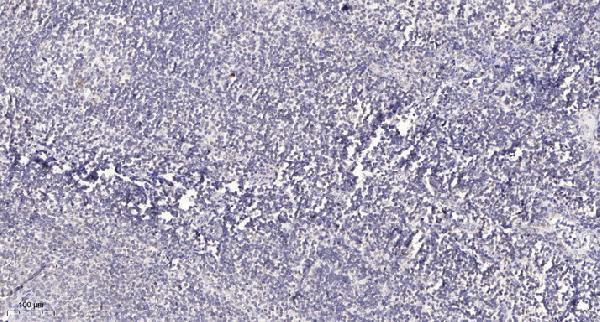
| Sample | Paraffin-embedded human tonsil tissue; fixative not specified (datasheet A06722-1) |
| Fixation | Image fixative and duration unreported (datasheet A06722-1); verify before use. |
| Sectioning | 4–5 µm sections on charged slides (standard) |
| Deparaffinisation | Xylene, graded ethanol series to water (standard) |
| Antigen retrieval | Heat retrieval: Tris-EDTA pH 9.0 (datasheet A06722-1); 20 min, 95–100 °C (standard) |
| Peroxidase block | 3% H2O2, 10 min, room temperature (standard) |
| Blocking | 10% normal serum of the secondary host, 30 min, room temperature (standard) |
| Primary antibody | Rabbit anti-BAG4, 1:100-1:300 (datasheet A06722-1) |
| Primary incubation | Overnight at 4 °C (standard) |
| Detection | HRP-polymer secondary, DAB chromogen 5–10 min (standard) |
| Counterstain | Hematoxylin, blue, dehydrate and mount (standard) |
| Expected result | BAG4-positive staining in glandular cells of adrenal gland (HPA tissue IHC: High). HPA tissue profile: General cytoplasmic expression. No signal in the no-primary control. |
BAG4 is expected mainly in the cytoplasm, consistent with its lack of a transmembrane segment (UniProt O95429: cytoplasm; no transmembrane segment). In paraffin sections, look for staining in glandular cells, kidney tubule cells and colon endothelial cells (HPA: High in these cells). HPA describes a general cytoplasmic pattern and rates its tissue IHC evidence Approved, with medium consistency between staining and RNA expression (HPA: tissue IHC).
| Cytoplasmic staining in glandular cells of appendix or duodenum (HPA: High in both). | This matches the reported compartment and cell pattern (HPA: general cytoplasmic expression; High in glandular cells). Compare with the same run's controls before scoring intensity (general IHC practice). |
| Predominantly nuclear staining with little cytoplasmic signal. | This conflicts with the reported main pattern (UniProt O95429: cytoplasm; HPA: general cytoplasmic expression). Treat it as suspect and check detection controls and staining conditions (general IHC practice). |
| Strong signal confined to an unexpected cell population while the documented population is unstained. | Consider cross-reactivity or endogenous detection activity (general IHC practice). BAG4 is broadly expressed, so an unlisted cell type alone does not prove a false positive (UniProt O95429: ubiquitous; HPA: low tissue specificity). |
| Diffuse color across multiple compartments and the surrounding section. | Poorly resolved staining prevents a reliable compartment call (general IHC practice). Background may reflect detection or blocking conditions; compare reagent controls before attributing it to BAG4 (general IHC practice). |
| No signal in kidney tubules or appendix glandular cells (HPA: High in both). | An absent signal in these reported high-staining populations weakens confidence in the run (HPA: tissue IHC). Check section quality, retrieval and detection controls before interpreting other areas as negative (general IHC practice). |
| Compartment and topology (UniProt O95429). | BAG4 is cytoplasmic and has no transmembrane segment; score the cytoplasm as the main expected compartment (UniProt O95429: subcellular location; topology). |
| Cell and tissue variation (HPA: tissue IHC). | Reported High staining includes adrenal and gallbladder glandular cells; glial cells in caudate and skeletal myocytes are Low (HPA: tissue IHC). Compare like cell populations when judging intensity (general IHC practice). |
| Ciliary exception (HPA: fallopian tube). | HPA also reports High staining at the cilia axoneme of fallopian tube ciliated cells (HPA: tissue IHC). Evaluate that finding separately from the general cytoplasmic profile (HPA: tissue IHC). |
| Isoforms and antibody recognition (UniProt O95429). | UniProt lists 2 isoforms (UniProt O95429: isoforms). The supplied evidence gives no antibody epitope or isoform coverage, so isoform-specific staining cannot be predicted. |
| Strength of tissue evidence (HPA: antibody validation). | HPA rates tissue staining Approved with medium RNA agreement and lists 2 IHC-Approved antibodies (HPA: tissue IHC; HPA018951, CAB013716). This does not establish an Enhanced validation rating. |
| Can IF/ICC provide a reference? (HPA: subcellular). | HPA supplies no ICC-IF images or main location assignment (HPA: subcellular). Use the UniProt cytoplasmic annotation as context, but do not infer a confirmed IF pattern (UniProt O95429: cytoplasm). |
| Situation | Likely cause | Next action |
|---|---|---|
| Kidney tubules lack stain although they are reported High (HPA: kidney). | The run may have lost sensitivity; this slide alone cannot identify why (general IHC practice). | Check section integrity, the run's positive control, retrieval conditions and detection reagents before calling the sample negative (general IHC practice). |
| Color is mainly nuclear rather than cytoplasmic (UniProt O95429: cytoplasm). | Non-specific signal or detection background is possible (general IHC practice). | Review a reagent control and assess whether cytoplasmic staining appears in a reported positive population (HPA: tissue IHC; general IHC practice). |
| Background obscures glandular cells reported High (HPA: appendix, duodenum). | Insufficiently controlled detection or blocking can obscure cellular boundaries (general IHC practice). | Compare reagent controls; adjust blocking, washing or detection conditions within the assay workflow (general IHC practice). |
| Colon endothelial cells stain strongly, but nearby cells differ (HPA: colon endothelial cells High). | Staining level can vary by cell population (HPA: tissue IHC). | Score endothelial cells separately and compare the observed compartment with the general cytoplasmic pattern (HPA: tissue IHC; general IHC practice). |
| Fallopian tube cilia are prominent while other compartments are faint (HPA: cilia axoneme High). | The ciliary finding is reported for this cell population (HPA: fallopian tube). | Record ciliary and cytoplasmic staining separately; avoid treating ciliary signal alone as proof that all other staining is specific (HPA: tissue IHC; general IHC practice). |
| Two sections give different staining intensity. | Biological variation or run conditions may contribute (general IHC practice); target-specific fixation sensitivity is unreported in the supplied evidence. | Compare matched cell populations and run controls under the same scoring criteria; record processing differences without assigning them a BAG4-specific effect (general IHC practice). |


Comprehensive Human Protein Atlas IHC scoring per tissue (reliability: Approved — Medium consistency between antibody staining and RNA expression data.). Rows are taken directly from the HPA tissue chart — click any row's HPA link to view the source.
| Tissue | Cell type | Level | Evidence | Source |
|---|---|---|---|---|
| Adrenal gland | Glandular cells | High | Protein (IHC) | HPA → |
| Appendix | Glandular cells | High | Protein (IHC) | HPA → |
| Colon | Endothelial cells | High | Protein (IHC) | HPA → |
| Duodenum | Glandular cells | High | Protein (IHC) | HPA → |
| Endometrium | Cells in endometrial stroma | Medium | Protein (IHC) | HPA → |
| Tissue | Cell type | Level | Evidence | Source |
|---|---|---|---|---|
| None in HPA: BAG4 is detected in all 45 scored tissues. Use a no-primary (secondary-only) and an isotype control instead. | ||||
Troubleshoot BAG4 staining in paraffin sections by checking retrieval, compartment, controls, and cell-specific scoring; IF/ICC requires separate validation.
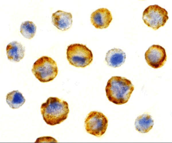
BAG4 antibodies have pictured IHC staining in paraffin-embedded human tonsil (A06722-1 image caption) and IF/ICC staining in HeLa cells (A06722 image captions); both list Human, Mouse and Rat reactivity (catalog).
A06722-1 lists IHC and IF applications and shows IHC staining in paraffin-embedded human tonsil (catalog; A06722-1 image caption). A06722 lists IF and ICC applications and shows staining in HeLa cells (catalog; A06722 image captions).
Which to pick: Choose A06722-1 for tissue IHC: its pictured protocol uses paraffin-embedded human tonsil, Tris-EDTA retrieval at pH 9.0 and primary antibody at 1:200 overnight at 4°C; the fixative is unreported (A06722-1 image caption). Choose A06722 for IF/ICC in cells because its images show HeLa staining; its application list does not include IHC (A06722 image captions; catalog). Both list Human, Mouse and Rat reactivity, but the pictured samples are human, so staining in Mouse or Rat samples needs validation (catalog; image captions).