This website uses cookies to ensure you get the best experience on our website.


- Table of Contents
Plan BAMBI IHC-P around cytoplasmic staining observed across several tissues (HPA tissue IHC) and its annotated membrane topology (UniProt). Lung macrophages show medium staining (HPA tissue IHC), but staining and RNA expression show low consistency (HPA tissue IHC).

Expected localisation, validated protocols, controls and antibodies — the at-a-glance facts below, then the full design guide.
| Expected localisation | Cytoplasmic staining observed; membrane location annotated (HPA tissue IHC; UniProt) | |
| Staining pattern | Cytoplasmic staining across several tissue types (HPA tissue IHC) | |
| Antigen retrieval | Citrate pH 6.0 HIER, 95–98 °C, 20 min (rule: cytoplasmic / membrane antigen) | |
| Positive control | Adipose tissue+4 more · see all | |
| Negative control | Cervix+4 more · see all |
| Fixation | Matched tissue-IHC evidence does not establish the fixation claim. Validate the specimen-specific method before use. (selected-SKU IHC image A07983) | |
| Caveat | Staining and RNA expression show low consistency (HPA tissue IHC) | |
| Regulation | Expression regulation is not annotated (UniProt) | |
| Isoform / epitope | No isoforms annotated; epitope side affects interpretation (UniProt) |
Compare the catalog antibody’s IHC-P protocol (datasheet) with four published BAMBI IHC protocols (PMC11504530; PMC5097819; PMC12194233; PMC2890648).
| Sample | Tissue sections; selected-image fixative not specified (standard IHC workflow) |
| Fixation | Image fixative and duration unreported (datasheet A07983); verify before use. |
| Sectioning | 4–5 µm sections on charged slides (standard) |
| Deparaffinisation | Xylene, graded ethanol series to water (standard) |
| Antigen retrieval | Heat-induced epitope retrieval in citrate buffer, pH 6.0, 20 min at 95–98 °C (standard rule: cytoplasmic / membrane antigen) |
| Peroxidase block | 3% H2O2, 10 min, room temperature (standard) |
| Blocking | 10% normal serum of the secondary host, 30 min, room temperature (standard) |
| Primary antibody | Rabbit anti-BAMBI, 5 μg/mL (datasheet A07983) |
| Primary incubation | Overnight at 4 °C (standard) |
| Detection | HRP-polymer secondary, DAB chromogen 5–10 min (standard) |
| Counterstain | Hematoxylin, blue, dehydrate and mount (standard) |
| Expected result | BAMBI-positive staining in adipocytes of adipose tissue (HPA tissue IHC: Medium). HPA tissue profile: Cytoplasmic expression in several different tissue types. No signal in the no-primary control. |
BAMBI is a transmembrane protein with an extracellular region at residues 21–152 and a cytoplasmic region at 174–260 (UniProt Q13145 topology). In tissue IHC, expect cytoplasmic staining in selected cells, including lung macrophages and bronchial respiratory epithelial cells (HPA tissue IHC: Medium). HPA rates its tissue staining Approved, while reporting low consistency with RNA data and pending external verification (HPA tissue IHC reliability).
| Selective, moderate cytoplasmic staining in lung macrophages or bronchial respiratory epithelial cells (HPA tissue IHC: Medium). | This matches the reported tissue IHC pattern (HPA tissue IHC: cytoplasmic expression). Assess the named cell population, since a positive area does not imply that every cell should stain. The membrane topology (UniProt Q13145) does not require a crisp membrane rim in chromogenic sections. |
| Predominantly nuclear staining, with little cytoplasmic signal, in a tissue section. | This differs from the reported tissue IHC profile (HPA tissue IHC: cytoplasmic expression). Check morphology and controls before calling it BAMBI. Approved nucleolar fibrillar center localisation comes from ICC-IF (HPA subcellular), so it cannot by itself validate nuclear chromogen in IHC. |
| Prominent staining in colon glandular cells or esophageal squamous epithelial cells (HPA tissue IHC: Not detected). | That conflicts with the reported cell-specific observations and raises possible cross-reactivity or endogenous detection activity (HPA tissue IHC). It is a reason to investigate, not proof of an artefact: the HPA IHC reliability note reports low consistency with RNA data and pending external verification. |
| Diffuse color across unrelated cells or tissue spaces, with poor cell boundaries. | A widespread haze does not resemble the selective cytoplasmic pattern (HPA tissue IHC). Consider nonspecific detection or background from the chromogenic workflow (general IHC practice). A no-primary control helps distinguish detection background from antibody-dependent staining. |
| No staining in lung macrophages or bronchial respiratory epithelial cells (HPA tissue IHC: Medium). | First check that the expected cells are present and identifiable. Review the IHC-validated antibody and detection controls, then repeat with appropriate assay controls if needed (general IHC practice). An HPA Medium observation is a useful comparison, not a guarantee for every specimen. |
| Compartment and epitope orientation | BAMBI has extracellular residues 21–152, a transmembrane segment at 153–173, and cytoplasmic residues 174–260 (UniProt Q13145 topology). Antibody epitope location is not supplied, so topology alone cannot predict the staining outline or a retrieval requirement. |
| Protein processing | UniProt lists a signal peptide at residues 1–20 and the mature chain at 21–260 (UniProt Q13145 processing). No propeptide or shedding event is annotated in the supplied record; do not use either to explain unexpected tissue staining. |
| Glycosylation | One glycosylation site is listed at residue 87 (UniProt Q13145 glycosylation). The supplied sources do not show that it changes recognition by the IHC-validated antibody, so use it as structural context rather than a cause of weak or absent staining. |
| Tissue and antibody evidence | Several named populations show Medium staining, while others are Not detected (HPA tissue IHC). The tissue IHC profile is Approved but has low RNA consistency and awaits external verification (HPA tissue IHC reliability). HPA010866 is IHC Approved; that status does not establish every new antibody or specimen as equivalent (HPA antibodies). |
| IF/ICC question: should puncta or nucleoli be expected in IHC? | HPA reports mainly vesicles, plus nucleoli fibrillar center and lipid droplets, in ICC-IF (HPA subcellular: approved locations). Those observations can guide review of an IF image, but the reported tissue IHC pattern is cytoplasmic (HPA tissue IHC); they do not establish an IHC puncta requirement. |
| Situation | Likely cause | Next action |
|---|---|---|
| The presumed positive section is blank. | The named HPA-positive cell population may be absent, or an assay step may have failed; neither conclusion follows from a blank slide alone (HPA tissue IHC: Medium in selected cells). | Locate the relevant cells on the counterstained section. Check a suitable positive assay control and the IHC-validated antibody's specified conditions, including retrieval and dilution if provided (general IHC practice). |
| Color covers most of the section. | Broad staining is unlike HPA's cytoplasmic signal in selected tissue populations (HPA tissue IHC); nonspecific antibody or detection background is possible (general IHC practice). | Compare a no-primary control and inspect whether color follows cell outlines. Review blocking, wash steps, antibody concentration, and chromogen development using the assay instructions (general IHC practice). |
| Strong signal appears in colon glandular cells. | HPA reports these cells as Not detected, so cross-reactivity or endogenous detection activity is possible; that comparison does not prove either cause (HPA tissue IHC: colon). | Check a no-primary control and compare cell morphology and the expected positive population. Seek independent antibody or orthogonal evidence before assigning that staining to BAMBI (general IHC practice). |
| Staining is mainly nuclear in tissue IHC. | Predominantly nuclear chromogen departs from the reported cytoplasmic tissue pattern (HPA tissue IHC). An ICC-IF nucleolar location does not establish this section's antibody specificity (HPA subcellular). | Check localisation against the counterstain and assay controls. Record the discrepancy and avoid scoring nuclear color as confirmed BAMBI solely from the ICC-IF finding. |
| A sharp membrane rim is absent despite cytoplasmic color. | Membrane topology (UniProt Q13145) and the cytoplasmic tissue IHC profile (HPA tissue IHC) describe different kinds of evidence; antibody epitope location is unavailable. | Judge the result against the reported cell-specific cytoplasmic pattern and controls. Do not reject an otherwise plausible IHC signal solely because it lacks a continuous membrane outline. |
| An IF image shows vesicular puncta while the IHC section looks broadly cytoplasmic. | The methods have separate observations: vesicles are the approved main ICC-IF location (HPA subcellular), and tissue IHC is described as cytoplasmic (HPA tissue IHC). | Interpret each image against its own reported pattern and controls. Do not require an ICC-IF vesicle pattern in chromogenic IHC or use the IF image alone to validate an IHC result. |


Comprehensive Human Protein Atlas IHC scoring per tissue (reliability: Approved — Low consistency between antibody staining and RNA expression data. Pending external verification.). Rows are taken directly from the HPA tissue chart — click any row's HPA link to view the source.
| Tissue | Cell type | Level | Evidence | Source |
|---|---|---|---|---|
| Adipose tissue | Adipocytes | Medium | Protein (IHC) | HPA → |
| Bone marrow | Hematopoietic cells | Medium | Protein (IHC) | HPA → |
| Bronchus | Respiratory epithelial cells | Medium | Protein (IHC) | HPA → |
| Cerebral cortex | Neuropil | Medium | Protein (IHC) | HPA → |
| Fallopian tube | Glandular cells | Medium | Protein (IHC) | HPA → |
| Tissue | Cell type | Level | Evidence | Source |
|---|---|---|---|---|
| Cervix | Glandular cells | Not detected | Protein (IHC) | HPA → |
| Colon | Glandular cells | Not detected | Protein (IHC) | HPA → |
| Duodenum | Glandular cells | Not detected | Protein (IHC) | HPA → |
| Esophagus | Squamous epithelial cells | Not detected | Protein (IHC) | HPA → |
| Oral mucosa | Squamous epithelial cells | Not detected | Protein (IHC) | HPA → |
Troubleshoot BAMBI staining in paraffin section IHC by checking retrieval, cell identity, and compartment before comparing staining intensity across specimens.
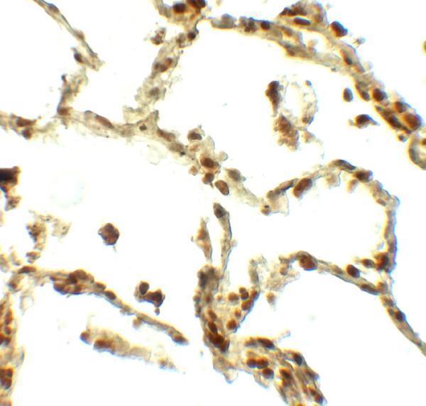
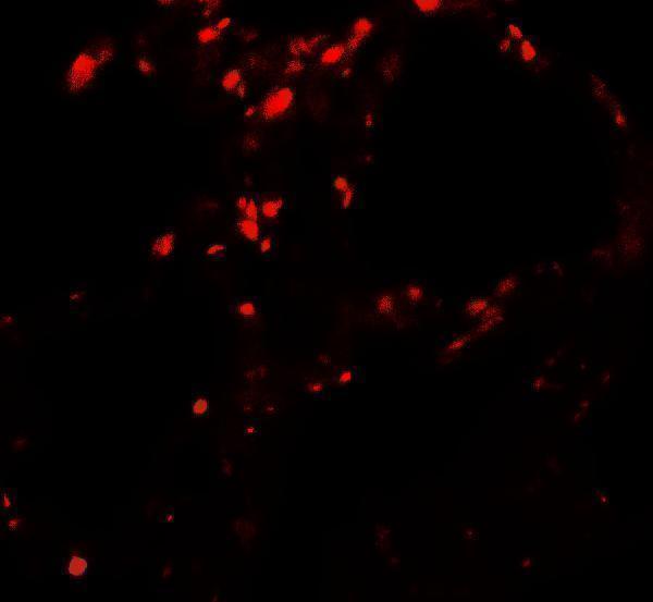
A07983 has IHC and IF images from human lung tissue (catalog: image captions), with listed reactivity to human, mouse and rat (catalog: reactivity).
A07983 is listed for IHC-P and IF, with an IHC image of human lung tissue at 5 μg/mL (catalog: applications; IHC image caption). Its IF image also shows human lung tissue at 20 μg/mL (catalog: IF image caption).
Which to pick: For tissue IHC, choose A07983 because IHC-P is listed and its IHC image shows human lung tissue; the caption does not report section processing or fixative (catalog: applications; IHC image caption). For IF, choose A07983 based on its human lung IF image; ICC is not listed as an application (catalog: IF image caption; applications). For work across species, A07983 lists human, mouse and rat reactivity, but the supplied IHC and IF images show human tissue only; it is rabbit hosted, with no clone reported (catalog: reactivity; image captions; host; clone).