CA12 / Carbonic anhydrase 12 · IHC design guide

Design Immunohistochemistry for CA12

Plan CA12 chromogenic IHC in paraffin sections using its membranous tissue pattern and cell-specific controls (HPA tissue IHC). The guide covers fixation consistency, antibody selection and interpretation of staining in intestinal cells and renal tubules (HPA tissue IHC).

Evidence assembled Oct 2026 · For research use; verify linked source records and product datasheet before use
Immunohistochemistry protocol sheet for CA12 (IHC for CA12): expected localisation Membranous staining in tissue sections (HPA tissue IHC), antibody A30588, validated IHC image, and IHC protocol steps
Printable CA12 IHC protocol sheet — expected localisation Membranous staining in tissue sections (HPA tissue IHC), antibody A30588, controls and protocol steps. Open the full CA12 IHC guide →

CA12 Immunohistochemistry Experimental Design Guide

Expected localisation, validated protocols, controls and antibodies — the at-a-glance facts below, then the full design guide.

Must know before staining
Expected localisation Membranous staining in tissue sections (HPA tissue IHC)
Staining pattern Membranous in intestinal cells and renal tubules (HPA tissue IHC)
Antigen retrieval Tris-EDTA pH 9.0 HIER, heat-mediated (datasheet A30588)
Positive control ⓘ Appendix+4 more · see all
Negative control ⓘ Adipose tissue+4 more · see all
Important caveats
Reasons your staining may differ from the expected pattern.
Fixation Matched tissue-IHC evidence does not establish the fixation claim. Validate the specimen-specific method before use. (selected-SKU IHC image A30588)
Caveat Intestinal endocrine staining varies by site (HPA tissue IHC)
Regulation Higher in renal cell cancers than normal kidney (UniProt)
Isoform / epitope 2 isoforms; map extracellular vs cytoplasmic epitope (UniProt)
Section 1

Recommended CA12 IHC & IF Protocols

Compare the catalog antibody’s IHC-P protocol (datasheet A30588) with published CA12 IHC methods for cervical cancer (PMC2990746) and follicular thyroid carcinoma (PMC12775590).

Recommended immunohistochemistry (IHC-P) protocol parameters
SampleParaffin-embedded human colon cancer tissue; fixative not specified (datasheet A30588)
FixationImage fixative and duration unreported (datasheet A30588); verify before use.
Sectioning4–5 µm sections on charged slides (standard)
DeparaffinisationXylene, graded ethanol series to water (standard)
Antigen retrievalHeat retrieval: Tris-EDTA pH 9.0 (datasheet A30588); 20 min, 95–100 °C (standard)
Peroxidase block3% H2O2, 10 min, room temperature (standard)
Blocking10% normal serum of the secondary host, 30 min, room temperature (standard)
Primary antibodyRabbit anti-CA12, 1:500 - 1:2000 (datasheet A30588)
Primary incubationOvernight at 4 °C (standard)
DetectionHRP-polymer secondary, DAB chromogen 5–10 min (standard)
CounterstainHematoxylin, blue, dehydrate and mount (standard)
Expected resultCA12-positive staining in endocrine cells of appendix (HPA tissue IHC: High). HPA tissue profile: Membranous expression in several cell types, most abundant in intestinal tract and renal tubules. No signal in the no-primary control.
💡Decision noteStart with heat-mediated Tris-EDTA retrieval at pH 9.0 (datasheet A30588). The cervical cancer study used a retrieval solution heated to 98°C for 15 minutes (PMC2990746).
Section 2

What Is the Expected CA12 Staining Pattern?

CA12 is a cell membrane protein with an extracellular domain spanning residues 25–301 and a transmembrane segment at 302–322 (UniProt O43570 topology). In paraffin sections, expect membranous staining, particularly in renal distal tubules and intestinal endocrine cells (HPA tissue IHC: High). HPA rates its tissue staining reliability Enhanced, citing consistency between antibody staining and RNA expression (HPA tissue IHC: Enhanced).

What am I looking at on my slide?
Distinct cell borders stain in renal distal tubules or colon endocrine cells, with limited staining between cells.This matches CA12 membrane localisation (UniProt O43570 topology) and the reported High staining in those cell populations (HPA tissue IHC: Kidney distal tubules; Colon endocrine cells). Judge the compartment and the identified cell type together; a strong signal alone does not establish a correct result (general IHC interpretation).
Nuclei or cytoplasm stain strongly while cell borders remain indistinct.Dominant nuclear or diffuse cytoplasmic staining conflicts with the expected membrane localisation (UniProt O43570 subcellular location; HPA tissue IHC: membranous profile). Treat it as a possible staining artefact or off-target signal, then compare a known-positive section and a no-primary control (general IHC practice).
Cells listed as Not detected stain as strongly as the intended positive cells.For example, HPA reports Not detected in adipocytes and adrenal glandular cells (HPA tissue IHC). Unexpected staining may reflect cross-reactivity or endogenous detection activity (general IHC practice). Confirm cell identity and inspect a no-primary control before attributing the signal to CA12; a negative HPA entry is specific to its sampled cell population.
Color spreads across tissue, including spaces outside cells, without crisp membrane outlines.This appearance does not reproduce the reported membranous pattern (HPA tissue IHC: profile). Diffuse background can obscure a genuine border signal (general IHC practice). Inspect the no-primary control, background around tissue, and counterstain; do not score broad haze as CA12-positive membrane staining.
A kidney section has no detectable staining in morphologically identified distal tubules.HPA reports High staining in kidney distal tubules (HPA tissue IHC: Kidney), making them a useful positive reference. First check that the sampled section contains those cells; then review the antibody's documented IHC conditions and the detection run (general IHC practice). Absence of signal in a reference section leaves a negative test section uninterpretable.
💡Expected CA12 appearanceCall a section positive when cell borders show clear membranous staining in the appropriate cells, such as High-staining renal distal tubules (UniProt O43570 topology; HPA tissue IHC: Kidney); diffuse haze or dominant nuclear color is suspect (general IHC interpretation).
How each factor affects the staining
Membrane topologyCA12 has an extracellular region at residues 25–301 and a cytoplasmic tail at 323–354 (UniProt O43570 topology). Expect a membrane-centered readout; the record does not identify the antibody epitope, so it cannot establish whether a particular stain depends on access to either side.
Cell-specific tissue patternHPA reports High staining in appendix, colon and rectum endocrine cells, kidney distal tubules, and pancreas exocrine glandular cells (HPA tissue IHC). Select reference cells by both tissue and cell identity: HPA also reports duodenal endocrine cells as Not detected (HPA tissue IHC).
Evidence and antibody validationThe tissue profile has Enhanced reliability (HPA tissue IHC). Among listed antibodies, HPA073203, CAB025181, CAB062549 and CAB068179 have Enhanced IHC status; HPA008773 is Supported (HPA antibodies). These ratings support the observed pattern but do not specify conditions for an unlisted antibody.
IF/ICC expectation?A membrane pattern is plausible from CA12 topology and the HPA subcellular summary (UniProt O43570 topology; HPA subcellular: Membrane). HPA supplies no ICC/IF image-bearing cell line or ICC validation for the listed antibodies (HPA subcellular; HPA antibodies), so this IHC evidence cannot establish an IF/ICC result.
Why is my staining missing, weak or wrong?
SituationLikely causeNext action
No staining appears in the test section or in kidney distal tubules.The positive reference has failed despite reported High distal-tubule staining (HPA tissue IHC: Kidney); the specific failed step is undetermined.Confirm distal tubules are present, then check the documented antibody dilution, antigen retrieval and detection steps for the IHC run (general IHC practice). Interpret test-section negatives only after a positive reference works.
A strong signal sits mainly in nuclei or fills cytoplasm.That compartment disagrees with CA12 membrane localisation (UniProt O43570 subcellular location; HPA tissue IHC: membranous profile).Compare a known-positive section and no-primary control; verify cell-border staining before scoring the signal as CA12 (general IHC practice).
Unexpected cells stain, including a population HPA lists as Not detected.Cross-reactivity or endogenous detection activity is possible (general IHC practice); HPA's negative designation applies to the named cell population (HPA tissue IHC).Verify morphology, compare the HPA cell-specific entry, and check a no-primary control. If that control stains, investigate detection background (general IHC practice).
The whole section looks hazy and membrane outlines are hard to resolve.Background staining may conceal the membranous pattern reported for CA12 (HPA tissue IHC: profile; general IHC practice).Inspect the no-primary control and review blocking, washes, antibody dilution and chromogen development using the documented IHC procedure (general IHC practice).
The expected positive population appears weak beside other stained cells.HPA levels refer to specified cell populations; they do not guarantee identical intensity across tissues or runs (HPA tissue IHC).Identify the cells before comparing intensity. Use the same validated run and scoring criteria for the positive reference and test section (general IHC practice).
A proposed IF/ICC result differs from the paraffin-section pattern.HPA gives a Membrane subcellular summary but no ICC/IF images or ICC validation for the listed antibodies (HPA subcellular; HPA antibodies).Treat the IHC pattern as a localisation expectation, not IF/ICC validation (UniProt O43570 topology; HPA tissue IHC). Consult the separate IF/ICC guide for assay-specific design.

Sample controls for CA12 IHC & IF

🧪Run kidney first and require strong staining in distal tubule cells (HPA: Kidney distal tubules High; UniProt O43570: cell membrane). Use adipose tissue adipocytes as the negative tissue (HPA: Adipocytes Not detected); on the kidney slide, assess neighboring structures for absence of the distal-tubule staining pattern without assuming they are CA12-free (HPA: Kidney distal tubules High).
Positive control tissue: Appendix (Endocrine cells, HPA High)
Negative control tissue: Adipose tissue (HPA Not detected)
ICC-IF cell lines (HPA subcellular resource): HPA carries no ICC-IF cell line for CA12; derive a cell-line control from the positive tissue's cell type (Endocrine cells) and confirm it by RNA or western blot first.
Technical controls: Include a no-primary, secondary-only control; an irrelevant isotype control matched to the primary antibody’s host species and, for a monoclonal antibody, subclass; and a CA12 knockout biological negative (standard IHC practice). For chromogenic kidney IHC, block endogenous peroxidase and check for endogenous biotin if using avidin–biotin detection (standard IHC practice).
⚠️Feasibility: A target-specific fixation window or fixation effect is unreported in the supplied evidence, and the selected A30588 colon cancer paraffin-section caption does not report a fixative (A30588 tissue-IHC caption). That caption used Tris-EDTA pH 9 retrieval and a 1:200 primary dilution overnight at 4 °C, but it does not establish that retrieval is required for kidney sections (A30588 tissue-IHC caption). The supplied evidence does not establish that frozen sections or IF are easier; kidney chromogenic staining should be checked for endogenous biotin if avidin–biotin detection is used (standard IHC practice).

HPA tissue IHC evidence for CA12

Comprehensive Human Protein Atlas IHC scoring per tissue (reliability: Enhanced — High consistency between antibody staining and RNA expression data.). Rows are taken directly from the HPA tissue chart — click any row's HPA link to view the source.

Positive expression · recommended positive controls

TissueCell typeLevelEvidenceSource
Appendix Endocrine cells High Protein (IHC) HPA →
Colon Endocrine cells High Protein (IHC) HPA →
Endometrium Ciliated epithelial cells High Protein (IHC) HPA →
Kidney Distal tubules High Protein (IHC) HPA →
Pancreas Exocrine glandular cells High Protein (IHC) HPA →

Undetected expression · recommended negative controls

TissueCell typeLevelEvidenceSource
Adipose tissue Adipocytes Not detected Protein (IHC) HPA →
Adrenal gland Glandular cells Not detected Protein (IHC) HPA →
Bone marrow Hematopoietic cells Not detected Protein (IHC) HPA →
Bronchus Respiratory epithelial cells Not detected Protein (IHC) HPA →
Caudate Glial cells Not detected Protein (IHC) HPA →
Section 3

Advanced CA12 IHC Tips

Troubleshoot CA12 staining in paraffin sections by checking retrieval, membrane localisation, controls and compartment-specific scoring (datasheet A30588; UniProt O43570 topology).

What retrieval should I try first when CA12 staining is weak?
Start with heat-mediated Tris-EDTA at pH 9.0 for CA12 paraffin-section IHC (datasheet A30588). The selected colon-cancer image used this retrieval, followed by primary antibody at 1:200 overnight at 4°C (caption A30588). If staining remains weak, check heating consistency, cooling, section adherence and antibody incubation before testing an alternative retrieval condition (standard IHC practice). Compare any retrieval change on adjacent sections with a positive control, such as renal distal tubules, where CA12 staining is reported as high (HPA: Kidney, distal tubules). Judge improvement by sharper membrane staining with limited background, rather than by total chromogen intensity alone (UniProt O43570 topology; standard IHC practice).
Can I infer the best fixation conditions from the CA12 tissue image?
No CA12-specific fixation comparison is supplied, so sensitivity to fixative choice or fixation duration is unknown (caption A30588: fixative unreported). The selected image identifies paraffin-embedded human colon cancer and pH 9.0 retrieval, but gives no fixative or fixation time (caption A30588). Its 4°C overnight step describes primary-antibody incubation, not tissue fixation (caption A30588). Record the actual fixative and processing history for each specimen, then compare sections processed alike when investigating weak staining (standard IHC practice). Use a consistently processed positive control to distinguish a processing problem from a specimen-specific result; do not treat the published image as fixation validation (standard IHC practice; caption A30588).
Which staining compartment should count as convincing CA12 signal?
Prioritise a crisp cell-membrane pattern: CA12 has an extracellular region at residues 25–301 and one transmembrane segment at 302–322 (UniProt O43570 topology). The tissue atlas reports membranous expression, especially in intestinal cell types and renal tubules (HPA: tissue IHC profile). Diffuse nuclear or uniformly cytoplasmic chromogen should trigger review of background and morphology before scoring (UniProt O43570 localisation; standard IHC practice). In kidney, examine distal tubules as a high-staining reference; in colon, the atlas specifically lists endocrine cells as high (HPA: Kidney and Colon tissue IHC). Compare cellular boundaries and the negative-control section at the same magnification and detection settings (standard IHC practice).
Could isoforms or glycosylation explain different CA12 staining patterns?
CA12 has 2 listed isoforms, but the supplied material does not identify the catalog antibody’s epitope or establish isoform selectivity (UniProt O43570 isoforms; supplied caption A30588). Its extracellular region spans residues 25–301, with listed glycosylation sites at 28, 80 and 162 (UniProt O43570 topology and glycosylation). Those features make epitope accessibility a question to test, not proof that processing caused a particular staining difference (UniProt O43570 topology; standard IHC interpretation). When sections disagree, keep retrieval at Tris-EDTA pH 9.0 while checking whether the antibody documentation specifies an immunogen or epitope (datasheet A30588; standard IHC practice). Interpret isoform-specific claims only after epitope-level validation (standard IHC practice).
How should I investigate CA12 by multiplex IF alongside tissue IHC?
Treat IF as a separate application: the supplied catalog image documents paraffin-section IHC, while the HPA subcellular record lists no ICC/IF images (caption A30588; HPA: subcellular record). Pair CA12 with a validated marker for the expected cell population, such as a distal-tubule marker in kidney, and inspect whether membrane outlines occur in the marked cells (HPA: Kidney distal tubules; UniProt O43570 topology; standard IF practice). Choose a far-red fluorophore when tissue autofluorescence obscures shorter-wavelength channels, and include single-colour controls for multiplex interpretation (standard IF practice). For an extracellular epitope in residues 25–301, assess staining without permeabilisation first; a cytoplasmic-tail epitope at 323–354 requires membrane access (UniProt O43570 topology; standard IF practice). Select permeabilisation only after confirming which side the antibody recognises (standard IF practice).
How can I separate CA12 staining from chromogenic background?
First inspect a section lacking primary antibody to reveal signal from the detection system and endogenous activity (standard IHC practice). Include a peroxidase block before DAB development and check whether precipitate persists in the no-primary control; these are general chromogenic IHC steps (standard IHC practice). The selected image reports secondary antibody at 1:200 for 45 minutes at room temperature, which provides a documented starting condition for that image (caption A30588). Reassess blocking, washes and chromogen development if background masks the expected membrane outline (UniProt O43570 localisation; standard IHC practice). Compare suspect deposits with high-staining distal tubules and a no-primary section before assigning them to CA12 (HPA: Kidney distal tubules; standard IHC practice).
What should I score when comparing CA12 across IHC sections? ⚠ ANSWER MARKED FOR VERIFICATION
Score the proportion of cells with interpretable membrane staining within a defined cell population, rather than pooling every chromogen-positive structure (UniProt O43570 localisation; standard IHC practice). An H-score combines percentage at intensity levels 0–3 into a 0–300 score; alternatively, report percentage of positive cells with the threshold stated (standard IHC scoring practice). Normalise counts to the number of eligible cells or annotated tissue area, and keep the compartment and cell-type rules constant across sections (standard IHC practice). For kidney, specify whether distal tubules are the denominator because they show high CA12 staining in the atlas (HPA: Kidney distal tubules). Record retrieval as Tris-EDTA pH 9.0 when comparing results against the selected catalog image (caption A30588).
When is a CA12-positive section convincing rather than artefactual?
A convincing result shows membrane staining in plausible cells, supported by a processed positive control and a clean no-primary control (UniProt O43570 localisation; HPA: tissue IHC profile; standard IHC practice). Renal distal tubules and colonic endocrine cells are reported as high, whereas adipocytes are reported as not detected in the atlas (HPA: Kidney, Colon and Adipose tissue IHC). Treat strong nuclear signal, staining confined to tissue edges or necrotic areas, and deposits reproduced without primary antibody as reasons to investigate artefact (UniProt O43570 localisation; standard IHC practice). Check endogenous peroxidase blocking and DAB development before assigning diffuse brown signal to CA12 (standard chromogenic IHC practice). Compare suspicious staining with the documented pH 9.0 retrieval condition and matched controls (caption A30588; standard IHC practice).
Boster reagents

Best CA12 / Carbonic anhydrase 12 IHC Antibodies

CA12 antibodies cover human and rat IHC and IF/ICC; A30588 also lists mouse reactivity (catalog: A04063-1, A30588). A30588 has a paraffin-section IHC figure (A30588 image caption); no IF figure is supplied (catalog image fields).

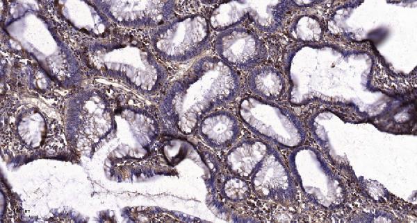
Real IHC data Immunohistochemical analysis of paraffin-embedded human colon cancer. 1, Tris-EDTA,pH9.0 was used for antigen retrieval. 2 Antibody was diluted at 1:200(4° overnight.3,Secondary antibody was diluted at 1:200(room temperature, 45min).
Anti-CA12/Ca Xii Antibody
Cat # A30588

A30588 will render with an IHC figure of human colon cancer in a paraffin section; its listed applications include IHC and IF/ICC, with human, mouse and rat reactivity (A30588 image caption; catalog: A30588). A04063-1 lists IHC and IF/ICC for human and rat, but has no supplied IHC or IF image (catalog: A04063-1).

Which to pick: For tissue IHC, pick A30588 when an illustrated paraffin-section example matters: its own caption specifies Tris-EDTA retrieval at pH 9 and primary antibody at 1:200 overnight at 4°C; the fixative is unreported (A30588 image caption). For IF/ICC, both SKUs list the application, but neither has a supplied IF image (catalog: A30588, A04063-1). For work spanning human, mouse and rat, pick A30588: it lists all three species and is described as polyclonal; A04063-1 lists human and rat, with clonality unreported (catalog: A30588 dilution_raw and reactivity; catalog: A04063-1 reactivity and clone).

Each figure is that product's own IHC / IF validation image from its datasheet.

References

  1. UniProt Consortium. UniProt entry O43570 (CAH12_HUMAN, Carbonic anhydrase 12).
  2. Human Protein Atlas. CA12 tissue IHC expression (reliability: Enhanced).
  3. Human Protein Atlas. CA12 subcellular location (ICC-IF): Membrane.
  4. Human Protein Atlas. CA12 antibody validation summary (5 antibodies).
  5. The Role of the miR-548au-3p/CA12 Axis in Tracheal Chondrogenesis in Congenital Pulmonary Airway Malformations. Oxidative medicine and cellular longevity 2023 — PMC9957630.
  6. Carbonic anhydrase XII expression is associated with histologic grade of cervical cancer and superior radiotherapy outcome. Radiation oncology (London, England) 2010 — PMC2990746.
  7. Carbonic Anhydrase 12 as a Novel Prognostic Biomarker and Therapeutic Target for High-Risk Follicular Thyroid Carcinoma. Cancer science 2026 — PMC12775590.
  8. Carbonic Anhydrase 12 Protects Endplate Cartilage From Degeneration Regulated by IGF-1/PI3K/CREB Signaling Pathway. Frontiers in cell and developmental biology 2020 — PMC7596245.
  9. PubMed PMID:9636197 — UniProt-cited evidence.
  10. PubMed PMID:9770531 — UniProt-cited evidence.
  11. PubMed PMID:14702039 — UniProt-cited evidence.