This website uses cookies to ensure you get the best experience on our website.


- Table of Contents
Plan CELF1 paraffin IHC around mainly nuclear tissue staining (HPA tissue IHC). This guide covers fixation consistency, antibody selection, and compartment-aware scoring, with six isoforms to consider when checking epitope coverage (UniProt).

Expected localisation, validated protocols, controls and antibodies — the at-a-glance facts below, then the full design guide.
| Expected localisation | Mainly nuclear in tissue IHC (HPA tissue IHC) | |
| Staining pattern | Mainly nuclear staining across tissues (HPA tissue IHC) | |
| Antigen retrieval | Tris-EDTA pH 9.0 HIER, heat-mediated (datasheet A30541) | |
| Positive control | Adrenal gland+4 more · see all | |
| Negative control | None in HPA (detected in all 45 tissues); use no-primary + isotype controls |
| Fixation | Keep fixation consistent across paraffin samples (standard IHC practice; not target-specific) | |
| Caveat | Antibody staining and RNA show medium consistency (HPA tissue IHC) | |
| Regulation | Low tissue specificity (HPA tissue RNA) | |
| Isoform / epitope | Six isoforms; check whether the epitope is shared (UniProt) |
The catalog antibody protocol uses Tris-EDTA pH 9.0 retrieval (datasheet A30541). The published CELF1 IHC protocols below use citrate retrieval (PMC3594286; PMC12646540).
| Sample | Paraffin-embedded human tonsil tissue; fixative not specified (datasheet A30541) |
| Fixation | Image fixative and duration unreported (datasheet A30541); verify before use. |
| Sectioning | 4–5 µm sections on charged slides (standard) |
| Deparaffinisation | Xylene, graded ethanol series to water (standard) |
| Antigen retrieval | Heat retrieval: Tris-EDTA pH 9.0 (datasheet A30541); 20 min, 95–100 °C (standard) |
| Peroxidase block | 3% H2O2, 10 min, room temperature (standard) |
| Blocking | 10% normal serum of the secondary host, 30 min, room temperature (standard) |
| Primary antibody | Rabbit anti-CELF1, 1:100 - 1:300 (datasheet A30541) |
| Primary incubation | Overnight at 4 °C (standard) |
| Detection | HRP-polymer secondary, DAB chromogen 5–10 min (standard) |
| Counterstain | Hematoxylin, blue, dehydrate and mount (standard) |
| Expected result | CELF1-positive staining in glandular cells of adrenal gland (HPA tissue IHC: High). HPA tissue profile: Mainly nuclear expression in all tissues. No signal in the no-primary control. |
In paraffin-section IHC, expect CELF1 mainly in nuclei across tissues (HPA tissue IHC: mainly nuclear expression in all tissues). Strong examples include glandular, hematopoietic, respiratory epithelial, glial, granular-layer and endothelial cells in the specified tissues (HPA tissue IHC: High). Cytoplasmic signal can occur (UniProt Q92879: nucleus and cytoplasm). CELF1 has no transmembrane segment (UniProt Q92879 topology). HPA rates its tissue IHC pattern Supported, with medium consistency between staining and RNA expression (HPA tissue IHC).
| Clearly stained nuclei in adrenal gland glandular cells or bone marrow hematopoietic cells (HPA tissue IHC: High). | This fits the reported cell types and mainly nuclear pattern (HPA tissue IHC). Judge the chromogenic signal against tissue structure and a counterstain so that stained nuclei can be assigned to the intended cells (general IHC practice). |
| Signal appears only at cell borders or in extracellular material, with no convincing nuclear staining. | A border-only pattern conflicts with the reported nuclear distribution and lack of a transmembrane segment (HPA tissue IHC; UniProt Q92879 topology). Review morphology and controls before calling it CELF1. Cytoplasmic staining alone is less decisive because CELF1 is also reported there (UniProt Q92879). |
| The strongest staining lies in an unexpected cell population while the listed High population is faint. | Check cell identification first: CELF1 is ubiquitous, so staining outside a listed High population is not automatically false (UniProt Q92879: ubiquitous; HPA tissue IHC: High examples). A discordant pattern may indicate cross-reactivity or endogenous chromogen activity; compare controls and another IHC-supported antibody (HPA antibodies; general IHC practice). |
| Brown signal spreads evenly over nuclei, cytoplasm, stroma and empty slide areas. | That distribution is difficult to reconcile with mainly nuclear cellular staining (HPA tissue IHC). Diffuse signal can arise from nonspecific antibody binding, incomplete blocking, excess detection reagent or endogenous detection activity (general IHC practice). Evaluate it against a no-primary control before scoring cells. |
| No convincing nuclear signal appears in a well-preserved reported High population (HPA tissue IHC: High). | An absent result is inconclusive until the run controls work. Confirm the expected cell population is present, then review the antibody’s IHC validation and the assay’s retrieval, detection and control performance (HPA antibodies; general IHC practice). HPA supplies no negative tissue reference for CELF1 (HPA tissue IHC: negative list empty). |
| Choice of reference tissue and cells | Use a reported High population, such as breast glandular cells, bronchial respiratory epithelial cells or caudate glial cells, as an expected-pattern reference (HPA tissue IHC: High). These are examples, not a ranked sensitivity panel: HPA reports no low or negative tissue entries in this payload (HPA tissue IHC). |
| Strength of tissue-pattern evidence | The overall tissue IHC reliability is Supported with medium staining–RNA consistency (HPA tissue IHC). Treat a match as supportive, not proof of antibody specificity in an individual section. The reported RNA specificity is low across tissues, and UniProt describes CELF1 as ubiquitous (HPA tissue IHC; UniProt Q92879). |
| Antibody validation | CAB016114 and CAB069989 are IHC Supported; HPA044597 has ICC Supported status but no IHC status in this payload (HPA antibodies). If comparing stains, keep validation tied to the application tested. No supplied antibody has an IHC Enhanced designation here (HPA antibodies). |
| IF/ICC Q&A: Where should the signal appear? | In ICC-IF, expect primarily nucleoplasmic signal, with cytosol also listed (HPA subcellular: Nucleoplasm enhanced; additional Cytosol). HPA lists images from A-431, U-251MG, U2OS, A-549, KOLF2.1J and NIH 3T3 (HPA subcellular). This localization evidence does not set IHC retrieval or dilution conditions. |
| Topology, processing and variants | CELF1 has no transmembrane segment or signal peptide and is annotated as one chain spanning residues 1–486 (UniProt Q92879 topology and processing). Six isoforms and several modified residues are listed (UniProt Q92879). Without an antibody epitope map, these facts do not predict which variants the catalog antibody detects. |
| Detection chemistry | Endogenous enzyme activity or nonspecific detection-reagent binding can add chromogen unrelated to the primary antibody (general IHC practice). Include an appropriate no-primary control and assess whether its color overlaps the proposed CELF1-positive cells (general IHC practice). HPA’s tissue pattern does not identify a target-specific detection artifact. |
| Situation | Likely cause | Next action |
|---|---|---|
| Nuclei are pale in an expected High population (HPA tissue IHC: High). | The target cells may be absent from the section, or the IHC run may have weak retrieval, primary-antibody or detection performance (general IHC practice). | Verify cell identity and section quality, check the run’s positive control, and review the validated IHC procedure before interpreting a negative result (HPA antibodies; general IHC practice). |
| Chromogen is strongest at membranes or along extracellular structures. | That distribution disagrees with mainly nuclear staining and a protein without a transmembrane segment (HPA tissue IHC; UniProt Q92879 topology). Nonspecific binding or detection background is possible (general IHC practice). | Inspect the no-primary control and tissue morphology; compare with an IHC-supported antibody if the pattern persists (HPA antibodies; general IHC practice). |
| Cytoplasm stains, but nuclear color is weak or absent. | Cytoplasmic CELF1 is plausible, yet HPA reports mainly nuclear tissue expression (UniProt Q92879; HPA tissue IHC). The discrepancy alone cannot establish either a biological shift or an artifact. | Score nuclear and cytoplasmic compartments separately, confirm cell identity, and compare a reported High reference population and assay controls (HPA tissue IHC; general IHC practice). |
| Many unrelated structures share a diffuse brown haze. | Background from nonspecific binding or endogenous detection activity may obscure the cellular pattern (general IHC practice). | Compare the no-primary control, review blocking and washing, and optimize primary and detection reagents using the applicable IHC procedure (general IHC practice). Do not infer a CELF1-specific fixation effect from this pattern. |
| A proposed positive cell type differs from the HPA High examples. | Cell identification may be uncertain; ubiquitous expression also makes staining elsewhere possible (UniProt Q92879: ubiquitous; HPA tissue IHC: High examples). | Recheck morphology and compartment, then compare controls and an independent IHC-supported antibody before calling cross-reactivity (HPA antibodies; general IHC practice). |
| Two antibody results disagree on the same tissue. | The supplied validation statuses establish application support, not identical epitope recognition or identical staining in every specimen (HPA antibodies). CELF1 has six annotated isoforms, but their relevance to either antibody is unknown (UniProt Q92879). | Confirm each antibody’s IHC status, compare the same cell populations and controls, and report the disagreement without assigning it to a particular isoform or modification (HPA antibodies; UniProt Q92879; general IHC practice). |


Comprehensive Human Protein Atlas IHC scoring per tissue (reliability: Supported — Medium consistency between antibody staining and RNA expression data.). Rows are taken directly from the HPA tissue chart — click any row's HPA link to view the source.
| Tissue | Cell type | Level | Evidence | Source |
|---|---|---|---|---|
| Adrenal gland | Glandular cells | High | Protein (IHC) | HPA → |
| Appendix | Glandular cells | High | Protein (IHC) | HPA → |
| Bone marrow | Hematopoietic cells | High | Protein (IHC) | HPA → |
| Breast | Glandular cells | High | Protein (IHC) | HPA → |
| Bronchus | Respiratory epithelial cells | High | Protein (IHC) | HPA → |
| Tissue | Cell type | Level | Evidence | Source |
|---|---|---|---|---|
| None in HPA: CELF1 is detected in all 45 scored tissues. Use a no-primary (secondary-only) and an isotype control instead. | ||||
Troubleshoot CELF1 staining in paraffin-section chromogenic IHC using the catalog antibody’s documented conditions and compartment-specific controls.
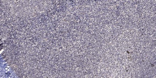
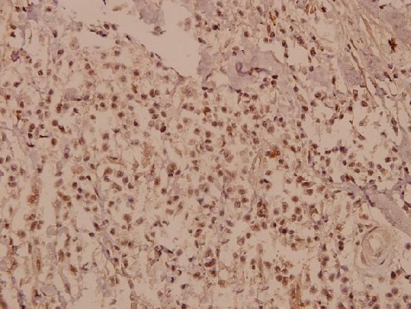
Two anti-CELF1 antibodies have IHC images from paraffin-embedded human tonsil and breast carcinoma (catalog: IHC captions). Both list human, mouse and rat reactivity (catalog: reactivity); A30541 also lists IF/ICC (catalog: applications).
A30541 shows IHC staining of paraffin-embedded human tonsil at 1:200 with Tris-EDTA pH 9.0 retrieval (A30541 image caption); it lists IHC, IF and ICC applications and human, mouse and rat reactivity (catalog: A30541). A02163 shows IHC staining of paraffin-embedded human breast carcinoma at 1:50 (A02163 image caption); it lists IHC and human, mouse and rat reactivity (catalog: A02163).
Which to pick: For tissue IHC, choose A30541 for the documented human tonsil example or A02163 for the documented human breast carcinoma example; both captions describe paraffin sections but leave the fixative unreported (catalog: IHC captions). For IF/ICC, choose A30541 because those applications are listed for it, while A02163 lists IHC only; both are rabbit polyclonals (catalog: applications, host and dilution_raw). Both list mouse and rat reactivity, but their IHC images show human tissue, so establish staining conditions in the intended species (catalog: reactivity and IHC captions).