This website uses cookies to ensure you get the best experience on our website.


- Table of Contents
Plan CHRM2 IHC-P using the catalog antibody at 1:25 (datasheet: M02733). Compare the observed cytoplasmic tissue staining (HPA tissue IHC) with the expected membrane location (UniProt), and interpret staining cautiously given its very low consistency with RNA expression (HPA tissue IHC).

Expected localisation, validated protocols, controls and antibodies — the at-a-glance facts below, then the full design guide.
| Expected localisation | Cytoplasmic tissue staining (HPA tissue IHC); membrane expected (UniProt) | |
| Staining pattern | Variable cytoplasmic staining in glandular and neuronal cells (HPA tissue IHC) | |
| Antigen retrieval | Citrate pH 6.0 HIER, 95–98 °C, 20 min (rule: cytoplasmic / membrane antigen) | |
| Positive control | Adrenal gland+4 more · see all | |
| Negative control | Adipose tissue |
| Fixation | Keep fixation consistent across sections (standard IHC practice; not target-specific) | |
| Caveat | Staining has very low consistency with RNA expression (HPA tissue IHC) | |
| Regulation | Agonist-driven internalization (UniProt) | |
| Isoform / epitope | No isoforms; extracellular versus cytoplasmic epitopes matter (UniProt) |
The catalog antibody protocol is accompanied by one published CHRM2 IHC protocol using mouse lung paraffin sections (PMC6791200).
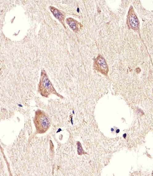
| Sample | Paraffin-embedded H. brain tissue; fixative not specified (datasheet M02733) |
| Fixation | Image fixative and duration unreported (datasheet M02733); verify before use. |
| Sectioning | 4–5 µm sections on charged slides (standard) |
| Deparaffinisation | Xylene, graded ethanol series to water (standard) |
| Antigen retrieval | Heat-induced epitope retrieval in citrate buffer, pH 6.0, 20 min at 95–98 °C (standard rule: cytoplasmic / membrane antigen) |
| Peroxidase block | 3% H2O2, 10 min, room temperature (standard) |
| Blocking | 10% normal serum of the secondary host, 30 min, room temperature (standard) |
| Primary antibody | Mouse monoclonal (clone 1424CT461.78.60) anti-CHRM2, 1:25 (datasheet M02733) |
| Primary incubation | Overnight at 4 °C (standard) |
| Detection | HRP-polymer secondary, DAB chromogen 5–10 min (standard) |
| Counterstain | Hematoxylin, blue, dehydrate and mount (standard) |
| Expected result | CHRM2-positive staining in glandular cells of adrenal gland (HPA tissue IHC: High). HPA tissue profile: Ubiquitous cytoplasmic expression in all tissues at variable levels. No signal in the no-primary control. |
CHRM2 is a seven-pass receptor at the cell and postsynaptic membranes (UniProt P08172 topology and subcellular location). In paraffin-section IHC, assess staining in the cell types HPA reports as high, including caudate neurons and cerebellar Purkinje cells (HPA: High). Interpret apparent positivity cautiously: HPA describes widespread cytoplasmic staining, but rates its IHC antibodies Approved with very low agreement between staining and RNA expression, pending external verification (HPA tissue IHC).
| Membrane-associated staining in caudate neurons or cerebellar Purkinje cells, with identifiable cells and limited surrounding stain. | This fits the UniProt cell-membrane location and HPA High staining in these cells (UniProt P08172; HPA tissue IHC). Record the membrane pattern and cellular distribution separately from overall intensity. HPA's low staining–RNA agreement means this appearance remains a provisional CHRM2 interpretation (HPA reliability description). |
| Strong, uniform cytoplasmic staining with little discernible membrane association. | HPA reports variable cytoplasmic staining across tissues, so cytoplasmic signal alone cannot be dismissed (HPA tissue IHC). It also cannot establish receptor localization: UniProt places CHRM2 at cell and postsynaptic membranes (UniProt P08172). Compare cellular boundaries and negative controls, then seek independent confirmation before calling diffuse cytoplasm specific. |
| Staining centered on adipocytes or equally intense across unrelated cells. | HPA reports CHRM2 as Not detected in adipocytes, making prominent adipocyte staining discordant with that reference (HPA tissue IHC). Widespread cell-independent color may reflect nonspecific binding or endogenous detection activity (general IHC practice). Check a no-primary control and the tissue's morphology before assigning cell-specific positivity. |
| Diffuse color over cells and extracellular areas, without a readable cellular pattern. | Treat this as background rather than a CHRM2 localization call (general IHC practice). The reported HPA profile is widespread cytoplasmic staining, but a diffuse field does not identify stained cells (HPA tissue IHC). Inspect the no-primary control and detection reagents; reassess staining only where cell boundaries and counterstain remain interpretable. |
| No convincing signal in a section containing caudate neurons or cerebellar Purkinje cells. | These are useful reference cells because HPA reports High staining in them (HPA tissue IHC), although its antibody evidence remains provisional (HPA reliability description). Review tissue preservation, assay controls, retrieval conditions and antibody use as general IHC troubleshooting. An absent signal alone cannot distinguish failed detection from a discrepancy in the HPA pattern. |
| Membrane topology and compartment | CHRM2 has seven transmembrane segments, with extracellular and cytoplasmic regions (UniProt P08172 topology). Assess cell borders and processes where morphology permits. The supplied record gives no antibody epitope, so topology cannot predict which retrieval condition or staining side a given IHC antibody requires. |
| Receptor internalization | Agonist-associated phosphorylation promotes CHRM2 internalization; phosphorylated receptor interacts with arrestins during internalization (UniProt P08172). Intracellular puncta can therefore be biologically plausible, but this record supplies no specimen treatment history. Do not identify puncta as internalized CHRM2 from appearance alone. |
| Tissue and cell selection | HPA reports High staining in bronchial respiratory epithelium and adrenal, appendix, colon and duodenal glandular cells, as well as specified neurons (HPA tissue IHC). It reports Low staining in ovarian stroma and skeletal-muscle myocytes, and Not detected in adipocytes (HPA tissue IHC). Use these as comparison patterns, with the HPA reliability caveat. |
| Strength of tissue evidence | Two listed rabbit antibodies, HPA029795 and CAB022338, have Approved IHC status; neither is listed as Enhanced (HPA antibodies). HPA also flags very low staining–RNA consistency and pending external verification (HPA tissue IHC). An Approved label should not be treated as independent confirmation of every stained cell or compartment. |
| IF/ICC: what pattern is supported? | HPA summarizes the subcellular location as Membrane but supplies no main location or cell-line ICC-IF images (HPA subcellular). UniProt also places CHRM2 at cell and postsynaptic membranes (UniProt P08172). These records support a membrane-location expectation, but provide no image-backed IF/ICC staining pattern or IF protocol. |
| Processing and target-specific fixation evidence | UniProt lists one CHRM2 chain, residues 1–466, with no signal peptide or propeptide and no annotated isoforms (UniProt P08172). The supplied UniProt and HPA records do not report a target-specific fixation effect. They cannot justify attributing a weak or absent IHC result specifically to CHRM2 fixation sensitivity. |
| Situation | Likely cause | Next action |
|---|---|---|
| Expected HPA High cells are unstained. | The assay may have failed, or this specimen may differ from the provisional HPA pattern (HPA tissue IHC, reliability description). | Confirm morphology and run appropriate assay controls; review retrieval and antibody conditions as general IHC practice. Interpret a repeat result against an independently supported reference. |
| Color is widespread but cell borders are unreadable. | Excess background or endogenous detection activity can obscure localization (general IHC practice); HPA also describes widespread cytoplasmic staining (HPA tissue IHC). | Inspect a no-primary control, blocking and chromogen development. Score CHRM2 only where cellular distribution can be distinguished from background. |
| Adipocytes appear strongly positive. | This conflicts with HPA's Not detected adipocyte result; nonspecific staining or endogenous activity is possible (HPA tissue IHC; general IHC practice). | Verify cell identity on the counterstain and examine a no-primary control. Seek independent target confirmation before reporting adipocyte CHRM2. |
| Signal is exclusively cytoplasmic or punctate. | HPA reports cytoplasmic staining, while UniProt places CHRM2 at membranes and notes receptor internalization (HPA tissue IHC; UniProt P08172). | Document the pattern without assigning a mechanism. Compare controls and an independent detection approach before calling the cytoplasmic signal specific. |
| A listed antibody produces an unexpected pattern. | Approved IHC status coexists with very low staining–RNA consistency and pending external verification (HPA antibodies; HPA reliability description). | Check the antibody identity and assay controls. If available, compare a second independently validated antibody or orthogonal evidence before accepting the cell-type assignment. |
| An IF/ICC result disagrees with the paraffin-section IHC result. | HPA gives a Membrane summary but no ICC-IF cell-line images, so the supplied evidence cannot resolve the difference (HPA subcellular). | Assess each assay with its own controls and record its compartment pattern. Avoid transferring an IHC intensity expectation to IF/ICC without separate validation. |


Comprehensive Human Protein Atlas IHC scoring per tissue (reliability: Approved — Very low consistency between antibody staining and RNA expression data. Pending external verification.). Rows are taken directly from the HPA tissue chart — click any row's HPA link to view the source.
| Tissue | Cell type | Level | Evidence | Source |
|---|---|---|---|---|
| Adrenal gland | Glandular cells | High | Protein (IHC) | HPA → |
| Appendix | Glandular cells | High | Protein (IHC) | HPA → |
| Bronchus | Respiratory epithelial cells | High | Protein (IHC) | HPA → |
| Caudate | Neuronal cells | High | Protein (IHC) | HPA → |
| Cerebellum | Purkinje cells | High | Protein (IHC) | HPA → |
| Tissue | Cell type | Level | Evidence | Source |
|---|---|---|---|---|
| Adipose tissue | Adipocytes | Not detected | Protein (IHC) | HPA → |
Troubleshoot CHRM2 staining in paraffin sections by checking retrieval, tissue preservation, compartment, detection background, and scoring controls (UniProt P08172; standard IHC practice).
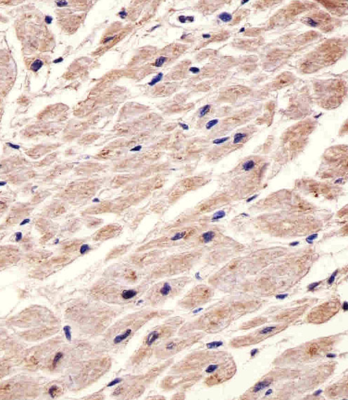
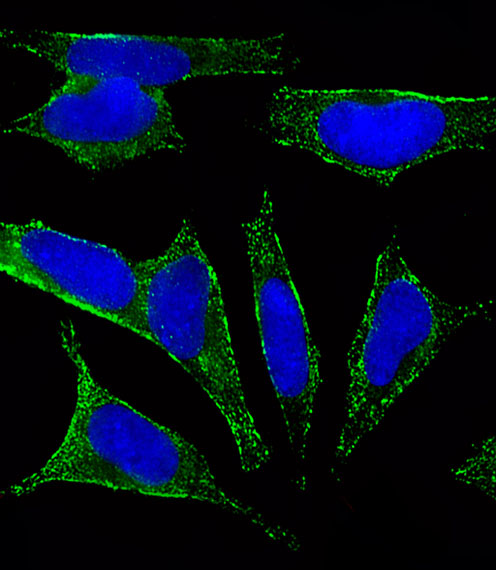
Validated anti-CHRM2 options show IHC in human brain and heart paraffin sections and rat brain tissue, plus IF in SH-SY5Y cells (catalog image captions: M02733, PA1325-1).
M02733 has IHC images from human brain and heart paraffin sections and an IF image from SH-SY5Y cells; its listed reactivity is human and mouse (M02733 image captions; catalog reactivity). PA1325-1 has a rat brain IHC(P) image and listed human, mouse and rat reactivity (PA1325-1 image caption; catalog reactivity).
Which to pick: For tissue IHC, choose mouse monoclonal M02733 for the documented human brain or heart paraffin-section examples, or rabbit PA1325-1 for the documented rat brain IHC(P) example (M02733 catalog host and IHC captions; PA1325-1 catalog host and IHC caption). For IF/ICC planning, M02733 lists IF and shows SH-SY5Y staining (M02733 catalog applications and IF caption). For human, mouse and rat coverage, PA1325-1 lists all three species (PA1325-1 catalog reactivity); neither SKU’s IHC caption reports the fixative (M02733 and PA1325-1 IHC captions).