CLK1 / Dual specificity protein kinase CLK1 · IHC design guide

Design Immunohistochemistry for CLK1

Plan CLK1 staining in paraffin sections using the catalog antibody’s IHC guidance. Compare cytoplasmic tissue staining (HPA tissue IHC) with the annotated nuclear location (UniProt), and account for the low consistency between staining and RNA data (HPA tissue IHC).

Evidence assembled Oct 2026 · For research use; verify linked source records and product datasheet before use
Immunohistochemistry protocol sheet for CLK1 (IHC for CLK1): expected localisation Cytoplasmic staining (HPA tissue IHC); nuclear location (UniProt), antibody A03271, validated IHC image, and IHC protocol steps
Printable CLK1 IHC protocol sheet — expected localisation Cytoplasmic staining (HPA tissue IHC); nuclear location (UniProt), antibody A03271, controls and protocol steps. Open the full CLK1 IHC guide →

CLK1 Immunohistochemistry Experimental Design Guide

Expected localisation, validated protocols, controls and antibodies — the at-a-glance facts below, then the full design guide.

Must know before staining
Expected localisation Cytoplasmic staining (HPA tissue IHC); nuclear location (UniProt)
Staining pattern Cytoplasmic staining in most tissues (HPA tissue IHC)
Antigen retrieval Tris-EDTA pH 8.0 HIER, heat-mediated (datasheet A03271)
Positive control ⓘ Appendix+4 more · see all
Negative control ⓘ Adipose tissue+4 more · see all
Important caveats
Reasons your staining may differ from the expected pattern.
Fixation Keep paraffin-section fixation consistent. (standard IHC practice; not target-specific)
Caveat Staining and RNA data show low consistency (HPA tissue IHC)
Regulation Low tissue specificity (HPA tissue RNA)
Isoform / epitope 3 isoforms; check epitope coverage for each (UniProt)
Section 1

Recommended CLK1 IHC & IF Protocols

The catalog antibody’s IHC-P protocol is accompanied by published FFPE tumor and pancreatic cancer tissue protocols (PMC6115463; PMC8045197).

Recommended immunohistochemistry (IHC-P) protocol parameters
SampleParaffin-embedded Human lung cancer tissue; fixative not specified (datasheet A03271)
FixationImage fixative and duration unreported (datasheet A03271); verify before use.
Sectioning4–5 µm sections on charged slides (standard)
DeparaffinisationXylene, graded ethanol series to water (standard)
Antigen retrievalHeat retrieval: Tris-EDTA pH 8.0 (datasheet A03271); 20 min, 95–100 °C (standard)
Peroxidase block3% H2O2, 10 min, room temperature (standard)
Blocking10% normal serum of the secondary host, 30 min, room temperature (standard)
Primary antibodyRabbit anti-CLK1, 1:100-1:300 (datasheet A03271)
Primary incubationOvernight at 4 °C (standard)
DetectionHRP-polymer secondary, DAB chromogen 5–10 min (standard)
CounterstainHematoxylin, blue, dehydrate and mount (standard)
Expected resultCLK1-positive staining in glandular cells of appendix (HPA tissue IHC: Medium). HPA tissue profile: Cytoplasmic expression in most tissues. No signal in the no-primary control.
💡Decision noteStart with heat-mediated Tris-EDTA pH 8.0 retrieval (datasheet A03271); optimize staining for the chosen tissue.
Section 2

What Is the Expected CLK1 Staining Pattern?

CLK1 is a nuclear, non-transmembrane kinase (UniProt P49759: location and topology). In paraffin-section IHC, HPA reports mainly cytoplasmic staining across tissues, including medium staining in kidney tubule cells and liver cholangiocytes (HPA: tissue IHC). Treat this compartment difference cautiously: HPA rates its IHC reliability Approved but reports low agreement between antibody staining and RNA expression, pending external verification (HPA: tissue IHC).

What am I looking at on my slide?
Medium cytoplasmic staining in kidney tubule cells or liver cholangiocytes, with visible tissue structure.This matches reported IHC observations in those cells (HPA: tissue IHC). Record the cytoplasmic compartment rather than calling it confirmed CLK1 localisation: UniProt places CLK1 in the nucleus, and HPA's IHC pattern awaits external verification (UniProt P49759: location; HPA: reliability).
Strong staining confined to an unexpected compartment, such as extracellular space, while expected cells show little signal.Investigate staining artefact or nonspecific binding (general IHC practice). A compartment mismatch alone is inconclusive because HPA tissue IHC reports cytoplasmic staining whereas UniProt and HPA ICC-IF support nuclear localisation (HPA: tissue IHC and subcellular; UniProt P49759: location).
Prominent staining in adipocytes or cardiomyocytes, especially with weak staining in a positive comparator.These cells are reported as not detected (HPA: tissue IHC). Consider antibody cross-reactivity or endogenous detection activity, then compare with the same-run negative control (general IHC practice). Absence in HPA is a reference observation, not proof that every specimen must be negative.
Diffuse colour across many cell types, tissue edges, or the whole section, obscuring cell boundaries.Interpret the distribution as background until controls clarify it (general IHC practice). HPA reports particular medium-staining populations rather than uniform, structure-free colour (HPA: tissue IHC). Examine a no-primary control and review blocking, washes and chromogen development (general IHC practice).
No discernible staining in kidney tubule cells or appendix glandular cells on an otherwise intact section.Both are reported at medium intensity (HPA: tissue IHC), so absence calls for a run-level check before a biological conclusion. Review the positive control, antibody suitability, retrieval conditions and detection reagents (general IHC practice); HPA's stated IHC uncertainty also limits any single-tissue inference (HPA: reliability).
💡Expected CLK1 appearanceA supported IHC reference result is discernible, medium cytoplasmic staining in kidney tubule cells or liver cholangiocytes (HPA: tissue IHC); widespread structure-free colour or strong staining in HPA-negative adipocytes raises concern for background or a false positive (HPA: tissue IHC; general IHC practice).
How each factor affects the staining
Compartment evidenceHPA tissue IHC reports cytoplasmic expression in most tissues, while UniProt lists the nucleus and HPA ICC-IF supports nucleoplasm and nuclear membrane (HPA: tissue IHC and subcellular; UniProt P49759: location). Keep assay-specific observations distinct when scoring.
Choice of comparison cellsKidney tubule cells, liver cholangiocytes and appendix glandular cells are medium IHC references; adipocytes, cardiomyocytes and skeletal myocytes are reported as not detected (HPA: tissue IHC). Include appropriate on-slide or same-run comparisons when available (general IHC practice).
Antibody validationThe listed IHC antibody CAB033141 is Approved; the listed ICC antibody HPA062405 is Approved for ICC, with no IHC status supplied (HPA: antibodies). HPA describes low agreement between tissue staining and RNA expression and pending external verification (HPA: tissue IHC).
IF/ICC Q&A: where should CLK1 signal appear?In ICC-IF, HPA supports nucleoplasm and nuclear membrane localisation and lists images from HeLa, PC-3 and U2OS (HPA: subcellular). This is an IF interpretation reference, not an IHC-P protocol or evidence that the IHC cytoplasmic pattern is resolved.
Sequence and epitope uncertaintyUniProt lists three CLK1 isoforms, a protein kinase domain at residues 161–477 and modified residues including phosphoserines 61 and 140 (UniProt P49759). The supplied sources do not map either antibody's epitope, so isoform or phosphorylation effects on staining cannot be assigned.
Why is my staining missing, weak or wrong?
SituationLikely causeNext action
A reported positive cell population is blank.The run may have inadequate target detection, or the specimen may differ from the HPA reference (general IHC practice; HPA: tissue IHC).Check a same-run positive section, then review retrieval, primary concentration, incubation and detection against the validated IHC workflow (general IHC practice). Do not infer a CLK1-specific fixation effect; none is supplied.
An HPA-negative cell population stains strongly.Possible nonspecific antibody binding or endogenous detection activity (general IHC practice); HPA reports adipocytes and cardiomyocytes as not detected (HPA: tissue IHC).Inspect no-primary and detection controls, then reassess blocking, washes and development time (general IHC practice). Interpret the result alongside a reported positive population.
Colour is diffuse and obscures cellular localisation.Background from incomplete blocking, insufficient washing or excessive development is possible (general IHC practice).Compare with a no-primary control and review the chromogenic workflow (general IHC practice). Score only interpretable cell-associated staining, keeping the HPA cytoplasmic observation and its reliability caveat in view (HPA: tissue IHC).
Nuclear staining appears where cytoplasmic IHC was expected.The result differs from HPA tissue IHC but agrees with UniProt's nuclear assignment and HPA ICC-IF localisation (HPA: tissue IHC and subcellular; UniProt P49759: location).Document the compartment and cell type, check controls, and avoid resolving the disagreement from localisation alone (general IHC practice). HPA states that the tissue IHC pattern awaits external verification (HPA: reliability).
The staining pattern changes between runs.Variation in section processing, retrieval, reagent handling or chromogen timing can change an IHC run (general IHC practice).Compare same-run controls and recorded workflow conditions before comparing biological specimens (general IHC practice). Use HPA's named positive and not-detected cell populations as references, with its low-consistency warning (HPA: tissue IHC).
A proposed antibody is supported only by ICC images.ICC validation does not establish IHC-P performance; HPA062405 is ICC Approved with no IHC status listed (HPA: antibodies).Use an antibody with stated IHC validation for paraffin-section interpretation; CAB033141 is listed as IHC Approved (HPA: antibodies). Keep the ICC nuclear pattern as a separate localisation reference (HPA: subcellular).

Sample controls for CLK1 IHC & IF

🧪Run appendix first and score glandular cells, which HPA rates Medium for CLK1 (HPA: Appendix, glandular cells, Medium). Use adipose tissue adipocytes as the negative comparator (HPA: Adipose tissue, adipocytes, Not detected); on the appendix slide, treat neighboring cells without visible staining as an internal background reference, without assuming they lack CLK1 (standard IHC practice).
Positive control tissue: Appendix (Glandular cells, HPA Medium)
Negative control tissue: Adipose tissue (HPA Not detected)
ICC-IF cell lines (HPA subcellular resource): HPA ICC-IF images show CLK1 in HeLa, PC-3, U2OS, with annotated localisation: Nucleoplasm (supported), Nuclear membrane (supported) (HPA subcellular).
Technical controls: Include a no-primary, secondary-only control and an isotype control matched to the primary antibody’s host species and clonality (standard IHC practice). Use CLK1 knockout material or peptide preabsorption as a biological specificity control (standard IHC practice; A03271 tissue-IHC caption: peptide preabsorption), and check appendix sections for endogenous peroxidase signal and chromogen trapped in gland lumens (standard chromogenic IHC practice).
⚠️Feasibility: A CLK1-specific fixation window or fixation effect is unreported, and the exact A03271 paraffin-section caption does not report a fixative (A03271 tissue-IHC caption). The caption uses high-pressure, high-temperature Tris-EDTA retrieval at pH 8.0 and antibody at 1:100 overnight at 4°C in lung cancer tissue; whether appendix staining depends on that retrieval is unreported (A03271 tissue-IHC caption). HPA shows nucleoplasm and nuclear membrane staining in ICC-IF images from HeLa, PC-3 and U2OS, but the supplied evidence does not establish that frozen sections or IF are easier for CLK1; inspect appendix gland lumens for trapped chromogen (HPA subcellular; standard chromogenic IHC practice).

HPA tissue IHC evidence for CLK1

Comprehensive Human Protein Atlas IHC scoring per tissue (reliability: Approved — Low consistency between antibody staining and RNA expression data. Pending external verification.). Rows are taken directly from the HPA tissue chart — click any row's HPA link to view the source.

Positive expression · recommended positive controls

TissueCell typeLevelEvidenceSource
Appendix Glandular cells Medium Protein (IHC) HPA →
Caudate Neuronal cells Medium Protein (IHC) HPA →
Cerebellum Purkinje cells Medium Protein (IHC) HPA →
Cerebral cortex Glial cells Medium Protein (IHC) HPA →
Epididymis Glandular cells Medium Protein (IHC) HPA →

Undetected expression · recommended negative controls

TissueCell typeLevelEvidenceSource
Adipose tissue Adipocytes Not detected Protein (IHC) HPA →
Bone marrow Hematopoietic cells Not detected Protein (IHC) HPA →
Heart muscle Cardiomyocytes Not detected Protein (IHC) HPA →
Hippocampus Glial cells Not detected Protein (IHC) HPA →
Oral mucosa Squamous epithelial cells Not detected Protein (IHC) HPA →
Section 3

Advanced CLK1 IHC Tips

Troubleshoot CLK1 staining in paraffin section IHC by checking retrieval, compartment, controls and cell specific scoring before interpreting signal.

Which retrieval conditions should I try first for weak CLK1 staining?
Start with heat mediated Tris-EDTA retrieval at pH 8.0 for paraffin section CLK1 IHC (datasheet A03271). The selected image used high pressure and temperature retrieval, followed by antibody at 1:100 overnight at 4°C (caption A03271). If staining is weak, compare a shorter and a longer heat exposure on adjacent sections while keeping antibody concentration and detection constant (standard IHC practice). Check whether improved signal appears in intact cells rather than section edges or damaged regions (standard IHC practice). Record retrieval conditions alongside compartment and cell type, because nuclear localisation and reported cytoplasmic tissue staining differ (UniProt P49759 localisation; HPA tissue IHC).
How should I investigate fixation related loss of CLK1 signal?
Target specific fixation sensitivity for CLK1 is unknown, and the selected paraffin section caption does not state a fixative (caption A03271). Document the actual fixative and fixation time for each specimen before comparing staining across blocks (standard IHC practice). If weak staining tracks with processing history, compare sections from blocks with documented, different fixation histories under identical pH 8.0 retrieval and detection conditions (datasheet A03271; standard IHC practice). Include a section from a consistently staining block in each run to detect run variation (standard IHC practice). Interpret differences cautiously until matched specimens separate fixation effects from biological variation (standard IHC practice).
Should CLK1 staining be nuclear or cytoplasmic in paraffin sections?
Expect a nuclear component when assessing CLK1, because UniProt places it in the nucleus and HPA subcellular imaging supports nucleoplasm and nuclear membrane localisation (UniProt P49759 localisation; HPA subcellular). HPA tissue IHC also reports cytoplasmic expression in most tissues, with low consistency between staining and RNA and external verification pending (HPA tissue IHC). Score nuclear and cytoplasmic signal separately against a counterstain, rather than merging them into one positive call (standard IHC practice). Compare the same cell type across test and control sections under identical development conditions (standard IHC practice). Diffuse staining without cellular boundaries warrants renewed review of morphology and controls (standard IHC practice).
Could CLK1 isoforms or epitope masking explain discordant staining?
CLK1 has 3 listed isoforms, but the supplied evidence does not locate the catalog antibody’s epitope or establish which isoforms it detects (UniProt P49759 isoforms; supplied A03271 caption). Ask for the immunogen sequence and map it against each isoform before assigning an isoform specific interpretation (standard antibody validation practice). CLK1 also has reported phosphorylation sites at residues 61, 138 and 140, without evidence here that they alter this antibody’s staining (UniProt P49759 modified residues). Compare retrieval and staining on adjacent sections, then use a separately validated epitope reagent if isoform coverage matters (standard IHC practice). Peptide preabsorption can support recognition of the immunogen but cannot alone establish isoform selectivity (caption A03271; standard antibody validation practice).
How can IF clarify an ambiguous CLK1 IHC pattern?
Use IF as a follow up to paraffin section IHC when chromogenic signal cannot resolve nuclear from cytoplasmic staining (standard microscopy practice; HPA tissue IHC). Multiplex CLK1 with an endothelial marker such as CD31 when testing endothelial cells, a reported CLK1 expressing cell type (UniProt P49759 tissue specificity; standard IF practice). Choose a fluorophore in a channel with low tissue autofluorescence and include single stain and no primary controls (standard IF practice). CLK1 has no transmembrane segment and has nuclear localisation, so use controlled permeabilisation to access its intracellular epitope while preserving nuclear morphology (UniProt P49759 topology and localisation; standard IF practice). Compare nuclear signal with the counterstain and the IHC cell type assignment (standard IF practice).
What controls help distinguish CLK1 signal from chromogenic background?
Run no primary and detection only controls to identify nonspecific secondary or chromogen deposition (standard IHC practice). Block endogenous peroxidase before peroxidase based DAB detection, and inspect pigment and tissue folds before calling brown signal positive (standard chromogenic IHC practice). The A03271 paraffin section image includes an immunogen peptide preabsorption control, which tests competition for antibody binding in that example (caption A03271). If background persists, compare a lower antibody concentration with the pictured 1:100 condition while holding retrieval and development constant (caption A03271; standard IHC practice). Require cell shaped staining with a plausible compartment before scoring, particularly when staining is strongest at section edges (UniProt P49759 localisation; standard IHC practice).
How should I score CLK1 across heterogeneous tissue regions? ⚠ ANSWER MARKED FOR VERIFICATION
Define the cell population and tissue region before scoring, then report nuclear and cytoplasmic CLK1 separately because the localisation sources differ (UniProt P49759 localisation; HPA tissue IHC). For cell based scoring, record percent positive cells and staining intensity to calculate an H-score from 0–300; use positive cells per mm² when cell density varies (standard IHC scoring practice). Normalise counts to the number of intact target cells or evaluable tissue area, and apply one threshold across the comparison set (standard IHC scoring practice). Exclude necrosis, folds and section edges using predefined morphology rules (standard IHC practice). Report cell type and compartment with each score so pooled signal does not obscure endothelial or other cellular contributions (UniProt P49759 tissue specificity; standard IHC scoring practice).
When is a CLK1 positive call biologically credible?
A credible call has reproducible, cell bounded staining in an identified cell type and an interpretable compartment, with controls free of comparable signal (standard IHC practice). Nuclear staining is consistent with UniProt and supported nucleoplasm and nuclear membrane localisation, while cytoplasmic tissue staining has been reported with low RNA concordance and pending external verification (UniProt P49759 localisation; HPA subcellular; HPA tissue IHC). Compare endothelial cells as one relevant population, while recognising that HPA also reports staining in other tissue cell types (UniProt P49759 tissue specificity; HPA tissue IHC). Reject edge accentuation, necrotic deposits and endogenous enzyme signal when they fail morphology or control checks (standard chromogenic IHC practice). Treat peptide competition as supporting antibody binding, not as conclusive proof of CLK1 specificity (caption A03271; standard antibody validation practice).
Boster reagents

Best CLK1 / Dual specificity protein kinase CLK1 IHC Antibodies

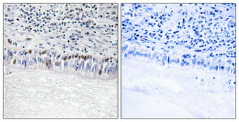
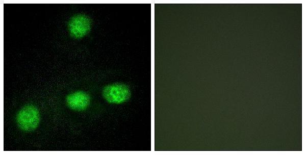
The IHC-validated antibody A03271 has paraffin-section data from human lung cancer (IHC image caption), IF data from HUVEC cells (IF image caption), and listed human and mouse reactivity (catalog).

Real IHC data Immunohistochemical analysis of paraffin-embedded Human lung cancer. Antibody was diluted at 1:100 (4° overnight). High-pressure and temperature Tris-EDTA, pH8.0 was used for antigen retrieval. Negetive contrl (right) obtaned from antibody was pre-absorbed by immunogen peptide.
Anti-CLK1 Antibody
Cat # A03271

A03271 will render with an IHC image of paraffin-embedded human lung cancer, stained at 1:100 (IHC image caption). A03271 also has an IF image of HUVEC cells (IF image caption), with human and mouse reactivity listed (catalog).

Which to pick: For tissue IHC, choose A03271: it is a polyclonal antibody listed for IHC, with an image from paraffin-embedded human lung cancer using Tris-EDTA retrieval at pH 8.0; the fixative is unreported (catalog; IHC image caption). For IF/ICC, A03271 is listed for IF and has HUVEC IF data; ICC-specific validation is unreported (catalog; IF image caption). For human or mouse samples, A03271 is the listed option by reactivity, although the supplied IHC and IF images do not show mouse samples (catalog; IHC and IF image captions).

Each figure is that product's own IHC / IF validation image from its datasheet.

References

  1. UniProt Consortium. UniProt entry P49759 (CLK1_HUMAN, Dual specificity protein kinase CLK1).
  2. Human Protein Atlas. CLK1 tissue IHC expression (reliability: Approved).
  3. Human Protein Atlas. CLK1 subcellular location (ICC-IF): Localized to the nucleoplasm and nuclear membrane..
  4. Human Protein Atlas. CLK1 antibody validation summary (2 antibodies).
  5. CLK1 is a potential tumor suppressor for NSCLC by regulating cell proliferation and immune infiltration. Frontiers in cell and developmental biology 2026 — PMC12926458.
  6. CLK1 Promotes Myeloid-Derived Suppressor Cell Trafficking and Reprograms the Tumor Microenvironment by Activating Hippo/YAP Signaling in Colorectal Cancer. Cancer immunology research 2026 — PMC13136879.
  7. In vivo phosphoproteomics reveals kinase activity profiles that predict treatment outcome in triple-negative breast cancer. Nature communications 2018 — PMC6115463.
  8. CLK1/SRSF5 pathway induces aberrant exon skipping of METTL14 and Cyclin L2 and promotes growth and metastasis of pancreatic cancer. Journal of hematology & oncology 2021 — PMC8045197.
  9. PubMed PMID:1704889 — UniProt-cited evidence.
  10. PubMed PMID:7990150 — UniProt-cited evidence.
  11. PubMed PMID:14702039 — UniProt-cited evidence.