CMA1 / Chymase · IHC design guide

Design Immunohistochemistry for CMA1

Plan chromogenic CMA1 IHC in paraffin sections around selective cytoplasmic staining in immune cells (HPA tissue IHC). This guide covers fixation consistency, the catalog antibody’s 0.5–1 μg/mL IHC range (datasheet PB9055), and interpretation of secreted chymase (UniProt).

Evidence assembled Oct 2026 · For research use; verify linked source records and product datasheet before use
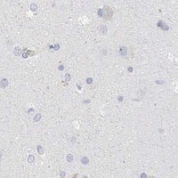
Immunohistochemistry protocol sheet for CMA1 (IHC for CMA1): expected localisation Cytoplasm in a subset of immune cells (HPA tissue IHC), antibody PB9055, validated IHC image, and IHC protocol steps
Printable CMA1 IHC protocol sheet — expected localisation Cytoplasm in a subset of immune cells (HPA tissue IHC), antibody PB9055, controls and protocol steps. Open the full CMA1 IHC guide →

CMA1 Immunohistochemistry Experimental Design Guide

Expected localisation, validated protocols, controls and antibodies — the at-a-glance facts below, then the full design guide.

Must know before staining
Expected localisation Cytoplasm in a subset of immune cells (HPA tissue IHC)
Staining pattern Selective cytoplasmic staining in immune cells (HPA tissue IHC)
Antigen retrieval EDTA pH 8.0 HIER, heat-mediated (datasheet PB9055)
Positive control ⓘ Bone marrow+2 more · see all
Negative control ⓘ Adipose tissue+4 more · see all
Important caveats
Reasons your staining may differ from the expected pattern.
Fixation Keep fixation consistent across sections (standard IHC practice; not target-specific)
Caveat Secreted CMA1 may appear away from cells that produce it (UniProt)
Regulation Tissue-dependent mast-cell expression (UniProt)
Isoform / epitope 2 isoforms; mature chain starts at residue 22—check the epitope (UniProt)
Section 1

Recommended CMA1 IHC & IF Protocols

The catalog antibody’s IHC-P protocol (datasheet: PB9055) is accompanied by published CMA1 staining of mouse ileum (PMC5913881) and airway tissue (PMC10945381).

Recommended immunohistochemistry (IHC-P) protocol parameters
SampleParaffin-embedded human placenta tissue; fixative not specified (datasheet PB9055)
FixationImage fixative and duration unreported (datasheet PB9055); verify before use.
Sectioning4–5 µm sections on charged slides (standard)
DeparaffinisationXylene, graded ethanol series to water (standard)
Antigen retrievalHeat retrieval: EDTA pH 8.0 (datasheet PB9055); 20 min, 95–100 °C (standard)
Peroxidase block3% H2O2, 10 min, room temperature (standard)
Blocking10% goat serum (datasheet PB9055)
Primary antibodyRabbit anti-CMA1, 0.5-1μg/ml (datasheet PB9055)
Primary incubationOvernight at 4 °C (datasheet PB9055)
DetectionStreptavidin-biotin complex (SABC), DAB chromogen (datasheet PB9055)
CounterstainHematoxylin, blue, dehydrate and mount (standard)
Expected resultCMA1-positive staining in hematopoietic cells of bone marrow (HPA tissue IHC: Medium). HPA tissue profile: Highly selective cytoplasmic expression in a subset of immune cells. No signal in the no-primary control.
💡Decision noteStart with heat-mediated EDTA pH 8.0 for the catalog antibody (datasheet: PB9055); the airway study used pH 6 with a different antibody (PMC10945381).
Section 2

What Is the Expected CMA1 Staining Pattern?

CMA1 is a secreted mast cell protease stored in cytoplasmic granules; it has no transmembrane segment (UniProt P23946). In paraffin section IHC, expect selective cytoplasmic staining in scattered immune cells, consistent with HPA’s highly selective profile (HPA tissue IHC). HPA rates its tissue IHC reliability Enhanced, while noting that secretion can complicate comparisons between RNA and protein location (HPA tissue IHC).

What am I looking at on my slide?
Discrete cytoplasmic staining in a subset of immune cells, with unstained neighboring cells.This matches HPA’s highly selective cytoplasmic profile and the granule location annotated for mast cell CMA1 (HPA tissue IHC; UniProt P23946). HPA reports medium staining in bone marrow hematopoietic cells and lymph node and tonsil non-germinal center cells; those labels do not identify every positive cell as a mast cell (HPA tissue IHC).
Predominantly nuclear staining or a crisp outline of the plasma membrane.Treat this as discordant with the expected cytoplasmic granule pattern and the absence of a transmembrane segment (UniProt P23946). Review the counterstain, morphology and detection controls before assigning CMA1 positivity (general IHC practice).
Strong staining across cell populations HPA reports as unstained, such as adipocytes or respiratory epithelial cells.HPA reports CMA1 as not detected in those specified cell populations, so broad staining warrants checks for antibody cross-reactivity or endogenous detection activity (HPA tissue IHC; general IHC practice). Those entries do not make the entire adipose or bronchus section a negative control (HPA tissue IHC).
Diffuse color over cells, extracellular space and section edges, without identifiable positive cells.The slide cannot establish selective cellular staining under those conditions (general IHC practice). Because CMA1 is secreted, extracellular signal alone is not proof of artifact; judge it alongside cell-associated staining, morphology and background controls (UniProt P23946; general IHC practice).
No cytoplasmic staining in a sampled population expected to be positive.First confirm that the section contains the relevant cells: HPA describes only a subset of immune cells and reports medium staining in its listed positive populations (HPA tissue IHC). If the cells are present, examine the IHC run and antibody controls (general IHC practice).
💡Expected CMA1 appearanceCall the result positive when identifiable immune cells show selective cytoplasmic signal, with medium staining in HPA’s listed positive populations; widespread nuclear or adipocyte staining is suspect (HPA tissue IHC; UniProt P23946).
How each factor affects the staining
Cell distribution and scoringScore the stained cell population and compartment, rather than averaging color across the section: HPA describes a subset of immune cells, with medium staining in selected bone marrow, lymph node and tonsil cells (HPA tissue IHC).
Granule storage and secretionCMA1 is annotated in mast cell granules and as secreted (UniProt P23946). Interpret extracellular color cautiously: HPA warns that the location of secreted protein and its RNA may differ (HPA tissue IHC).
Protein processingThe precursor has a signal peptide at residues 1–19 and a propeptide at 20–21; the annotated chymase chain spans 22–247 (UniProt P23946). The payload gives no antibody epitope, so it cannot predict recognition of each processed form.
Isoforms and antibody evidenceUniProt lists 2 isoforms without a staining comparison (UniProt P23946). HPA rates IHC for HPA052634 and CAB000363 Enhanced; that validation supports the reported pattern but does not specify isoform recognition (HPA antibodies).
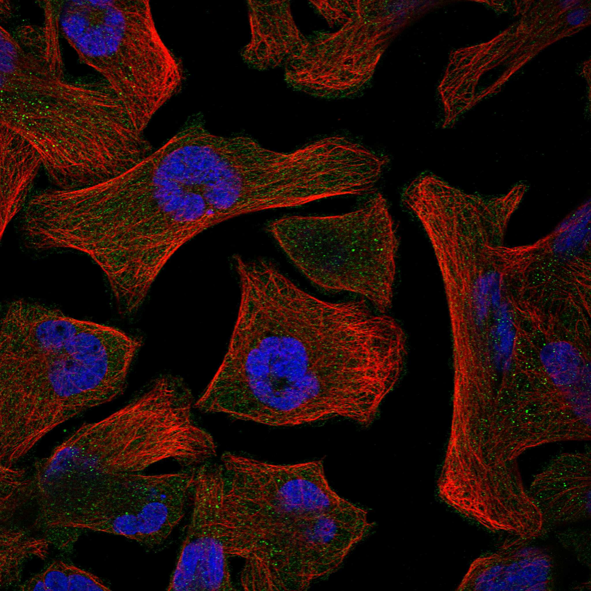
IHC and IF localisation evidenceHPA reports cytoplasmic bodies as the supported main ICC-IF location and cytosol as an approved additional location; its images are from Rh30, SuSa and U2OS (HPA subcellular). These observations should be read separately from its tissue IHC profile (HPA tissue IHC; HPA subcellular).
Why is my staining missing, weak or wrong?
SituationLikely causeNext action
The intended positive area has no signal.The field may miss the positive immune cell subset, or the staining run may have failed (HPA tissue IHC; general IHC practice).Check cell morphology and examine another area containing the reported positive population; then verify the run with an appropriate positive control (HPA tissue IHC; general IHC practice).
Nuclei or cell borders dominate the staining.That pattern conflicts with cytoplasmic granule localisation and the lack of a transmembrane segment (UniProt P23946).Compare signal with the counterstain and a negative detection control; score only cells with convincing cytoplasmic staining (general IHC practice; UniProt P23946).
Many adipocytes or respiratory epithelial cells appear positive.Those particular populations are listed as not detected by HPA; cross-reactivity or endogenous detection activity is possible (HPA tissue IHC; general IHC practice).Check the no-primary control and the detection-system controls, then compare with the IHC-validated antibody’s reported cell pattern (general IHC practice; HPA tissue IHC).
Brown color is widespread and obscures cell boundaries.Diffuse detection background can prevent assignment of a positive cell; CMA1 secretion also permits caution about extracellular signal (general IHC practice; UniProt P23946).Inspect the no-primary control and assess background before scoring; require identifiable cell-associated cytoplasmic staining for a cellular call (general IHC practice; HPA tissue IHC).
The section contains staining, but RNA and protein maps seem inconsistent.HPA notes that at least one variant is secreted, so RNA and protein tissue locations can differ and correlation is complex (HPA tissue IHC).Interpret the observed cell type and compartment against the tissue IHC profile; do not infer antibody failure solely from RNA location (HPA tissue IHC; general IHC practice).
Can an ICC-IF cytoplasmic body image define the paraffin IHC result?ICC-IF images represent cell line localisation, while this guide’s expected tissue pattern comes from paraffin section IHC (HPA subcellular; HPA tissue IHC).Use HPA tissue IHC to judge tissue staining. The ICC-IF evidence supports cytoplasmic bodies and additional cytosol; HPA052634 is ICC Supported (HPA subcellular; HPA antibodies).

Sample controls for CMA1 IHC & IF

🧪Run bone marrow first and score staining in hematopoietic cells (HPA: Medium in bone marrow hematopoietic cells). Use adipose tissue adipocytes as the negative comparison (HPA: Not detected in adipocytes); cells outside the designated hematopoietic compartment on the bone marrow slide should lack specific granular staining (HPA: bone marrow positive cell designation; UniProt P23946: mast cell granules).
Positive control tissue: Bone marrow (Hematopoietic cells, HPA Medium)
Negative control tissue: Adipose tissue (HPA Not detected)
ICC-IF cell lines (HPA subcellular resource): HPA ICC-IF images show CMA1 in Rh30, SuSa, U2OS, with annotated localisation: Cytoplasmic bodies (supported) (HPA subcellular).
Technical controls: Include a no-primary, secondary-only control and a host-species- and clonality-matched isotype control (caption: rabbit anti-CMA1 primary; clonality unreported); use CMA1-knockout tissue, if available, as a biological negative (standard IHC practice). Block endogenous peroxidase in bone marrow and check endogenous biotin background when using the caption’s biotin-based SABC/DAB detection (caption: biotinylated secondary, SABC and DAB; standard IHC practice).
⚠️Feasibility: The selected PB9055 paraffin-section caption reports heat retrieval in EDTA at pH 8.0, but does not report a fixative; a target-specific fixation window or fixation effect is unreported (PB9055 caption). No matched evidence establishes whether frozen sections or IF are easier than paraffin IHC for this target (supplied application evidence). Bone marrow can show endogenous peroxidase background, so interpret chromogenic staining against the no-primary control (standard IHC practice).

HPA tissue IHC evidence for CMA1

Comprehensive Human Protein Atlas IHC scoring per tissue (reliability: Enhanced — High consistency between antibody staining and RNA expression data. At least one protein variant secreted, tissue location of RNA and protein might differ and correlation is complex.). Rows are taken directly from the HPA tissue chart — click any row's HPA link to view the source.

Positive expression · recommended positive controls

TissueCell typeLevelEvidenceSource
Bone marrow Hematopoietic cells Medium Protein (IHC) HPA →
Lymph node Non-germinal center cells Medium Protein (IHC) HPA →
Tonsil Non-germinal center cells Medium Protein (IHC) HPA →

Undetected expression · recommended negative controls

TissueCell typeLevelEvidenceSource
Adipose tissue Adipocytes Not detected Protein (IHC) HPA →
Adrenal gland Glandular cells Not detected Protein (IHC) HPA →
Appendix Endocrine cells Not detected Protein (IHC) HPA →
Breast Adipocytes Not detected Protein (IHC) HPA →
Bronchus Respiratory epithelial cells Not detected Protein (IHC) HPA →
Section 3

Advanced CMA1 IHC Tips

Troubleshoot CMA1 staining in paraffin sections by checking retrieval, granular cytoplasmic localisation, controls and cell based scoring (UniProt P23946; HPA tissue IHC).

How should I retrieve CMA1 in paraffin sections with weak staining?
Start with heat mediated retrieval in EDTA at pH 8.0 for paraffin sections (datasheet PB9055). The documented placenta stain used that retrieval, followed by 1 μg/ml primary antibody overnight at 4°C (datasheet PB9055). If staining is weak, confirm that sections stayed covered by retrieval solution, then compare retrieval durations on matched sections while keeping detection and exposure conditions constant (standard IHC practice). Examine tissue morphology alongside signal, because excessive heating can damage sections and complicate interpretation (standard IHC practice). The caption does not report a fixative, so retrieval performance cannot establish fixation tolerance (datasheet PB9055).
Could fixation explain variable CMA1 staining between specimens?
Target specific fixation sensitivity is unknown from the supplied evidence; the catalog image identifies a paraffin section but does not state its fixative (datasheet PB9055). Record the fixative, fixation duration and processing history for each specimen before comparing staining intensity (standard IHC practice). If those conditions differ, stain matched sections in one run and include a known positive section to assess whether the assay performed consistently (standard IHC practice). Keep heat mediated EDTA retrieval at pH 8.0 as the documented starting condition for this antibody (datasheet PB9055). Do not assign a fixation effect from tissue staining patterns alone (HPA tissue IHC).
Where should convincing CMA1 staining appear?
Look for signal in the cytoplasm of a subset of immune cells, with a granular distribution consistent with mast cell granules (HPA tissue IHC; UniProt P23946 subcellular location). HPA also describes cytoplasmic bodies as the main location and cytosol as an additional location in its subcellular data (HPA subcellular). CMA1 is secreted and has no transmembrane segment, so a crisp cell surface outline would need independent validation before being scored as expected localisation (UniProt P23946 topology and subcellular location). Review the cellular pattern at high magnification and compare it with a mast cell marker on an adjacent or multiplexed section (standard IHC practice; UniProt P23946 tissue specificity).
Could processing or isoforms change which CMA1 cells are detected?
CMA1 has 2 annotated isoforms, and its precursor includes a signal peptide at residues 1–19 and propeptide at 20–21 (UniProt P23946). The annotated chymase chain spans residues 22–247, with glycosylation sites at 80 and 103 (UniProt P23946). The supplied antibody caption does not identify its epitope or establish equal recognition of both isoforms and processed forms (datasheet PB9055). Check the antibody’s documented immunogen or epitope before interpreting a negative cell as lacking all CMA1 forms (standard IHC practice). Compare staining with an independent assay when isoform or processing state matters to the study (standard IHC practice).
How can I check CMA1 localisation by multiplex immunofluorescence?
Pair CMA1 with a marker for mast cells and check whether the signals occupy the same cells, because mast cells are an annotated CMA1 source (UniProt P23946 tissue specificity; standard IF practice). Select fluorophores after inspecting unstained tissue autofluorescence; a spectrally separated far red channel can help when shorter wavelengths show strong background (standard IF practice). Use permeabilisation appropriate for an intracellular granule epitope, since CMA1 is annotated in cytoplasmic granules and has no transmembrane segment (UniProt P23946 subcellular location and topology). Include single stain and no primary controls to assess bleed through and nonspecific signal (standard IF practice). Optimise this IF workflow independently of the paraffin IHC caption (datasheet PB9055; standard IF practice).
What should I check when DAB background obscures CMA1 positive cells?
First inspect a no primary control to separate detection background from primary antibody staining (standard IHC practice). The documented method used 10% goat serum, a biotinylated secondary antibody and streptavidin biotin detection with DAB (datasheet PB9055). For a peroxidase based chromogenic assay, check the peroxidase block and consider a biotin control if background follows the tissue rather than the expected cells (standard IHC practice). Compare a primary antibody dilution series while holding retrieval, detection and DAB development constant (standard IHC practice). Score only well defined cellular signal that matches the expected cytoplasmic immune cell pattern (HPA tissue IHC; standard IHC practice).
How should I quantify CMA1 in sections with sparse positive cells? ⚠ ANSWER MARKED FOR VERIFICATION
Count CMA1 positive cells per mm² of evaluable tissue and report the sampled area, because expression is highly selective within a subset of immune cells (HPA tissue IHC; standard IHC practice). When cell identity is established, also report the percentage of marker identified mast cells that stain for CMA1 (UniProt P23946 tissue specificity; standard IHC practice). An H score can summarise intensity and fraction using intensity grades 0–3, but whole section values may be dominated by CMA1 negative cells (standard IHC practice; HPA tissue IHC). Apply one positivity threshold and exclude folds, edges and necrotic areas consistently across cases (standard IHC practice).
How do I distinguish true CMA1 positivity from staining artefacts?
A credible positive shows cytoplasmic staining in an appropriate immune cell, ideally with a granular pattern and agreement with a mast cell marker (HPA tissue IHC; UniProt P23946 subcellular location and tissue specificity). Diffuse staining limited to section edges or damaged and necrotic tissue should prompt review before scoring (standard IHC practice). Strong nuclear signal or a continuous membrane outline does not match the supplied CMA1 localisation and needs independent support (UniProt P23946 topology and subcellular location; HPA subcellular). In DAB assays, use no primary and peroxidase block controls to assess endogenous enzyme or detection signal (standard IHC practice). Interpret extracellular staining cautiously because CMA1 is secreted (UniProt P23946 subcellular location).
Boster reagents

Best CMA1 / Chymase IHC Antibodies

The catalog lists two anti-CMA1 antibodies for IHC, with a human placenta paraffin-section image for PB9055; neither has IF/ICC data (catalog applications; PB9055 IHC image caption).

Real IHC data IHC analysis of CMA1 using anti-CMA1 antibody (PB9055). CMA1 was detected in a paraffin-embedded section of human placenta tissue. Heat mediated antigen retrieval was performed in EDTA buffer (pH 8.0, epitope retrieval solution). The tissue section was blocked with 10% goat serum. The tissue section was then incubated with 1 μg/ml rabbit anti-CMA1 Antibody (PB9055) overnight at 4°C. Biotinylated goat anti-rabbit IgG was used as secondary antibody and incubated for 30 minutes at 37°C. The tissue section was developed using Strepavidin-Biotin-Complex (SABC) (Catalog # SA1022) with DAB as the chromogen.
Anti-Mast Cell Chymase/CMA1 Antibody ®
Cat # PB9055

PB9055 lists IHC and human, mouse and rat reactivity; its IHC image shows a human placenta paraffin section (catalog applications/reactivity; PB9055 IHC image caption). M03446-1 lists human IHC but has no IHC image (catalog applications/reactivity; catalog image records).

Which to pick: Choose PB9055 for human paraffin-section IHC: its caption documents EDTA retrieval at pH 8.0 and 1 μg/ml primary antibody, but does not report the fixative (PB9055 IHC image caption). For other species, PB9055 lists mouse and rat reactivity, although its pictured IHC result is human only; M03446-1 is a rabbit monoclonal listed for human IHC without an IHC image (catalog reactivity; PB9055 IHC image caption; M03446-1 catalog record). Neither SKU lists IF/ICC or provides an IF image, so the payload supports no IF/ICC pick (catalog applications and image records).

Each figure is that product's own IHC / IF validation image from its datasheet.

References

  1. UniProt Consortium. UniProt entry P23946 (CMA1_HUMAN, Chymase).
  2. Human Protein Atlas. CMA1 tissue IHC expression (reliability: Enhanced).
  3. Human Protein Atlas. CMA1 subcellular location (ICC-IF): Mainly localized to the cytoplasmic bodies. In addition localized to the cytosol..
  4. Human Protein Atlas. CMA1 antibody validation summary (2 antibodies).
  5. Oral sensitization to whey proteins induces age- and sex-dependent behavioral abnormality and neuroinflammatory responses in a mouse model of food allergy: a potential role of mast cells. Journal of neuroinflammation 2018 — PMC5913881.
  6. Differential expression of mast cells in the small airways and alveolar septa of current smokers and patients with small airway disease and COPD. ERJ open research 2024 — PMC10945381.
  7. Higher Mast Cell Accumulation in Human Adipose Tissues Defines Clinically Favorable Obesity Sub-Phenotypes. Cells 2020 — PMC7349306.
  8. Chymase-producing cells of the innate immune system are required for decidual vascular remodeling and fetal growth. Scientific reports 2017 — PMC5361184.
  9. PubMed PMID:2071582 — UniProt-cited evidence.
  10. PubMed PMID:1894611 — UniProt-cited evidence.
  11. PubMed PMID:8144971 — UniProt-cited evidence.