CNP / 2',3'-cyclic-nucleotide 3'-phosphodiesterase · IHC design guide

Design Immunohistochemistry for CNP

Plan paraffin-section CNP IHC around the glial and neuropil staining reported in CNS tissue (HPA tissue IHC). This guide covers controls, detection and interpretation of regional staining intensity.

Evidence assembled Oct 2026 · For research use; verify linked source records and product datasheet before use
Immunohistochemistry protocol sheet for CNP (IHC for CNP): expected localisation Glia and neuropil observed; membrane association expected (HPA tissue IHC; UniProt), antibody A01017-4, validated IHC image, and IHC protocol steps
Printable CNP IHC protocol sheet — expected localisation Glia and neuropil observed; membrane association expected (HPA tissue IHC; UniProt), antibody A01017-4, controls and protocol steps. Open the full CNP IHC guide →

CNP Immunohistochemistry Experimental Design Guide

Expected localisation, validated protocols, controls and antibodies — the at-a-glance facts below, then the full design guide.

Must know before staining
Expected localisation Glia and neuropil observed; membrane association expected (HPA tissue IHC; UniProt)
Staining pattern CNS glial cells, granular-layer processes and neuropil (HPA tissue IHC)
Antigen retrieval EDTA pH 8.0 HIER, heat-mediated (datasheet A01017-4)
Positive control ⓘ Caudate+4 more · see all
Negative control ⓘ Adipose tissue+4 more · see all
Important caveats
Reasons your staining may differ from the expected pattern.
Fixation Keep fixation consistent across sections (standard IHC practice; not target-specific). Selected-image fixative and duration unreported (datasheet A01017-4); verify before use.
Caveat Staining varies by region: high in CNS, low in peripheral nerve (HPA tissue IHC)
Regulation Brain-enriched expression (HPA tissue IHC)
Isoform / epitope 2 isoforms (CNPI and CNPII); epitope coverage unknown (UniProt)
Section 1

Recommended CNP IHC & IF Protocols

The catalog antibody’s IHC-P protocol is paired with a published CNP myelin-protein study of rat medial prefrontal cortex (PMC12160429).

Recommended immunohistochemistry (IHC-P) protocol parameters
SampleParaffin-embedded mouse brain tissue; fixative not specified (datasheet A01017-4)
FixationImage fixative and duration unreported (datasheet A01017-4); verify before use.
Sectioning4–5 µm sections on charged slides (standard)
DeparaffinisationXylene, graded ethanol series to water (standard)
Antigen retrievalHeat retrieval: EDTA pH 8.0 (datasheet A01017-4); 20 min, 95–100 °C (standard)
Peroxidase block3% H2O2, 10 min, room temperature (standard)
Blocking10% goat serum (datasheet A01017-4)
Primary antibodyRabbit anti-CNP, 1-2 μg/ml (datasheet A01017-4)
Primary incubationOvernight at 4 °C (datasheet A01017-4)
DetectionHRP-conjugated secondary, DAB chromogen (datasheet A01017-4)
CounterstainHematoxylin, blue, dehydrate and mount (standard)
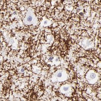
Expected resultCNP-positive staining in glial cells of caudate (HPA tissue IHC: High). HPA tissue profile: Highly specific expression in oligodendrocytes in CNS and peripheral nerves. No signal in the no-primary control.
💡Decision noteStart with heat-mediated EDTA pH 8.0 retrieval (datasheet A01017-4); the emitted study does not specify retrieval conditions (PMC12160429).
Section 2

What Is the Expected CNP Staining Pattern?

CNP should stain oligodendrocytes and their processes in the CNS, with lower signal in peripheral nerves (HPA: Enhanced tissue IHC; oligodendrocyte profile). Expect staining associated with membrane rich structures, including brain white matter; CNP has no transmembrane segment (UniProt P09543: subcellular location and topology). HPA also reports an approved plasma membrane location by ICC-IF, which is separate evidence from paraffin section IHC (HPA: subcellular ICC-IF).

What am I looking at on my slide?
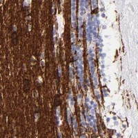
Strong glial cell and process staining in brain, with a structured neuropil pattern.This fits high staining in caudate and hippocampal glial cells, cerebellar granular layer processes, and cerebral cortex neuropil (HPA: tissue IHC). Interpret the stained structures alongside tissue morphology; HPA does not assign every stained process to an individual cell.
Predominantly nuclear staining, or a uniform stain across unrelated cell compartments.This does not match the reported membrane association or approved plasma membrane ICC-IF location (UniProt P09543: subcellular location; HPA: subcellular ICC-IF). Review morphology and detection controls before assigning it to CNP (general IHC practice).
Prominent staining of respiratory epithelium or marrow hematopoietic cells.These cell types are reported as not detected (HPA: bronchus respiratory epithelial cells; bone marrow hematopoietic cells). Consider antibody cross-reactivity or endogenous chromogenic detection activity; a no-primary control helps distinguish the latter (general IHC practice).
Haze spreads across the section and obscures individual cells or processes.The result cannot be scored against the reported cell and process pattern (HPA: tissue IHC). Review blocking, antibody concentration, washing, and chromogen development, then compare with a no-primary control (general IHC practice).
No signal in an otherwise evaluable caudate or hippocampus section.Both have high glial cell staining in the HPA tissue data (HPA: caudate and hippocampus). Check that glial cells are present, then review antibody suitability and the detection run using a known-positive section (general IHC practice).
💡Expected CNP appearanceCall the stain positive when glial cells or processes show distinct membrane associated signal in CNS regions with high reported staining (HPA: tissue IHC; UniProt P09543: membrane association); broad nuclear or unrelated epithelial staining is discordant (HPA: subcellular ICC-IF and bronchus tissue IHC).
How each factor affects the staining
Tissue and cell selectionBrain is RNA enriched, and several CNS glial or process patterns stain high; peripheral nerve stains low (HPA: tissue IHC and RNA specificity). Use the named CNS structures for a clearer positive comparison.
Membrane associationCNP is firmly bound to brain white matter membrane structures but has no transmembrane segment (UniProt P09543: subcellular location and topology). Assess cell and process morphology; a diffuse stain alone cannot establish the expected location.
Antibody validationFive listed antibodies have Enhanced IHC status (HPA: antibody validation). That supports the reported tissue pattern, but does not establish that another antibody, dilution, or retrieval condition will reproduce it.
IF/ICC Q: What location is reported?A: Plasma membrane is the approved ICC-IF location (HPA: subcellular ICC-IF). This helps interpret an IF image; it does not supply a paraffin IHC protocol or prove identical staining in every tissue.
Why is my staining missing, weak or wrong?
SituationLikely causeNext action
Brain positive control has no stain.The expected high glial signal is absent (HPA: caudate and hippocampus tissue IHC); the cause remains undetermined.Confirm glial tissue is represented, then inspect antibody dilution and detection controls and repeat with a known-positive section (general IHC practice).
Peripheral nerve appears weak beside brain.Lower peripheral nerve staining is reported (HPA: soft tissue peripheral nerve, Low; CNS sites, High).Compare matched tissue structures and avoid scoring weak peripheral nerve staining as assay failure solely because brain is stronger (HPA: tissue IHC).
Nuclei dominate the chromogenic signal.The pattern conflicts with reported membrane association and approved plasma membrane ICC-IF location (UniProt P09543; HPA: subcellular ICC-IF).Inspect the counterstain and no-primary control, then reassess the chromogen separately from nuclear colour (general IHC practice).
Stain appears in HPA-negative cell types.Respiratory epithelial and marrow hematopoietic cells are not detected (HPA: bronchus and bone marrow tissue IHC); cross-reactivity or endogenous activity is possible.Run a no-primary control and check endogenous enzyme blocking before assigning that signal to CNP (general IHC practice).
Diffuse colour hides glial processes.Background prevents comparison with the reported CNS process pattern (HPA: cerebellum and cerebral cortex tissue IHC).Review blocking and washes; adjust antibody concentration or chromogen development while retaining a positive tissue control (general IHC practice).
An IF image looks more sharply membrane outlined than the paraffin IHC slide.Approved plasma membrane localization comes from ICC-IF, while tissue IHC reports glial cells and processes (HPA: subcellular ICC-IF; tissue IHC).Score each preparation against its own reported evidence and morphology; avoid requiring an identical outline across methods (HPA: ICC-IF and tissue IHC; general IHC practice).

Sample controls for CNP IHC & IF

🧪Run caudate first and expect staining in glial cells (HPA: High in caudate glial cells). Use adipose tissue as the negative tissue, where adipocytes should lack detectable staining (HPA: Not detected in adipocytes); on the caudate slide, assess non-glial cells for background signal rather than assuming they are CNP-negative.
Positive control tissue: Caudate (Glial cells, HPA High)
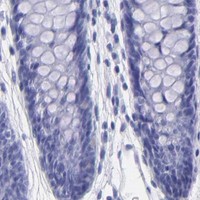
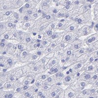
Negative control tissue: Adipose tissue (HPA Not detected)
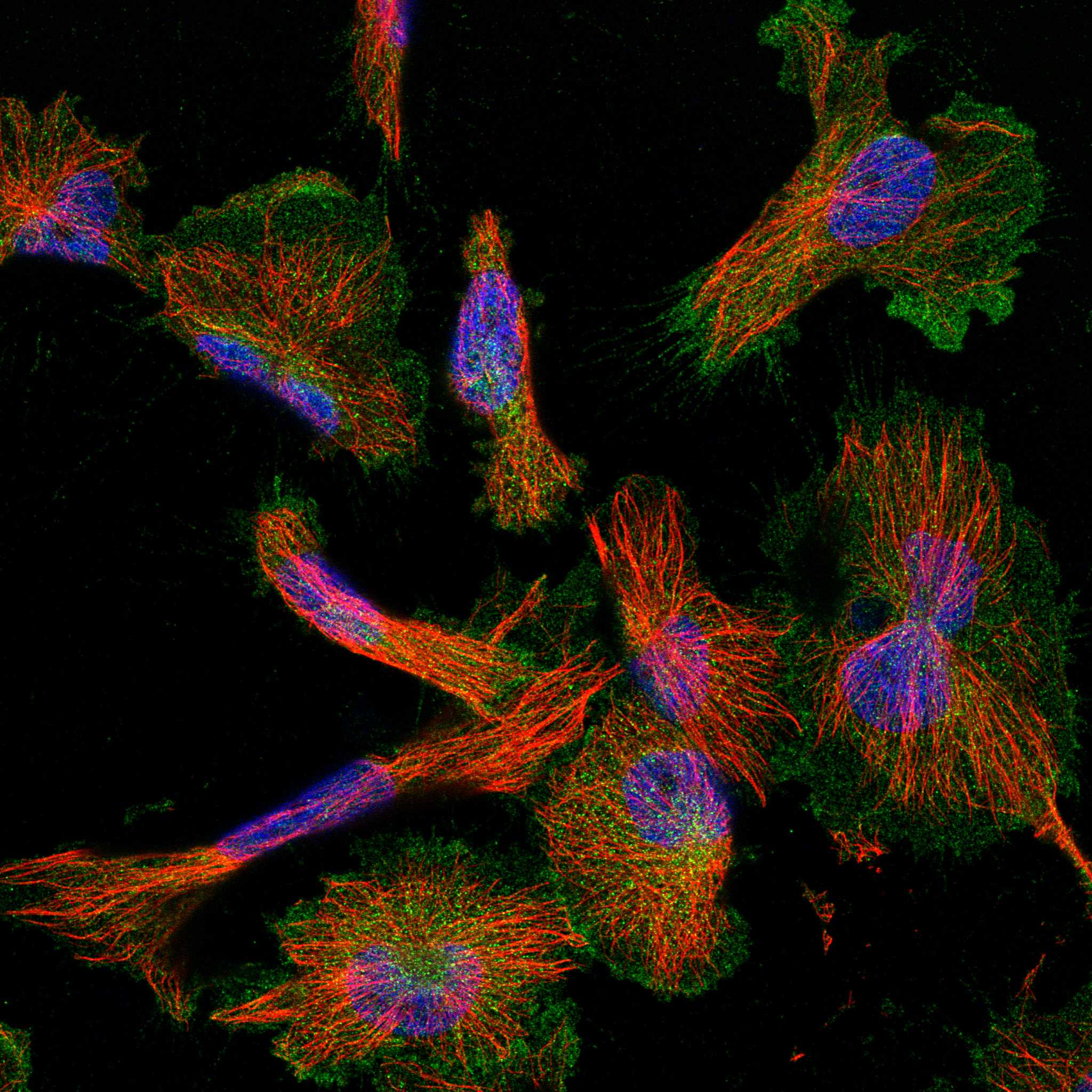
ICC-IF cell lines (HPA subcellular resource): HPA ICC-IF images show CNP in U-251MG, U2OS, with annotated localisation: Plasma membrane (approved) (HPA subcellular).
Technical controls: Include a no-primary, secondary-only control and a concentration-matched rabbit IgG isotype control for the rabbit primary (caption: rabbit anti-CNP antibody; standard IHC practice). Use CNP-knockout tissue or a validated immunizing-peptide block as a biological specificity control, and quench endogenous peroxidase before DAB detection (standard IHC practice; caption: HRP/DAB detection).
⚠️Feasibility: A target-specific fixation window or fixation effect is unreported, and the selected paraffin-section caption does not state the fixative (caption: fixative not stated). Heat retrieval in EDTA at pH 8.0 was used for mouse brain IHC, but retrieval dependence has not been established (caption: heat-mediated EDTA retrieval); whether frozen sections or IF are easier is unreported. In brain sections, assess any diffuse signal against the controls because CNP is firmly bound to membrane structures of white matter (UniProt P09543: subcellular location).

HPA tissue IHC evidence for CNP

Comprehensive Human Protein Atlas IHC scoring per tissue (reliability: Enhanced — High consistency between antibody staining and RNA expression data. Antibody staining in cells/structures not annotated, view images.). Rows are taken directly from the HPA tissue chart — click any row's HPA link to view the source.

Positive expression · recommended positive controls

TissueCell typeLevelEvidenceSource
Caudate Glial cells High Protein (IHC) HPA →
Cerebellum Processes in granular layer High Protein (IHC) HPA →
Cerebral cortex Neuropil High Protein (IHC) HPA →
Hippocampus Glial cells High Protein (IHC) HPA →
Soft tissue Peripheral nerve Low Protein (IHC) HPA →

Undetected expression · recommended negative controls

TissueCell typeLevelEvidenceSource
Adipose tissue Adipocytes Not detected Protein (IHC) HPA →
Adrenal gland Glandular cells Not detected Protein (IHC) HPA →
Appendix Glandular cells Not detected Protein (IHC) HPA →
Bone marrow Hematopoietic cells Not detected Protein (IHC) HPA →
Breast Adipocytes Not detected Protein (IHC) HPA →
Section 3

Advanced CNP IHC Tips

Use the paraffin-section CNP stain as the starting point, then assess signal against expected glial and nerve-associated patterns.

How should I optimize retrieval when CNP staining is weak in paraffin sections?
Start with heat-mediated retrieval in EDTA at pH 8.0 (datasheet A01017-4). Keep section thickness, heating time, and cooling conditions consistent across the first comparison, then adjust heating in small increments if signal remains weak (standard IHC practice). The selected mouse brain image used this retrieval before incubation with 2 μg/ml primary antibody overnight at 4°C (datasheet A01017-4). Compare signal in glial cells and processes with a no-primary control and check whether stronger retrieval also increases diffuse tissue staining (HPA: glial and process staining; standard IHC practice). Avoid treating a brighter background as improved CNP detection (standard IHC practice).
Can fixation explain weak or uneven CNP staining?
The selected mouse brain caption identifies a paraffin section but does not state its fixative (datasheet A01017-4). Target-specific fixation sensitivity is therefore unknown from the supplied evidence; neither the tissue pattern nor CNP topology establishes which fixation conditions preserve its epitope (HPA: tissue profile; UniProt P09543 topology). For a controlled comparison, document fixative, fixation duration, section thickness, and storage history, and stain matched sections in the same run (standard IHC practice). Check morphology and a no-primary control before attributing uneven signal to fixation, since section damage, retrieval variation, and nonspecific detection can also alter a chromogenic image (standard IHC practice).
Which staining compartments should I expect for CNP?
In tissue, assess staining along oligodendrocyte-associated processes and white matter structures, while considering the sampled region (HPA: highly specific oligodendrocyte expression in CNS and peripheral nerves; UniProt P09543: firmly bound to brain white matter membranes). HPA reports high signal in caudate glial cells, cerebellar granular-layer processes, cerebral-cortex neuropil, and hippocampal glial cells (HPA: tissue IHC). A membrane-associated pattern is plausible even though CNP has no annotated transmembrane segment (UniProt P09543 topology; HPA: approved plasma-membrane location). Compare those structures with adjacent morphology on the counterstained section; a uniform nuclear stain or broad signal in unrelated cells warrants investigation (HPA: tissue profile; standard IHC practice).
Could isoforms or epitope accessibility change the CNP pattern?
CNP has 2 annotated isoforms, CNPI and CNPII, but the supplied product caption does not identify which isoform its antibody recognizes (UniProt P09543 isoforms; datasheet A01017-4). It also gives no epitope sequence, so do not assign an isoform-specific tissue pattern from this stain (datasheet A01017-4). CNP lacks an annotated transmembrane segment and has reported phosphorylation and acetylation sites; these facts alone do not establish antibody sensitivity to a modification (UniProt P09543 topology and modified residues). If an unexpected pattern persists, obtain the antibody's immunogen or epitope information and compare validated reagents on serial sections under matched retrieval and detection conditions (standard IHC practice).
How can I investigate the CNP pattern with multiplex immunofluorescence?
Use IF as a separate assay and pair CNP with a validated oligodendrocyte marker to assess whether stained cells fit its reported tissue distribution (HPA: oligodendrocyte expression; standard IF practice). Select well-separated fluorophores after checking unstained tissue autofluorescence, especially before interpreting faint signal in processes (standard IF practice; HPA: high signal in cerebellar processes). Optimize permeabilisation against the antibody's mapped epitope and its accessibility on the relevant side of a membrane; an epitope orientation is not provided here (UniProt P09543: no transmembrane segment; standard IF practice). Include single-label, no-primary, and secondary-only controls to assess channel bleed-through and background before comparing IF with chromogenic IHC (standard IF practice).
How do I distinguish CNP staining from chromogenic background?
Start with a no-primary control and inspect whether diffuse brown signal follows tissue edges, damaged areas, or structures outside the expected glial and nerve-associated pattern (HPA: tissue IHC; standard IHC practice). The selected mouse brain image used 10% goat serum blocking, a peroxidase-conjugated secondary, and DAB detection (datasheet A01017-4). Apply an endogenous peroxidase block and verify washing and secondary-antibody specificity as general chromogenic controls (standard IHC practice). If background persists, compare shorter development or a primary-antibody titration while keeping retrieval and section handling matched; preserve glial-process detail when judging improvement (HPA: glial and process staining; standard IHC practice).
How should I quantify CNP across tissue sections? ⚠ ANSWER MARKED FOR VERIFICATION
Define regions before scoring and keep section thickness, retrieval, illumination, and DAB development consistent across groups (standard IHC practice). For identifiable glial cell bodies, report percent positive cells or an H-score with stated intensity thresholds; for process-rich regions, measure positive area or process density per mm² (HPA: glial cells and processes; standard IHC practice). Normalize cell counts to the number of eligible cells and area measures to the annotated tissue area, excluding folds, tears, and empty spaces by a prespecified rule (standard IHC practice). Keep anatomical regions separate because HPA describes different CNP-positive structures across caudate, cerebellum, cortex, and hippocampus (HPA: tissue IHC).
What would make a CNP-positive IHC result convincing?
A convincing result follows the sampled anatomy: glial cells, neuropil, or processes in the reported brain regions support interpretation when morphology agrees (HPA: caudate, cortex, cerebellum, and hippocampus staining; standard IHC practice). Membrane-associated signal is compatible with CNP, but a dominant nuclear pattern requires scrutiny (UniProt P09543: membrane association and no transmembrane segment; HPA: approved plasma-membrane location). Compare suspicious staining at cut edges or necrotic areas with intact tissue and a no-primary control (standard IHC practice). Check endogenous peroxidase contribution before calling widespread DAB deposition positive, then confirm the pattern across independently stained sections under matched conditions (standard IHC practice).
Boster reagents

Best CNP / 2',3'-cyclic-nucleotide 3'-phosphodiesterase IHC Antibodies

A01017-4 has paraffin-section IHC images from human, mouse, and rat brain (catalog IHC image captions), plus IF images from mouse and rat brain (catalog IF image captions).

Real IHC data IHC analysis of CNPase/CNP using anti-CNPase/CNP antibody (A01017-4). CNPase/CNP was detected in a paraffin-embedded section of mouse brain tissue. Heat mediated antigen retrieval was performed in EDTA buffer (pH 8.0, epitope retrieval solution). The tissue section was blocked with 10% goat serum. The tissue section was then incubated with 2 μg/ml rabbit anti-CNPase/CNP Antibody (A01017-4) overnight at 4°C. Peroxidase Conjugated Goat Anti-rabbit IgG was used as secondary antibody and incubated for 30 minutes at 37°C. The tissue section was developed using HRP Conjugated Rabbit IgG Super Vision Assay Kit (Catalog # SV0002) with DAB as the chromogen.
Anti-CNPase/CNP Antibody ®
Cat # A01017-4

The sole rendered SKU, A01017-4, lists IHC and IF applications and human, mouse, and rat reactivity (catalog applications/reactivity). Its IHC images show paraffin-embedded brain from all three species; its IF images show paraffin-embedded mouse and rat brain (catalog IHC/IF image captions).

Which to pick: For tissue IHC, choose A01017-4: its own captions show paraffin sections with EDTA pH 8.0 retrieval, 2 μg/ml primary antibody, and DAB detection; the fixative is unreported (catalog IHC image captions). For IF, the same rabbit antibody has mouse and rat brain section images at 5 μg/ml; ICC validation and clonality are unreported (catalog IF image captions; catalog applications/host/clone). For cross-species IHC, its images cover human, mouse, and rat brain (catalog IHC image captions).

Each figure is that product's own IHC / IF validation image from its datasheet.

References

  1. UniProt Consortium. UniProt entry P09543 (CN37_HUMAN, 2',3'-cyclic-nucleotide 3'-phosphodiesterase).
  2. Human Protein Atlas. CNP tissue IHC expression (reliability: Enhanced).
  3. Human Protein Atlas. CNP subcellular location (ICC-IF): Localized to the plasma membrane..
  4. Human Protein Atlas. CNP antibody validation summary (5 antibodies).
  5. Impact of obesity on the expression profile of natriuretic peptide system in a rat experimental model. PloS one 2013 — PMC3756951.
  6. Combined enriched environment and fluoxetine enhance myelin protein expression in the prefrontal cortex of a chronic unpredictable stress depression model. Behavioral and brain functions : BBF 2025 — PMC12160429.
  7. The Local CNP/GC-B system in growth plate is responsible for physiological endochondral bone growth. Scientific reports 2015 — PMC5395013.
  8. Natriuretic Peptides in Post-mortem Brain Tissue and Cerebrospinal Fluid of Non-demented Humans and Alzheimer's Disease Patients. Frontiers in neuroscience 2018 — PMC6275179.
  9. PubMed PMID:1385234 — UniProt-cited evidence.
  10. PubMed PMID:2835044 — UniProt-cited evidence.
  11. PubMed PMID:8392017 — UniProt-cited evidence.