CYP3A4 / Cytochrome P450 3A4 · IHC design guide

Design Immunohistochemistry for CYP3A4

Plan chromogenic CYP3A4 IHC in paraffin sections using hepatocyte and intestinal endocrine cell staining as reference patterns (HPA tissue IHC). This guide covers fixation consistency, staining controls, and titration of the catalog antibody at 0.5–1 µg/mL (datasheet PB10055).

Evidence assembled Oct 2026 · For research use; verify linked source records and product datasheet before use
Immunohistochemistry protocol sheet for CYP3A4 (IHC for CYP3A4): expected localisation Cytoplasmic staining (HPA tissue IHC); ER and microsome membrane protein (UniProt), antibody PB10055, validated IHC image, and IHC protocol steps
Printable CYP3A4 IHC protocol sheet — expected localisation Cytoplasmic staining (HPA tissue IHC); ER and microsome membrane protein (UniProt), antibody PB10055, controls and protocol steps. Open the full CYP3A4 IHC guide →

CYP3A4 Immunohistochemistry Experimental Design Guide

Expected localisation, validated protocols, controls and antibodies — the at-a-glance facts below, then the full design guide.

Must know before staining
Expected localisation Cytoplasmic staining (HPA tissue IHC); ER and microsome membrane protein (UniProt)
Staining pattern Cytoplasmic hepatocytes and intestinal endocrine cells (HPA tissue IHC)
Antigen retrieval EDTA pH 8.0 HIER, heat-mediated (datasheet PB10055)
Positive control ⓘ Colon+3 more · see all
Negative control ⓘ Adipose tissue+4 more · see all
Important caveats
Reasons your staining may differ from the expected pattern.
Fixation Keep formalin fixation consistent across sections (standard IHC practice; not target-specific)
Caveat Hepatic endogenous peroxidase may increase DAB background (standard IHC practice)
Regulation Specific expression regulators are unreported (UniProt)
Isoform / epitope No isoforms annotated; chain spans residues 2–503 (UniProt)
Section 1

Recommended CYP3A4 IHC & IF Protocols

The catalog antibody has an IHC-P protocol (datasheet: PB10055). Two published CYP3A4 IHC protocols provide additional conditions for liver and breast cancer sections (PMC4933826; PMC5503538).

Recommended immunohistochemistry (IHC-P) protocol parameters
SampleParaffin-embedded human liver cancer tissue; fixative not specified (datasheet PB10055)
FixationImage fixative and duration unreported (datasheet PB10055); verify before use.
Sectioning4–5 µm sections on charged slides (standard)
DeparaffinisationXylene, graded ethanol series to water (standard)
Antigen retrievalHeat retrieval: EDTA pH 8.0 (datasheet PB10055); 20 min, 95–100 °C (standard)
Peroxidase block3% H2O2, 10 min, room temperature (standard)
Blocking10% goat serum (datasheet PB10055)
Primary antibodyRabbit anti-CYP3A4, 0.5-1μg/ml (datasheet PB10055)
Primary incubationOvernight at 4 °C (datasheet PB10055)
DetectionStreptavidin-biotin complex (SABC), DAB chromogen (datasheet PB10055)
CounterstainHematoxylin, blue, dehydrate and mount (standard)
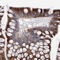
Expected resultCYP3A4-positive staining in endocrine cells of colon (HPA tissue IHC: High). HPA tissue profile: Selective cytoplasmic expression in liver, small intestine and duodenum. No signal in the no-primary control.
💡Decision noteStart with heat-mediated EDTA pH 8.0 for the catalog antibody (datasheet: PB10055). Citrate pH 6.0 is a published alternative using a different antibody (PMC5503538).
Section 2

What Is the Expected CYP3A4 Staining Pattern?

CYP3A4 is expected to stain the cytoplasm of hepatocytes and intestinal endocrine cells (HPA tissue IHC). UniProt places it in endoplasmic reticulum and microsomal membranes, with a transmembrane segment at residues 2–22 (UniProt P08684 topology). HPA rates its tissue IHC evidence Enhanced, citing high consistency between antibody staining and RNA expression (HPA tissue IHC).

What am I looking at on my slide?
Strong cytoplasmic staining in liver hepatocytes, with staining in intestinal endocrine cells.This matches HPA's High staining in hepatocytes and endocrine cells of the duodenum, small intestine and colon (HPA tissue IHC). Score the named cell populations and their cytoplasm; staining elsewhere in a section does not establish the same pattern (general IHC practice).
Predominantly nuclear, extracellular or sharply cell-surface staining instead of cytoplasmic staining.That distribution conflicts with HPA's cytoplasmic tissue profile and UniProt's endoplasmic reticulum and microsomal membrane annotation (HPA tissue IHC; UniProt P08684 subcellular location). Treat it as suspect and compare with a positive tissue and a no-primary control before assigning specificity (general IHC practice).
Strong staining in adipocytes or bone-marrow hematopoietic cells.HPA reports CYP3A4 as Not detected in those specific cell populations (HPA tissue IHC). Consider cross-reactivity or endogenous chromogenic detection activity, particularly if staining survives omission of the primary antibody; a negative HPA observation does not prove universal absence (general IHC practice; HPA tissue IHC).
Diffuse color across tissue and empty slide areas, with little cell-specific contrast.This is not the selective cytoplasmic pattern described by HPA (HPA tissue IHC). Background can arise from detection reagents, endogenous enzyme activity or inadequate washing; a no-primary control helps separate detection-related color from staining that requires the primary antibody (general IHC practice).
No detectable staining in hepatocytes on a liver section.HPA reports High hepatocyte staining, so first investigate the IHC run, antibody conditions and tissue quality (HPA tissue IHC; general IHC practice). One blank section alone cannot establish CYP3A4 absence; compare a positive reference section processed in the same run (general IHC practice).
💡Expected CYP3A4 appearanceCall a result positive when staining is predominantly cytoplasmic in hepatocytes or the specified intestinal endocrine cells, with High signal expected in HPA's positive populations; isolated nuclear, extracellular or broadly indiscriminate color is suspect (HPA tissue IHC; UniProt P08684 subcellular location; general IHC practice).
How each factor affects the staining
Membrane topologyCYP3A4 has one transmembrane segment at residues 2–22 and is assigned to endoplasmic reticulum and microsomal membranes (UniProt P08684 topology and subcellular location). This supports a cytoplasmic compartment call at light-microscope resolution; it does not predict a distinctive visible reticular pattern (general IHC interpretation).
Cell and tissue choiceHPA lists High staining in liver hepatocytes and endocrine cells of the duodenum, small intestine and colon (HPA tissue IHC). Use those named cells as positive references; HPA's Not detected calls apply to the specified negative cell populations, not automatically to every cell in those tissues (HPA tissue IHC).
Antibody and retrieval evidenceHPA lists Enhanced IHC validation for antibody CAB033671 (HPA antibodies). The supplied records give no mapped epitope, retrieval condition or target-specific fixation sensitivity; choose retrieval and working dilution from the IHC-validated antibody's IHC-P instructions, then judge the resulting pattern against HPA (supplied evidence scope; general IHC practice).
IF/ICC Q: Should the IF localization settle an ambiguous IHC result?A: HPA calls cytosol approved and vesicles uncertain in ICC-IF, while cautioning that the subcellular summary uses antibodies targeting proteins from multiple genes (HPA subcellular). Use it as context, not as proof that a vesicular IHC signal is specific (HPA subcellular; general assay interpretation).
Why is my staining missing, weak or wrong?
SituationLikely causeNext action
Liver hepatocytes are blank.The run may have lost sensitivity or the section may be unsuitable; hepatocytes are High in HPA tissue IHC (HPA tissue IHC; general IHC practice).Run a positive liver reference alongside the sample; check the catalog antibody's IHC-P retrieval, dilution and detection instructions and confirm that the counterstained section contains intact hepatocytes (general IHC practice).
Color appears in the no-primary control.The color does not require the primary antibody; endogenous enzyme activity or detection reagents may contribute in chromogenic IHC (general IHC practice).Check the enzyme-blocking and secondary-detection steps, then repeat the no-primary control before interpreting tissue staining (general IHC practice).
The whole section has diffuse color.Broad background obscures HPA's selective cytoplasmic pattern; incomplete washing or nonspecific reagent binding are possible (HPA tissue IHC; general IHC practice).Compare no-primary and positive-reference sections, then review blocking, antibody dilution and wash steps under the antibody's IHC-P instructions (general IHC practice).
Staining is mainly nuclear or extracellular.Those compartments do not fit the HPA cytoplasmic tissue profile or UniProt membrane localization (HPA tissue IHC; UniProt P08684 subcellular location).Inspect the positive reference and no-primary control; withhold a CYP3A4-positive call until the expected cell-specific cytoplasmic pattern is reproduced (general IHC practice).
Adipocytes or bone-marrow hematopoietic cells stain strongly.HPA marks these cell populations Not detected; cross-reactivity or endogenous detection activity is possible (HPA tissue IHC; general IHC practice).Check whether color persists without primary antibody and whether positive-reference hepatocytes show the expected pattern; reassess specificity before scoring those cells (general IHC practice; HPA tissue IHC).
Vesicular IF/ICC signal is used to justify an unusual IHC pattern.HPA labels vesicular ICC-IF localization uncertain and cautions that its summary draws on antibodies targeting proteins from multiple genes (HPA subcellular).Interpret the paraffin-section result against HPA tissue IHC and the IHC antibody's validation; do not upgrade an ambiguous IHC pattern solely from the vesicular IF observation (HPA tissue IHC; HPA antibodies; HPA subcellular).

Sample controls for CYP3A4 IHC & IF

🧪Run liver first and expect cytoplasmic staining in hepatocytes (HPA: High in hepatocytes; UniProt P08684: endoplasmic reticulum membrane). Run adipose tissue as a negative comparator, focusing on adipocytes (HPA: Not detected in adipocytes); on the liver slide, use cells lacking hepatocyte-like staining as an internal background reference, without assuming every nonhepatocyte is CYP3A4-negative.
Positive control tissue: Colon (Endocrine cells, HPA High)
Negative control tissue: Adipose tissue (HPA Not detected)
ICC-IF cell lines (HPA subcellular resource): HPA ICC-IF images show CYP3A4 in Hep-G2, U2OS, A-549, CACO-2, with annotated localisation: Cytosol (approved) (HPA subcellular).
Technical controls: Include a no-primary (secondary-only) slide, a concentration-matched rabbit IgG isotype control, and a CYP3A4 knockout sample or peptide-blocked primary antibody as a biological specificity control (selected PB10055 caption: rabbit primary antibody). For liver DAB staining, block endogenous peroxidase and check for endogenous biotin before using the biotin-based detection system (selected PB10055 caption: biotinylated secondary, streptavidin–biotin complex and DAB).
⚠️Feasibility: A target-specific fixation window or fixation effect is unreported, and the selected PB10055 tissue-IHC caption does not state a fixative. Heat retrieval in EDTA at pH 8.0 was used on a paraffin-embedded liver cancer section (selected PB10055 caption), but the supplied evidence does not establish that retrieval is required or that frozen sections or IF are easier. In liver, endogenous peroxidase or biotin can complicate interpretation of the reported biotin-based DAB method (selected PB10055 caption: streptavidin–biotin complex and DAB).

HPA tissue IHC evidence for CYP3A4

Comprehensive Human Protein Atlas IHC scoring per tissue (reliability: Enhanced — High consistency between antibody staining and RNA expression data.). Rows are taken directly from the HPA tissue chart — click any row's HPA link to view the source.

Positive expression · recommended positive controls

TissueCell typeLevelEvidenceSource
Colon Endocrine cells High Protein (IHC) HPA →
Duodenum Endocrine cells High Protein (IHC) HPA →
Liver Hepatocytes High Protein (IHC) HPA →
Small intestine Endocrine cells High Protein (IHC) HPA →

Undetected expression · recommended negative controls

TissueCell typeLevelEvidenceSource
Adipose tissue Adipocytes Not detected Protein (IHC) HPA →
Adrenal gland Glandular cells Not detected Protein (IHC) HPA →
Appendix Endocrine cells Not detected Protein (IHC) HPA →
Bone marrow Hematopoietic cells Not detected Protein (IHC) HPA →
Breast Adipocytes Not detected Protein (IHC) HPA →
Section 3

Advanced CYP3A4 IHC Tips

Use the catalog antibody’s paraffin-section IHC conditions as the starting point, then judge staining against CYP3A4’s expected cell types and location.

What should I change if CYP3A4 staining is weak in paraffin sections?
Start with heat-mediated antigen retrieval in EDTA at pH 8.0 (datasheet PB10055). The catalog antibody stained a paraffin-embedded human liver cancer section after this retrieval, followed by 1 μg/ml primary antibody overnight at 4°C (datasheet PB10055). If signal is weak, compare heating durations on adjacent sections while keeping antibody concentration and chromogen development constant (standard IHC practice). Excessive retrieval can damage section morphology or increase background, so inspect tissue integrity alongside signal (standard IHC practice). Include a liver hepatocyte control, where CYP3A4 staining is reported as high, to distinguish failed retrieval from a low-expressing specimen (HPA: Liver hepatocytes High).
Could fixation explain absent CYP3A4 staining?
Target-specific fixation sensitivity is unknown: the catalog image identifies a paraffin-embedded section but does not state its fixative (datasheet PB10055: fixative not stated). Record each specimen’s fixative and fixation duration before comparing staining across cases (standard IHC practice). If fixation histories differ, run sections together using EDTA at pH 8.0 and the same detection conditions, then assess morphology and signal in matched regions (datasheet PB10055; standard IHC practice). A high-staining liver hepatocyte control can show whether the run worked, but it cannot establish which fixation condition preserves this antibody’s epitope (HPA: Liver hepatocytes High; standard IHC practice).
Where should convincing CYP3A4 staining appear within a positive cell?
Expect predominantly cytoplasmic staining at light-microscope resolution, consistent with the selective cytoplasmic tissue pattern reported for CYP3A4 (HPA: tissue IHC profile). CYP3A4 is assigned to the endoplasmic reticulum and microsome membranes, with a transmembrane segment at residues 2–22 (UniProt P08684: subcellular location and topology). A granular or reticular cytoplasmic pattern can therefore be plausible, but routine chromogenic IHC cannot establish membrane-side orientation (UniProt P08684; standard IHC practice). Compare positive cells with adjacent tissue architecture rather than treating isolated nuclear or extracellular deposits as expected localisation (standard IHC practice). HPA’s subcellular cytosol assignment carries a multiple-gene antibody caution and should not alone override the UniProt membrane annotation (HPA: subcellular caution; UniProt P08684).
How can I assess whether the antibody recognises CYP3A4 specifically?
Check the catalog antibody’s immunogen or mapped epitope before interpreting cross-reactivity; neither is supplied in this payload (datasheet PB10055: epitope not provided). UniProt lists 0 annotated isoforms and 0 annotated glycosylation sites for CYP3A4, but those annotations do not establish antibody specificity (UniProt P08684). Its single transmembrane segment spans residues 2–22; epitope accessibility cannot be inferred without the epitope sequence and its membrane orientation (UniProt P08684; standard IHC practice). Compare staining with expected positive liver hepatocytes and an appropriate negative tissue control, then seek independent specificity evidence before assigning unexpected staining to CYP3A4 (HPA: Liver hepatocytes High; standard IHC practice).
How should I adapt these IHC findings when planning CYP3A4 immunofluorescence?
Treat IF as a separate validation task: the supplied catalog image documents chromogenic IHC in a paraffin-embedded section, with no IF result or fixative reported (datasheet PB10055). Multiplex CYP3A4 with a hepatocyte identity marker when examining liver, since hepatocytes show high tissue-IHC staining (HPA: Liver hepatocytes High; standard IF practice). Choose spectrally separated fluorophores and inspect unstained tissue, favoring a far-red channel if tissue autofluorescence obscures shorter wavelengths (standard IF practice). Compare permeabilised and minimally permeabilised preparations because CYP3A4 has a membrane anchor at residues 2–22, while this antibody’s epitope and membrane-facing side are unspecified (UniProt P08684; datasheet PB10055: epitope not provided).
What should I check when CYP3A4 DAB staining is diffuse or widespread?
First compare a no-primary section with the stained section to identify signal from the detection system or tissue rather than the primary antibody (standard IHC practice). The catalog example used biotinylated goat anti-rabbit secondary antibody, an avidin-biotin detection complex, and DAB, so a detection-control section is particularly useful (datasheet PB10055). Apply a peroxidase block and check whether DAB development is excessive; these are general chromogenic IHC controls, not demonstrated CYP3A4-specific requirements (standard IHC practice). Reassess blocking and primary concentration against the reported 10% goat serum block and 1 μg/ml primary condition (datasheet PB10055). Interpret widespread deposits cautiously when they obscure the expected cytoplasmic cell pattern (HPA: tissue IHC profile).
How should I quantify CYP3A4 staining across paraffin sections? ⚠ ANSWER MARKED FOR VERIFICATION
Define the cell population and region before scoring, then record either percent positive cells with intensity, an H-score, or stained area per mm² (standard IHC practice). For liver, score hepatocytes separately from other cells because HPA reports high staining in hepatocytes (HPA: Liver hepatocytes High). Normalize positive counts or stained area to the corresponding viable, assessable cell count or tissue area, and exclude folds and necrotic regions consistently (standard IHC practice). Keep retrieval, 1 μg/ml primary concentration, and DAB development comparable across sections so processing differences do not masquerade as biological differences (datasheet PB10055; standard IHC practice). Report the scoring threshold and control performance alongside each comparison (standard IHC practice).
How do I distinguish true CYP3A4 staining from artefact?
Favor cytoplasmic staining in an expected cell population, such as liver hepatocytes, over isolated nuclear or extracellular signal (HPA: tissue IHC profile and Liver hepatocytes High; standard IHC practice). Check section edges, folds, and necrotic regions for staining that follows tissue damage rather than intact cell boundaries (standard IHC practice). A no-primary control and a peroxidase-block control help assess detection background and endogenous enzyme activity in a DAB workflow (standard IHC practice). Compare an appropriate negative tissue control: HPA reports adipose adipocytes as not detected, although a negative stain alone cannot validate specificity (HPA: Adipose adipocytes Not detected; standard IHC practice). Unexpected positives warrant independent antibody or orthogonal expression evidence before biological interpretation (standard IHC practice).
Boster reagents

Best CYP3A4 / Cytochrome P450 3A4 IHC Antibodies

The catalog antibodies have real IHC images from paraffin-embedded human liver cancer tissue (PB10055 image caption) and human liver carcinoma tissue (A00339-1 image caption); IF is listed for A00339-1 (catalog applications).

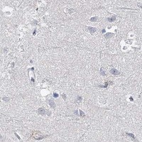
Real IHC data IHC analysis of CYP3A4 using anti-CYP3A4 antibody (PB10055). CYP3A4 was detected in a paraffin-embedded section of human liver cancer tissue. Heat mediated antigen retrieval was performed in EDTA buffer (pH 8.0, epitope retrieval solution). The tissue section was blocked with 10% goat serum. The tissue section was then incubated with 1 μg/ml rabbit anti-CYP3A4 Antibody (PB10055) overnight at 4°C. Biotinylated goat anti-rabbit IgG was used as secondary antibody and incubated for 30 minutes at 37°C. The tissue section was developed using Strepavidin-Biotin-Complex (SABC) (Catalog # SA1022) with DAB as the chromogen.
Anti-Cytochrome P450 3A4/CYP3A4 Antibody ®
Cat # PB10055
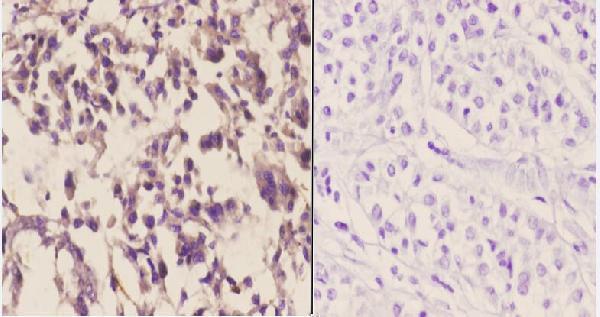
Real IHC data Immunohistochemistry (IHC) analyzes of CYP3A4/5 (F386) pAb in paraffin-embedded human liver carcinoma tissue at 1:50.showing cytoplasmic staining. Negative control (the right)Using PBS instead of primary antibody, secondary antibody is Goat Anti-Rabbit IgG-biotin followed by avidin-peroxidase.
Anti-CYP3A4/5 (F386) Antibody
Cat # A00339-1

PB10055 will render with IHC staining of a paraffin-embedded human liver cancer section at 1 μg/ml (PB10055 image caption). A00339-1 will render with IHC staining of paraffin-embedded human liver carcinoma tissue at 1:50, alongside a PBS primary-omission control (A00339-1 image caption).

Which to pick: For human paraffin-section IHC, choose PB10055 when its documented EDTA pH 8.0 retrieval and DAB workflow fit the experiment (PB10055 image caption). For IF/ICC or planned human, mouse, or rat work, consider A00339-1: IF and IHC are listed applications, and all three species are listed as reactive, although its supplied image documents only human paraffin-section IHC (A00339-1 catalog applications and reactivity; A00339-1 image caption). The fixative is unreported in both IHC image captions (PB10055 image caption; A00339-1 image caption).

Each figure is that product's own IHC / IF validation image from its datasheet.

References

  1. UniProt Consortium. UniProt entry P08684 (CP3A4_HUMAN, Cytochrome P450 3A4).
  2. Human Protein Atlas. CYP3A4 tissue IHC expression (reliability: Enhanced).
  3. Human Protein Atlas. CYP3A4 subcellular location (ICC-IF): Mainly localized to the cytosol. In addition localized to vesicles. Caution: Based on antibodies targeting proteins from multiple genes..
  4. Human Protein Atlas. CYP3A4 antibody validation summary (3 antibodies).
  5. Immunohistochemical markers of CYP3A4 and CYP3A7: a new tool towards personalized pharmacotherapy of hepatocellular carcinoma. European journal of histochemistry : EJH 2016 — PMC4933826.
  6. The associations of genetic polymorphisms in CYP1A2 and CYP3A4 with clinical outcomes of breast cancer patients in northern China. Oncotarget 2017 — PMC5503538.
  7. A Promising Approach to Treat Psoriasis: Inhibiting Cytochrome P450 3A4 Metabolism to Enhance Desoximetasone Therapy. Pharmaceutics 2023 — PMC10458942.
  8. The Intervention and Mechanism of Action for Aloin against Subchronic Aflatoxin B1 Induced Hepatic Injury in Rats. International journal of molecular sciences 2021 — PMC8584096.
  9. PubMed PMID:3460094 — UniProt-cited evidence.
  10. PubMed PMID:3267210 — UniProt-cited evidence.
  11. PubMed PMID:3464943 — UniProt-cited evidence.