DAXX / Death domain-associated protein 6 · IHC design guide

Design Immunohistochemistry for DAXX

Plan DAXX paraffin IHC around its general nuclear tissue staining and high signal in germinal center cells and round or early spermatids (HPA tissue IHC). This guide covers positive controls, consistent fixation and scoring of nuclear staining.

Evidence assembled Oct 2026 · For research use; verify linked source records and product datasheet before use
Immunohistochemistry protocol sheet for DAXX (IHC for DAXX): expected localisation Tissue: nuclear ; cytoplasm also annotated (HPA tissue IHC; UniProt), antibody PB9550, validated IHC image, and IHC protocol steps
Printable DAXX IHC protocol sheet — expected localisation Tissue: nuclear ; cytoplasm also annotated (HPA tissue IHC; UniProt), antibody PB9550, controls and protocol steps. Open the full DAXX IHC guide →

DAXX Immunohistochemistry Experimental Design Guide

Expected localisation, validated protocols, controls and antibodies — the at-a-glance facts below, then the full design guide.

Must know before staining
Expected localisation Tissue: nuclear; cytoplasm also annotated (HPA tissue IHC; UniProt)
Staining pattern Nuclear staining; distinct in epididymis, testis and lymphoid tissue (HPA tissue IHC)
Antigen retrieval EDTA pH 8.0 HIER, heat-mediated (datasheet PB9550)
Positive control ⓘ Appendix+4 more · see all
Negative control ⓘ None in HPA (detected in all 45 tissues); use no-primary + isotype controls
Important caveats
Reasons your staining may differ from the expected pattern.
Fixation Keep fixation consistent across sections (standard IHC practice; not target-specific); Matched tissue-IHC evidence does not establish the fixation claim. Validate the specimen-specific method before use. (selected-SKU IHC image PB9550)
Caveat Glucose deprivation or oxidative stress can shift DAXX to cytoplasm (UniProt)
Regulation SPOP promotes DAXX degradation (UniProt)
Isoform / epitope Five isoforms; no extracellular domain; check epitope coverage (UniProt)
Section 1

Recommended DAXX IHC & IF Protocols

The catalog antibody’s IHC-P protocol (datasheet: PB9550) is accompanied by published DAXX IHC protocols for gastrointestinal stromal tumors (PMC4695492) and canine prostate and bladder samples (PMC12737483).

Recommended immunohistochemistry (IHC-P) protocol parameters
SampleParaffin-embedded rat intestine tissue; fixative not specified (datasheet PB9550)
FixationImage fixative and duration unreported (datasheet PB9550); verify before use.
Sectioning4–5 µm sections on charged slides (standard)
DeparaffinisationXylene, graded ethanol series to water (standard)
Antigen retrievalHeat retrieval: EDTA pH 8.0 (datasheet PB9550); 20 min, 95–100 °C (standard)
Peroxidase block3% H2O2, 10 min, room temperature (standard)
Blocking10% goat serum (datasheet PB9550)
Primary antibodyRabbit anti-DAXX, 0.5-1μg/ml (datasheet PB9550)
Primary incubationOvernight at 4 °C (datasheet PB9550)
DetectionStreptavidin-biotin complex (SABC), DAB chromogen (datasheet PB9550)
CounterstainHematoxylin, blue, dehydrate and mount (standard)
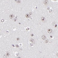
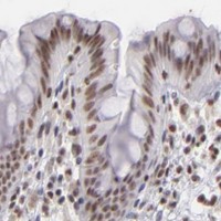
Expected resultDAXX-positive staining in lymphoid tissue of appendix (HPA tissue IHC: High). HPA tissue profile: General nuclear expression, distinct expression in epididymis, testis and lymphoid tissue. No signal in the no-primary control.
💡Decision noteStart with heat-mediated EDTA pH 8.0 retrieval for the catalog antibody (datasheet: PB9550); published protocols use different retrieval buffers and should be followed with their respective antibodies (PMC4695492; PMC12737483).
Section 2

What Is the Expected DAXX Staining Pattern?

DAXX should stain predominantly in nuclei across many tissues (HPA: general nuclear expression; UniProt Q9UER7: ubiquitous). Stronger IHC signal is reported in appendix lymphoid tissue, lymph node germinal center cells, round or early spermatids, tonsil non-germinal center cells, and urothelial cells (HPA: High). HPA rates its tissue IHC reliability Enhanced, with medium consistency between antibody staining and RNA expression (HPA: tissue IHC). DAXX has no transmembrane segment (UniProt Q9UER7: topology).

What am I looking at on my slide?
Nuclear chromogenic staining in the expected cells, with some nuclei appearing punctate.This fits the reported general nuclear IHC pattern (HPA: tissue IHC). DAXX occupies the nucleoplasm and nuclear bodies (UniProt Q9UER7: subcellular location; HPA: ICC-IF); discrete bodies may be easier to resolve by IF than in a chromogenic section (general microscopy practice). Score the nuclear signal and its cell distribution, without requiring visible puncta in every positive nucleus.
Predominantly cytoplasmic staining with little nuclear signal in an otherwise expected positive tissue.This departs from the usual nuclear pattern (HPA: tissue IHC). Check whether the signal follows cell boundaries, pigment, or uneven detection before calling it DAXX (general IHC practice). Cytoplasmic puncta and stress-associated nuclear-to-cytoplasmic movement are reported (UniProt Q9UER7: subcellular location), so cytoplasmic signal alone cannot establish artefact.
Strong signal appears in cells expected to stain weakly, while expected positive cells do not stain.Compare cell identity and compartment before interpreting intensity: glial cells in hippocampus and caudate, cholangiocytes, prostate glandular cells, endometrial stromal cells, and cardiomyocytes are listed as Low, not negative (HPA: tissue IHC). An unexpected distribution warrants checks for cross-reactivity or endogenous detection activity (general IHC practice); it does not prove either cause.
Diffuse color covers nuclei, cytoplasm, and tissue spaces without a clear cellular pattern.This obscures the predominantly nuclear DAXX distribution (HPA: tissue IHC). Review the negative control and detection background, then adjust blocking, washing, or detection conditions as indicated by those controls (general IHC practice). Diffuse color cannot be scored as DAXX merely because the section contains a reported positive cell population.
No nuclear signal is visible in a section containing a reported high-staining cell population.Appendix lymphoid tissue, lymph node germinal center cells, round or early spermatids, tonsil non-germinal center cells, and urothelial cells are reported High (HPA: tissue IHC). Confirm that the intended cells are present and assess the run controls before inferring loss of DAXX (general IHC practice). HPA staining levels are observations, not a guarantee for every specimen or antibody.
💡Expected DAXX appearanceCall a convincing positive result predominantly nuclear staining in the reported cell population, with strong signal plausible in HPA High groups (HPA: tissue IHC); widespread compartment-free color is suspect (general IHC practice).
How each factor affects the staining
Tissue and cell selectionDAXX is described as ubiquitous (UniProt Q9UER7: tissue specificity), but observed IHC intensity varies by cell population (HPA: High, Medium, and Low tissue entries). Use the named cell population when choosing and reading a control; a Low entry is not a negative control (HPA: tissue IHC).
Antibody-specific evidenceIHC validation differs among listed antibodies: CAB002224 and CAB025546 are Enhanced, while HPA008736 is Supported (HPA: antibody validation). Those ratings describe the listed antibodies; they do not transfer automatically to another reagent or establish the pattern in every specimen.
Subcellular context and stressNucleoplasm and nuclear bodies are supported by ICC-IF (HPA: subcellular); UniProt also reports nucleoli, cytoplasmic puncta, and movement to cytoplasm after glucose deprivation or oxidative stress (UniProt Q9UER7: subcellular location). Record the experimental context when evaluating an atypical cytoplasmic pattern.
Isoforms and epitope coverageFive isoforms are listed (UniProt Q9UER7: isoforms). The supplied record gives no epitope position or isoform coverage for the antibody, so it cannot predict whether staining represents every isoform. Check reagent-specific epitope information before making an isoform claim (general IHC practice).
Why is my staining missing, weak or wrong?
SituationLikely causeNext action
Expected positive cells show no nuclear staining.The section may lack the intended cell population, or the IHC run may have failed (general IHC practice); the HPA High designation alone does not diagnose the cause (HPA: tissue IHC).Verify the cells on the counterstain, inspect run controls, and review the antibody dilution, antigen retrieval, and detection steps against the reagent's validated IHC instructions (general IHC practice).
Nuclear staining is faint but the run control works.The sampled cells may belong to a lower-staining population: HPA reports Medium and Low as well as High cell groups (HPA: tissue IHC). Section and detection conditions can also affect visible contrast (general IHC practice).Identify the cells being scored, compare like cell populations, and optimize detection conditions within the antibody's validated IHC instructions (general IHC practice). Do not score a Low HPA entry as necessarily absent.
Most color is cytoplasmic.This is atypical for the general nuclear IHC profile (HPA: tissue IHC), although UniProt reports cytoplasmic puncta and stress-associated redistribution (UniProt Q9UER7: subcellular location).Check nuclear and cytoplasmic boundaries, review negative controls and the experimental stress context, then repeat with controlled detection conditions if the compartment remains uncertain (general IHC practice).
Signal appears in an unexpected cell population.Cross-reactivity or endogenous detection activity may mimic immunostaining (general IHC practice). HPA Low populations can still stain, so unexpected intensity alone does not identify a false positive (HPA: tissue IHC).Confirm cell identity, compare compartment and negative control, and assess endogenous detection background using the laboratory's routine controls (general IHC practice).
Background makes nuclear scoring difficult.Diffuse detection background can conceal the nuclear pattern (general IHC practice; HPA: general nuclear expression). The supplied sources do not identify a DAXX-specific background mechanism.Inspect the negative control and tissue spaces; adjust blocking, washing, or detection exposure according to the control pattern (general IHC practice). Score only cellular signal that remains distinguishable from background.
IF/ICC: what pattern should be expected?HPA reports nucleoplasm and nuclear bodies as enhanced locations, with Enhanced ICC validation for HPA008797 and HPA065779 (HPA: subcellular; HPA: antibody validation).Look for nucleoplasmic signal with nuclear bodies and assess the compartment against controls (HPA: subcellular; general IF practice). Use the separate IF/ICC guide for procedure choices; these ICC findings do not supply an IHC protocol.

Sample controls for DAXX IHC & IF

🧪Run lymph node first and assess staining in germinal center cells (HPA: High in lymph node germinal center cells). HPA detects DAXX in all 45 scored tissues, so there is no validated negative tissue or negative cell population within the positive slide; no-primary and isotype controls carry the negative comparison, with acellular areas checked for background (HPA: no negative tissue rows; UniProt Q9UER7: ubiquitous).
Positive control tissue: Appendix (Lymphoid tissue, HPA High)
Negative control tissue: None in HPA: DAXX is detected in all 45 scored tissues. Use a no-primary (secondary-only) and an isotype control instead.
ICC-IF cell lines (HPA subcellular resource): HPA ICC-IF images show DAXX in HEK293, SH-SY5Y, U2OS, A-431, U-251MG, HeLa BAC 4662, HeLa , with annotated localisation: Nucleoplasm (enhanced), Nuclear bodies (enhanced) (HPA subcellular).
Technical controls: Include a no-primary, secondary-only section and a concentration-matched rabbit IgG isotype control for the rabbit catalog antibody, plus a DAXX knockout sample as a biological negative (selected IHC caption: rabbit anti-DAXX). For the biotin-based DAB method, block endogenous peroxidase and assess endogenous biotin background in the lymph node section (selected IHC caption: biotinylated secondary, streptavidin-biotin detection and DAB).
⚠️Feasibility: A target-specific fixation window or fixation effect is unreported, and the exact PB9550 paraffin-section caption does not state a fixative (selected IHC caption). That example used heat retrieval in EDTA at pH 8.0; whether DAXX staining requires retrieval is unreported (selected IHC caption). Frozen-section ease relative to paraffin IHC is unreported; IF/ICC has image evidence for enhanced nucleoplasmic and nuclear-body localization, while the biotin-based DAB method calls for background checks in lymph node tissue (HPA: subcellular ICC-IF; selected IHC caption: biotin-based DAB detection).

HPA tissue IHC evidence for DAXX

Comprehensive Human Protein Atlas IHC scoring per tissue (reliability: Enhanced — Medium consistency between antibody staining and RNA expression data.). Rows are taken directly from the HPA tissue chart — click any row's HPA link to view the source.

Positive expression · recommended positive controls

TissueCell typeLevelEvidenceSource
Appendix Lymphoid tissue High Protein (IHC) HPA →
Lymph node Germinal center cells High Protein (IHC) HPA →
Testis Round or early spermatids High Protein (IHC) HPA →
Tonsil Non-germinal center cells High Protein (IHC) HPA →
Urinary bladder Urothelial cells High Protein (IHC) HPA →

Undetected expression · recommended negative controls

TissueCell typeLevelEvidenceSource
None in HPA: DAXX is detected in all 45 scored tissues. Use a no-primary (secondary-only) and an isotype control instead.
Section 3

Advanced DAXX IHC Tips

Troubleshoot DAXX staining by checking retrieval, nuclear localisation, controls and scoring in matched paraffin sections (datasheet PB9550; HPA subcellular).

How should I retrieve DAXX in paraffin sections when nuclear staining is weak?
Start with heat-mediated antigen retrieval in EDTA at pH 8.0 for PB9550 paraffin sections (datasheet PB9550). Keep the retrieval run consistent across comparison sections, then assess whether viable nuclei show the expected nucleoplasmic signal (standard IHC practice; HPA subcellular). If staining remains weak, test a shorter or longer heating interval on adjacent sections before considering citrate at pH 6.0 as a fallback (standard IHC practice). Compare those trials using the same 1 μg/ml primary concentration and overnight incubation at 4°C reported for the catalog antibody (datasheet PB9550). Excessive tissue damage or diffuse DAB staining makes a stronger retrieval condition difficult to interpret (standard IHC practice).
Could fixation explain variable DAXX staining across my paraffin blocks?
The PB9550 paraffin-section caption does not report a fixative, so DAXX-specific fixation sensitivity is unknown (caption PB9550). Record each block’s fixative and fixation duration before comparing DAXX intensity across cases (standard IHC practice). For a controlled comparison, process matched specimens with documented fixation conditions, identical EDTA retrieval at pH 8.0, and the same staining run (standard IHC practice; datasheet PB9550). Examine tissue preservation and nuclear detail alongside signal, since poorly preserved areas can distort chromogenic scoring (standard IHC practice). Do not infer a DAXX fixation requirement from its nuclear localisation, modified residues, or the HPA staining profile (UniProt Q9UER7; HPA tissue IHC).
Should DAXX appear only as a uniform nuclear stain?
Assess nucleoplasmic staining and discrete nuclear bodies as expected DAXX patterns in viable cells (HPA subcellular). DAXX can also occupy nucleoli and a subset of interphase centromeres, so document puncta separately from diffuse nuclear signal (UniProt Q9UER7 subcellular). A cytoplasmic component deserves context because DAXX has been detected in cytoplasmic puncta and can redistribute after glucose deprivation or oxidative stress (UniProt Q9UER7 subcellular). In chromogenic sections, compare signal with nuclear counterstain and tissue morphology before assigning a punctum to a nucleus (standard IHC practice). Score nuclear and cytoplasmic compartments separately, using matched processing and exposure to chromogen across specimens (standard IHC practice).
Can this stain distinguish DAXX isoforms or modified protein?
DAXX has 5 listed isoforms: 1, 2, 3, beta and gamma (UniProt Q9UER7 isoforms). The supplied PB9550 caption does not identify the antibody epitope, so staining cannot be assigned to a particular isoform from this evidence (caption PB9550). DAXX also has annotated modified residues, including phosphoserines at positions 25 and 178, but their effect on antibody binding is unreported (UniProt Q9UER7 modified residues; caption PB9550). Request an epitope map or compare independently validated antibodies before claiming isoform or modification specificity (standard IHC practice). Report a positive chromogenic signal as DAXX immunoreactivity unless that additional validation supports a narrower interpretation (standard IHC practice).
How should I investigate DAXX by multiplex IF after IHC?
Plan IF as a separate validation experiment: the supplied PB9550 tissue evidence describes chromogenic staining of a paraffin section (caption PB9550). Pair DAXX with a validated cell identity marker when examining germinal center cells, where tissue IHC reports high signal (HPA tissue IHC; standard IF practice). Select spectrally separated fluorophores and test an unstained section for autofluorescence before assigning faint nuclear puncta to DAXX (standard IF practice; HPA subcellular). Because the expected signal is nucleoplasmic and in nuclear bodies, optimise post-fixation permeabilisation for access to nuclear epitopes (HPA subcellular; standard IF practice). Include single-stain controls to assess channel bleed-through and compare the DAXX channel with a nuclear counterstain (standard IF practice).
What controls help separate DAXX signal from chromogenic background?
The PB9550 example used 10% goat serum blocking, a biotinylated secondary antibody and streptavidin-biotin detection with DAB (caption PB9550). Run a no-primary section through the same detection steps to reveal secondary, endogenous biotin or enzyme-related background (standard IHC practice). Include an appropriate peroxidase block before DAB development and assess whether additional biotin blocking improves the control section (standard IHC practice). Check section edges, folds and damaged areas before interpreting concentrated brown deposits as DAXX nuclear bodies (standard IHC practice; HPA subcellular). If background persists, optimise detection and blocking on matched sections while retaining the documented 1 μg/ml primary condition as a comparison point (caption PB9550; standard IHC practice).
How should I score DAXX across heterogeneous tissue sections? ⚠ ANSWER MARKED FOR VERIFICATION
Define viable regions and cell populations before scoring, then use the nuclear counterstain to identify evaluable nuclei (standard IHC practice). Record the percentage of DAXX-positive nuclei and an intensity score, or combine intensity and percentage into a nuclear H-score from 0–300 (standard IHC practice). If punctate nuclear bodies are the endpoint, report positive nuclei or puncta per mm² of viable tissue and state the counting rule (standard IHC practice; HPA subcellular). Normalise cell counts to all evaluable cells of the same population, and area-based counts to viable tissue area (standard IHC practice). Keep cytoplasmic staining separate because DAXX localisation can change under stress (UniProt Q9UER7 subcellular).
When is an apparent DAXX-positive cell likely an artefact?
Give greatest weight to staining aligned with intact nuclei and the expected nucleoplasmic or nuclear-body pattern (HPA subcellular; standard IHC practice). A positive call is more credible when it occurs in a defined cell population and survives comparison with no-primary and matched-run controls (standard IHC practice). Treat signal confined to cut edges, folds or necrotic tissue cautiously, especially when brown deposits do not align with the counterstained nucleus (standard IHC practice). Check for endogenous enzyme activity if DAB appears in the no-primary control (standard IHC practice). Cytoplasmic staining alone is inconclusive: DAXX can redistribute during stress, so document morphology and compartment before assigning a biological explanation (UniProt Q9UER7 subcellular; standard IHC practice).
Boster reagents

Best DAXX / Death domain-associated protein 6 IHC Antibodies

The catalog includes IHC data from paraffin-embedded rat intestine and IF/ICC data from HeLa and U2OS cells (catalog figures: PB9550, A00439, PA1809).

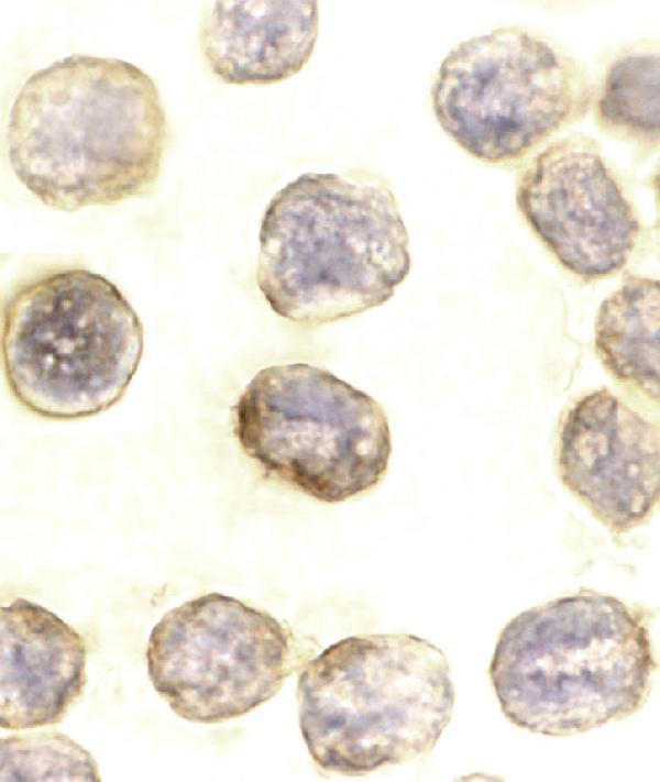
Real IHC data IHC analysis of DAXX using anti-DAXX antibody (PB9550). DAXX was detected in a paraffin-embedded section of rat intestine tissue. Heat mediated antigen retrieval was performed in EDTA buffer (pH 8.0, epitope retrieval solution). The tissue section was blocked with 10% goat serum. The tissue section was then incubated with 1 μg/ml rabbit anti-DAXX Antibody (PB9550) overnight at 4°C. Biotinylated goat anti-rabbit IgG was used as secondary antibody and incubated for 30 minutes at 37°C. The tissue section was developed using Strepavidin-Biotin-Complex (SABC) (Catalog # SA1022) with DAB as the chromogen.
Anti-Daxx Antibody ®
Cat # PB9550
Real IF data Immunocytochemistry of DAXX in HeLa cells with DAXX antibody at 10 μg/mL.
Anti-Daxx Antibody
Cat # A00439
Real IF data IF analysis of DAXX using anti-DAXX antibody (PA1809) and anti-Beta Tubulin antibody (M01857-3). DAXX was detected in immunocytochemical section of U2OS cell. Enzyme antigen retrieval was performed using IHC enzyme antigen retrieval reagent (AR0022) for 15 mins. The cells were blocked with 10% goat serum. And then incubated with 5 μg/mL rabbit anti-DAXX Antibody (PA1809) and mouse anti-Beta Tubulin antibody (M01857-3) overnight at 4°C. DyLight®488 Conjugated Goat Anti-Rabbit IgG (BA1127) and Cy3 Conjugated Goat Anti-Mouse IgG (BA1031) were used as secondary antibody at 1:500 dilution and incubated for 30 minutes at 37°C. Visualize using a fluorescence microscope and filter sets appropriate for the label used.
Anti-Daxx Antibody ®
Cat # PA1809

PB9550 shows IHC staining in paraffin-embedded rat intestine, while A00439 shows ICC staining in HeLa cells (catalog figures: PB9550, A00439). PA1809 shows IF staining in U2OS cells (catalog figure: PA1809).

Which to pick: For tissue IHC, choose PB9550: its own image documents paraffin-embedded rat intestine, EDTA retrieval at pH 8.0, and 1 μg/mL primary antibody; the fixative is unreported (catalog figure: PB9550). For IF/ICC, choose A00439 for HeLa cells or PA1809 for U2OS cells, as shown in their respective figures (catalog figures: A00439, PA1809). For cross-species planning, PB9550 lists Human, Mouse, and Rat reactivity with IHC and IF applications, while PA1809 lists those species with IF and ICC applications; confirm staining in each intended sample because PB9550’s cited IHC figure shows rat intestine (catalog applications/reactivity: PB9550, PA1809; catalog figure: PB9550).

Each figure is that product's own IHC / IF validation image from its datasheet.

References

  1. UniProt Consortium. UniProt entry Q9UER7 (DAXX_HUMAN, Death domain-associated protein 6).
  2. Human Protein Atlas. DAXX tissue IHC expression (reliability: Enhanced).
  3. Human Protein Atlas. DAXX subcellular location (ICC-IF): Localized to the nucleoplasm and nuclear bodies..
  4. Human Protein Atlas. DAXX antibody validation summary (5 antibodies).
  5. Performance of DAXX Immunohistochemistry as a Screen for DAXX Mutations in Pancreatic Neuroendocrine Tumors. Pancreas 2019 — PMC6414083.
  6. Real-World-Feasible Immunohistochemistry of ATRX, DAXX, and Menin Identifies a Subgroup of Non-Functioning Pancreatic Neuroendocrine Tumors with low Recurrence Risk to Guide De-Escalating Surveillance. Endocrine pathology 2026 — PMC13526144.
  7. TERT promoter mutations are a rare event in gastrointestinal stromal tumors. SpringerPlus 2015 — PMC4695492.
  8. DAXX and ATRX Expression in Canine Prostate and Bladder Cancer Identified by Immunohistochemistry-A Digital Quantitative Pilot Study. Veterinary sciences 2025 — PMC12737483.
  9. PubMed PMID:9215629 — UniProt-cited evidence.
  10. PubMed PMID:9407001 — UniProt-cited evidence.
  11. PubMed PMID:9645950 — UniProt-cited evidence.