This website uses cookies to ensure you get the best experience on our website.


- Table of Contents
Plan chromogenic DBI IHC in paraffin sections using cytoplasmic staining as the tissue pattern (HPA tissue IHC). Compare hepatocytes and kidney tubules with documented low or undetected cell populations, and account for isoform-dependent expression (HPA tissue IHC; UniProt).

Expected localisation, validated protocols, controls and antibodies — the at-a-glance facts below, then the full design guide.
| Expected localisation | Cytoplasmic in tissue (HPA tissue IHC); ER/Golgi (UniProt) | |
| Staining pattern | Cytoplasmic staining in hepatocytes and kidney tubules (HPA tissue IHC) | |
| Antigen retrieval | Citrate pH 6 HIER, heat-mediated (datasheet A01267) | |
| Positive control | Appendix+4 more · see all | |
| Negative control | Bone marrow+4 more · see all |
| Fixation | Keep fixation consistent across sections. (standard IHC practice; not target-specific) | |
| Caveat | Placenta image differs from HPA decidual-cell result (datasheet A01267; HPA tissue IHC) | |
| Regulation | Isoform 2 is high in liver and adipose tissue (UniProt) | |
| Isoform / epitope | Six isoforms; confirm epitope coverage across variants (UniProt) |
The catalog antibody’s IHC-P protocol (datasheet A01267) is accompanied by published DBI staining protocols for cartilage and mouse knee joints (PMC12326017) and tumors (PMC11378439).
| Sample | Paraffin-embedded human placenta tissues; fixative not specified (datasheet A01267) |
| Fixation | Image fixative and duration unreported (datasheet A01267); verify before use. |
| Sectioning | 4–5 µm sections on charged slides (standard) |
| Deparaffinisation | Xylene, graded ethanol series to water (standard) |
| Antigen retrieval | Heat retrieval: Citrate pH 6, 20 min (datasheet A01267) |
| Peroxidase block | 3% H2O2, 10 min, room temperature (standard) |
| Blocking | 10% goat serum (datasheet A01267) |
| Primary antibody | Rabbit anti-DBI, 0.5-1μg/ml (datasheet A01267) |
| Primary incubation | Overnight at 4 °C (datasheet A01267) |
| Detection | Streptavidin-biotin complex (SABC), DAB chromogen (datasheet A01267) |
| Counterstain | Hematoxylin, blue, dehydrate and mount (standard) |
| Expected result | DBI-positive staining in glandular cells of appendix (HPA tissue IHC: High). HPA tissue profile: General cytoplasmic expression. No signal in the no-primary control. |
DBI should show predominantly cytoplasmic staining in paraffin sections, including hepatocytes, kidney tubule cells and several glandular cell populations with high HPA staining (HPA: tissue IHC). Its annotated locations are the endoplasmic reticulum and Golgi apparatus, with Golgi localisation dependent on ligand binding; DBI has no transmembrane segment (UniProt P07108). HPA rates the tissue IHC pattern Approved, with medium consistency against RNA expression (HPA: tissue IHC).
| Cytoplasmic stain is strong in hepatocytes or kidney tubule cells, while nuclei remain relatively clear. | This fits the reported high staining in those cells and the general cytoplasmic profile (HPA: tissue IHC). Compare cell identity and compartment before scoring intensity; a brown deposit anywhere in the section is not, by itself, a DBI-positive cell (general IHC practice). |
| Staining is concentrated in nuclei, or appears only as a sharp cell-surface outline. | Neither pattern matches HPA's general cytoplasmic profile or UniProt's endoplasmic reticulum and Golgi annotations (HPA: tissue IHC; UniProt P07108). Treat it as a localisation mismatch and check controls and detection conditions before assigning it to DBI; the sources do not provide a nuclear or membrane IHC pattern. |
| Strong stain appears in bone-marrow hematopoietic cells or smooth muscle cells. | Those cell populations were reported as not detected in the HPA tissue IHC record (HPA: tissue IHC). Unexpected stain could reflect nonspecific antibody binding or endogenous detection activity (general IHC practice). It is a reason to investigate, rather than proof that every stained cell is a false positive. |
| Brown colour covers stroma, lumina and multiple cell types without clear cell boundaries. | A widespread deposit is difficult to reconcile with the reported cell-associated cytoplasmic pattern (HPA: tissue IHC). Review the no-primary control and background across the section; inadequate blocking, residual detection reagents or chromogen development can obscure a specific signal (general IHC practice). |
| A liver section has no convincing hepatocyte signal. | HPA reports high staining in hepatocytes, so an entirely blank result is unexpected under a working assay (HPA: tissue IHC). Verify that the section contains intact hepatocytes, then review antibody, retrieval and detection controls (general IHC practice). A blank slide alone does not establish absent DBI expression. |
| Compartment and topology | HPA describes general cytoplasmic staining; UniProt annotates endoplasmic reticulum and Golgi localisation and no transmembrane segment (HPA: tissue IHC; UniProt P07108). A surface-only IHC pattern therefore needs independent scrutiny. UniProt notes that Golgi localisation depends on ligand binding, so a discrete Golgi accent is not required in every cell (UniProt P07108). |
| Tissue and cell context | HPA reports high staining in hepatocytes, kidney tubule cells, type II alveolar cells and specified glandular and glial cells, but not detected staining in several other listed populations (HPA: tissue IHC). These are useful comparisons within identified cell types; HPA also reports low tissue RNA specificity, so an unlisted tissue is not automatically negative (HPA: tissue IHC). |
| Isoforms and epitope coverage | UniProt lists 6 DBI isoforms, with isoform-specific expression descriptions, but the supplied record gives no antibody epitope or isoform coverage (UniProt P07108). Differences among tissues cannot be assigned to isoform recognition from these data. The annotated protein chain spans residues 2–87; this does not establish which sequence the antibody recognises (UniProt P07108). |
| Validation and IF/ICC Q&A | Q: Does this IHC pattern establish an IF/ICC localisation? A: No. HPA lists antibody HPA051428 as IHC Approved, with no ICC status, no ICC-IF images and no available main subcellular location in its ICC-IF record (HPA: antibodies; HPA: subcellular). Use the IHC pattern as context, without treating it as IF/ICC validation. |
| Fixation and retrieval evidence | The supplied HPA and UniProt records do not report DBI-specific fixation sensitivity or an antigen-retrieval setting (HPA: tissue IHC; UniProt P07108). Retrieval can be optimised as a general paraffin-section IHC step, but these sources do not support a claim that fixation or a particular retrieval method changes DBI staining (general IHC practice). |
| Situation | Likely cause | Next action |
|---|---|---|
| Known-positive hepatocytes are blank. | An assay or section issue is possible because hepatocytes are HPA high; the blank result does not identify which step failed (HPA: tissue IHC). | Confirm hepatocyte preservation and run the assay controls. Review the antibody application, retrieval, detection reagents and chromogen steps without assuming a DBI-specific retrieval requirement (general IHC practice). |
| All tissue compartments stain brown, including cell-free areas. | The pattern exceeds HPA's general cytoplasmic description and may reflect detection background (HPA: tissue IHC; general IHC practice). | Inspect the no-primary control, blocking and reagent rinses; reassess chromogen development before scoring any cell as positive (general IHC practice). |
| Signal is nuclear or restricted to cell outlines. | The compartment conflicts with HPA's cytoplasmic profile and UniProt's endoplasmic reticulum and Golgi locations (HPA: tissue IHC; UniProt P07108). | Check morphology and counterstain, compare a known-positive tissue and review background controls. Record the mismatch rather than scoring it as the expected DBI pattern (general IHC practice). |
| Bone-marrow hematopoietic cells stain as strongly as hepatocytes. | HPA reports hematopoietic cells as not detected and hepatocytes as high; nonspecific binding or endogenous detection activity is possible (HPA: tissue IHC; general IHC practice). | Compare matched controls and cell identity, then reassess blocking and the detection system. Do not use the contrast alone to declare biological expression or a validated negative result (general IHC practice). |
| A listed positive tissue shows only weak staining. | HPA levels describe observed staining, not a guaranteed intensity in every section; its IHC reliability is Approved with medium RNA concordance (HPA: tissue IHC). | Score the correct cell population and cytoplasmic compartment first. Compare assay controls and section quality before changing interpretation or optimising routine IHC conditions (general IHC practice). |
| An IF/ICC image is requested to confirm the paraffin-section result. | The supplied HPA record has no ICC status, ICC-IF images or main ICC-IF location for DBI (HPA: antibodies; HPA: subcellular). | Report the IHC result with its tissue and compartment evidence. Treat any IF/ICC result as a separate validation question; the supplied sources provide no IF/ICC protocol or image benchmark (HPA: tissue IHC; HPA: subcellular). |

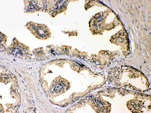
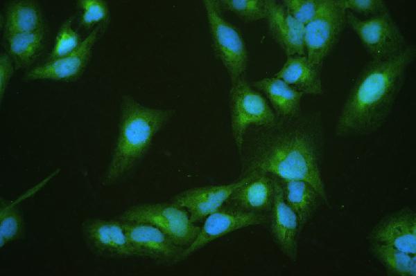


Comprehensive Human Protein Atlas IHC scoring per tissue (reliability: Approved — Medium consistency between antibody staining and RNA expression data.). Rows are taken directly from the HPA tissue chart — click any row's HPA link to view the source.
| Tissue | Cell type | Level | Evidence | Source |
|---|---|---|---|---|
| Appendix | Glandular cells | High | Protein (IHC) | HPA → |
| Breast | Glandular cells | High | Protein (IHC) | HPA → |
| Caudate | Glial cells | High | Protein (IHC) | HPA → |
| Epididymis | Glandular cells | High | Protein (IHC) | HPA → |
| Gallbladder | Glandular cells | High | Protein (IHC) | HPA → |
| Tissue | Cell type | Level | Evidence | Source |
|---|---|---|---|---|
| Bone marrow | Hematopoietic cells | Not detected | Protein (IHC) | HPA → |
| Fallopian tube | Glandular cells | Not detected | Protein (IHC) | HPA → |
| Oral mucosa | Squamous epithelial cells | Not detected | Protein (IHC) | HPA → |
| Placenta | Decidual cells | Not detected | Protein (IHC) | HPA → |
| Smooth muscle | Smooth muscle cells | Not detected | Protein (IHC) | HPA → |
These DBI troubleshooting answers use the selected paraffin section IHC example, the protein record, and tissue staining data to guide interpretation.
A01267 has IHC images from human placenta and prostatic cancer paraffin sections, plus an IF/ICC image from U20S cells (catalog image captions).
A01267 was shown on human placenta and prostatic cancer paraffin sections using IHC (A01267 IHC image captions). A01267 was also shown in U20S cells using IF/ICC and is listed as human-reactive (A01267 IF image caption; catalog applications and reactivity).
Which to pick: Choose A01267 for paraffin-section IHC: its captions show staining in human placenta and prostatic cancer at 1 μg/ml after citrate retrieval; the fixative is unreported (A01267 IHC image captions). The same SKU is listed for IF/ICC at 2 μg/ml and has a U20S cell IF image (catalog applications and dilution; A01267 IF image caption). For other species, the payload provides no validated choice because A01267 lists human reactivity only (catalog reactivity).