DGKA / Diacylglycerol kinase alpha · IHC design guide

Design Immunohistochemistry for DGKA

Plan DGKA IHC in paraffin sections using cytoplasmic staining as the expected pattern (HPA tissue IHC). Tonsil non-germinal center cells and splenic white pulp cells show high staining, though staining and RNA levels have low consistency (HPA tissue IHC).

Evidence assembled Oct 2026 · For research use; verify linked source records and product datasheet before use
Immunohistochemistry protocol sheet for DGKA (IHC for DGKA): expected localisation Cytoplasmic in most tissues (HPA tissue IHC), antibody A06354, validated IHC image, and IHC protocol steps
Printable DGKA IHC protocol sheet — expected localisation Cytoplasmic in most tissues (HPA tissue IHC), antibody A06354, controls and protocol steps. Open the full DGKA IHC guide →

DGKA Immunohistochemistry Experimental Design Guide

Expected localisation, validated protocols, controls and antibodies — the at-a-glance facts below, then the full design guide.

Must know before staining
Expected localisation Cytoplasmic in most tissues (HPA tissue IHC)
Staining pattern Cytoplasmic staining across most tissues (HPA tissue IHC)
Antigen retrieval Tris-EDTA pH 9.0 HIER, heat-mediated (datasheet A06354)
Positive control ⓘ Spleen+4 more · see all
Negative control ⓘ Adipose tissue+4 more · see all
Important caveats
Reasons your staining may differ from the expected pattern.
Fixation Keep fixation consistent across sections. (standard IHC practice; not target-specific)
Caveat Staining shows low consistency with RNA levels (HPA tissue IHC)
Regulation Lymphoid tissue-enhanced RNA (HPA tissue RNA)
Isoform / epitope 3 isoforms; check antibody epitope coverage (UniProt)
Section 1

Recommended DGKA IHC & IF Protocols

The catalog antibody has an IHC-P protocol (datasheet A06354). The published protocols below cover nasopharyngeal carcinoma, ovarian cancer and intrahepatic cholangiocarcinoma (PMC12271027; PMC7367757; PMC10876206).

Recommended immunohistochemistry (IHC-P) protocol parameters
SampleParaffin-embedded human tonsil tissue; fixative not specified (datasheet A06354)
FixationImage fixative and duration unreported (datasheet A06354); verify before use.
Sectioning4–5 µm sections on charged slides (standard)
DeparaffinisationXylene, graded ethanol series to water (standard)
Antigen retrievalHeat retrieval: Tris-EDTA pH 9.0 (datasheet A06354); 20 min, 95–100 °C (standard)
Peroxidase block3% H2O2, 10 min, room temperature (standard)
Blocking10% normal serum of the secondary host, 30 min, room temperature (standard)
Primary antibodyRabbit anti-DGKA, 1:100-1:300 (datasheet A06354)
Primary incubationOvernight at 4 °C (standard)
DetectionHRP-polymer secondary, DAB chromogen 5–10 min (standard)
CounterstainHematoxylin, blue, dehydrate and mount (standard)
Expected resultDGKA-positive staining in cells in white pulp of spleen (HPA tissue IHC: High). HPA tissue profile: Cytoplasmic expression in most tissues. No signal in the no-primary control.
💡Decision noteStart with heat-mediated Tris-EDTA retrieval at pH 9.0 for the catalog antibody (datasheet A06354); citrate at pH 8.0 was reported with another DGKA antibody (PMC12271027).
Section 2

What Is the Expected DGKA Staining Pattern?

DGKA is cytosolic and has no transmembrane segment (UniProt P23743: subcellular location and topology). In paraffin-section IHC, expect mainly cytoplasmic staining, with stronger signal in spleen white-pulp cells and tonsil non-germinal-center cells (HPA: tissue IHC). HPA rates its tissue IHC Approved but reports low consistency between antibody staining and RNA expression (HPA: reliability description).

What am I looking at on my slide?
Cytoplasmic staining is strong in spleen white-pulp cells or tonsil non-germinal-center cells.This fits the reported high-staining cell populations (HPA: tissue IHC) and cytosolic location (UniProt P23743). Judge the cells and compartment together; intensity alone is insufficient.
Predominantly nuclear staining replaces cytoplasmic staining in an IHC section.This diverges from the reported tissue IHC pattern (HPA: cytoplasmic expression in most tissues). Check staining specificity and slide interpretation. Additional nucleoplasmic localization was observed by ICC-IF, a different application (HPA: subcellular ICC-IF).
Adipocytes or liver cholangiocytes stain strongly.HPA reports DGKA as not detected in these specific cell types (HPA: tissue IHC). Consider cross-reactivity or endogenous chromogen-generating activity; confirm the cell identity and review controls before calling the stain positive (general IHC practice).
Brown signal covers cells and surrounding tissue without clear cellular boundaries.Diffuse background prevents a reliable cytoplasmic call (UniProt P23743: cytosolic location). Review a no-primary control, blocking, washes and chromogen development to identify nonspecific or endogenous signal (general IHC practice).
No signal appears in spleen white pulp or tonsil non-germinal-center cells.These are reported high-staining populations (HPA: tissue IHC). First check that the expected cells are present, then review the staining run and its controls; a blank section alone cannot establish absent DGKA (general IHC practice).
💡Expected DGKA appearanceCall the IHC result positive when spleen white-pulp or tonsil non-germinal-center cells show strong cytoplasmic staining (HPA: High; UniProt P23743: cytosol); dominant nuclear or diffuse tissue-wide color warrants review (HPA: tissue IHC; general IHC practice).
How each factor affects the staining
Cell population chosen for scoringHPA reports high staining in spleen white-pulp and tonsil non-germinal-center cells, medium staining in several other listed populations, and no detection in adipocytes (HPA: tissue IHC). Score the named cells, not the whole organ.
Compartment and membrane statusDGKA is cytosolic and has no transmembrane segment (UniProt P23743: location and topology). A membrane-only IHC pattern does not match that annotation; review it alongside morphology and controls (general IHC practice).
Antibody evidence and isoformsThe listed IHC antibody is Approved, while HPA reports low staining–RNA consistency (HPA: antibodies and reliability). UniProt lists three isoforms (UniProt P23743: isoforms); the supplied record gives no epitope map or isoform-specific IHC pattern.
Fixation and retrievalTarget-specific fixation sensitivity and retrieval requirements are unreported in the supplied UniProt and HPA records. Use the catalog antibody's validated IHC-P conditions as the starting point, and assess any adjustment with controls (general IHC practice).
IF/ICC Q: Should an IF image match the paraffin IHC pattern?A: HPA reports mainly vesicular ICC-IF signal, with additional nucleoplasmic and mitochondrial localization (HPA: subcellular ICC-IF). Its listed ICC antibody differs from its IHC antibody (HPA: antibodies); interpret IF on its own guide page.
Why is my staining missing, weak or wrong?
SituationLikely causeNext action
Positive-control cells are blank.The expected cells may be absent from the section, or the IHC run may have failed (HPA: high staining in spleen white pulp and tonsil non-germinal-center cells; general IHC practice).Verify cell identity and tissue preservation, then check the catalog antibody's IHC-P instructions, detection reagents and run controls (general IHC practice).
The entire section develops brown color.Endogenous detection activity, nonspecific reagent binding or excessive chromogen development can obscure the expected cellular pattern (general IHC practice).Compare a no-primary control; review endogenous-enzyme blocking, reagent blocking, washes and development time (general IHC practice).
Nuclei dominate the IHC signal.The pattern conflicts with predominantly cytoplasmic tissue staining (HPA: tissue IHC), although ICC-IF reports an additional nucleoplasmic location (HPA: subcellular ICC-IF).Recheck compartment assignment against a counterstain and compare controls; do not transfer the ICC-IF pattern directly to IHC scoring (general IHC practice).
Adipocytes or cholangiocytes appear positive.Those populations are reported as not detected (HPA: tissue IHC); cross-reactivity or endogenous detection activity is possible (general IHC practice).Confirm the stained cell type, inspect the no-primary control and evaluate signal in a reported high-staining population from the same run (HPA: tissue IHC; general IHC practice).
Only faint signal appears in a proposed positive tissue.Staining level depends on the sampled cell population: HPA reports high, medium and low populations within its tissue survey (HPA: tissue IHC).Identify and score the specified cells before changing conditions. If a high-staining population is faint, review the IHC-P run and controls (HPA: tissue IHC; general IHC practice).
Staining varies between specimens.HPA reports low consistency between antibody staining and RNA expression, so RNA abundance alone cannot settle a discordant IHC result (HPA: reliability description).Compare the same cell population and compartment across sections, document controls, and treat unexplained discordance as unresolved (HPA: tissue IHC; general IHC practice).

Sample controls for DGKA IHC & IF

🧪Run tonsil first: non-germinal-center cells should stain strongly (HPA: High in tonsil non-germinal-center cells; catalog antibody caption: tonsil IHC). Use adipose tissue as the negative, focusing on adipocytes (HPA: Not detected in adipocytes); on the tonsil slide, internal negative cells should retain a visible counterstained nucleus but lack specific chromogenic deposit (standard IHC interpretation).
Positive control tissue: Spleen (Cells in white pulp, HPA High)
Negative control tissue: Adipose tissue (HPA Not detected)
ICC-IF cell lines (HPA subcellular resource): HPA ICC-IF images show DGKA in BJ [Human fibroblast], RT-4, U2OS, with annotated localisation: Vesicles (approved) (HPA subcellular).
Technical controls: Include a no-primary (secondary-only) control, an irrelevant antibody matched to the primary antibody’s host and isotype or immunoglobulin class, and a DGKA knockout specimen or validated peptide-block control (standard IHC control practice). Block endogenous peroxidase in tonsil for chromogenic detection, and check endogenous biotin if using a biotin-based system (standard IHC practice).
⚠️Feasibility: The tonsil paraffin-section caption reports Tris-EDTA retrieval at pH 9.0 and a primary dilution of 1:200, but its fixative is unreported (catalog antibody caption: A06354). A target-specific fixation window or fixation effect is unreported in the supplied evidence, and the available ICC-IF images do not establish that frozen sections or tissue IF are easier (HPA: ICC-IF images). Endogenous peroxidase in tonsil leukocytes can cause chromogenic background, making the peroxidase block and no-primary control useful checks (standard IHC practice).

HPA tissue IHC evidence for DGKA

Comprehensive Human Protein Atlas IHC scoring per tissue (reliability: Approved — Low consistency between antibody staining and RNA expression data.). Rows are taken directly from the HPA tissue chart — click any row's HPA link to view the source.

Positive expression · recommended positive controls

TissueCell typeLevelEvidenceSource
Spleen Cells in white pulp High Protein (IHC) HPA →
Tonsil Non-germinal center cells High Protein (IHC) HPA →
Breast Glandular cells Medium Protein (IHC) HPA →
Cerebellum Cells in granular layer Medium Protein (IHC) HPA →
Cerebral cortex Neuropil Medium Protein (IHC) HPA →

Undetected expression · recommended negative controls

TissueCell typeLevelEvidenceSource
Adipose tissue Adipocytes Not detected Protein (IHC) HPA →
Adrenal gland Glandular cells Not detected Protein (IHC) HPA →
Cervix Glandular cells Not detected Protein (IHC) HPA →
Epididymis Glandular cells Not detected Protein (IHC) HPA →
Fallopian tube Glandular cells Not detected Protein (IHC) HPA →
Section 3

Advanced DGKA IHC Tips

Troubleshooting paraffin-section DGKA IHC starts with the catalog antibody’s tonsil conditions, then checks cell type, staining pattern and controls.

Which retrieval conditions should I try first for weak DGKA staining?
Start with heat-mediated Tris-EDTA at pH 9.0 for paraffin sections (datasheet A06354). The catalog antibody’s tonsil image used primary antibody at 1:200, 4°C overnight, followed by secondary antibody at 1:200 for 30 minutes at room temperature (caption A06354). If staining remains weak, compare heating duration on adjacent sections while keeping antibody incubation and detection constant (standard IHC practice). Include a no-primary control to reveal signal from the detection system (standard IHC practice). Evaluate whether any gain occurs in tonsil non-germinal center cells, where staining is reported as high, rather than accepting a uniform rise in background (HPA: tonsil tissue IHC).
Could fixation explain weak or uneven DGKA staining?
Target-specific fixation sensitivity is unknown: the selected image identifies paraffin-embedded tonsil but does not state its fixative (caption A06354). Record the fixative, fixation duration and processing history before changing retrieval or antibody concentration, because these variables can alter epitope accessibility in routine IHC (standard IHC practice). If comparable specimens with different fixation histories are available, stain them together using Tris-EDTA at pH 9.0 and the same detection run (datasheet A06354; standard IHC practice). Examine well-preserved interior tissue as well as exposed edges, and compare morphology with signal (standard IHC practice). Do not assign a DGKA-specific fixation effect without a controlled comparison.
Should DGKA staining be diffuse or punctate in paraffin sections?
Expect a predominantly cytoplasmic tissue-IHC pattern, while assessing each cell population separately (HPA: cytoplasmic expression in most tissues). DGKA is annotated in the cytosol and has no transmembrane segment (UniProt P23743: subcellular location and topology). Puncta can be examined as a possible pattern, since ICC/IF reports vesicles as the main location, but that observation alone does not validate punctate chromogenic staining in paraffin tissue (HPA: subcellular ICC/IF). Check whether puncta occur within intact cell boundaries and persist across neighboring, well-preserved sections (standard IHC practice). Treat isolated nuclear staining cautiously: nucleoplasm is an additional ICC/IF location, whereas the tissue-IHC profile is cytoplasmic (HPA: subcellular ICC/IF and tissue IHC).
How can I assess whether an unexpected pattern reflects epitope or isoform recognition?
Map the catalog antibody’s documented immunogen or epitope against all 3 DGKA isoforms before interpreting a missing cell population (UniProt P23743: isoforms; standard antibody-validation practice). The record identifies EF-hand regions at residues 110–145 and 155–190, and a DAGKc region at 372–506, but supplies no antibody epitope (UniProt P23743: domains). It also lists acetyllysine at residue 484; whether that modification changes this antibody’s staining is unknown (UniProt P23743: modified residues). Compare staining from an independently validated antibody with a documented, distinct epitope on adjacent sections (standard IHC practice). Keep retrieval and detection matched so differences can be investigated as reagent-dependent rather than processing-dependent (standard IHC practice).
How should I check DGKA localisation with multiplex immunofluorescence?
Use IF as a separate validation experiment alongside the paraffin-section chromogenic result, since the catalog caption provides an IHC image rather than IF conditions (caption A06354). Pair DGKA with a validated lymphocyte marker, such as CD3 for T cells, when examining lymphoid tissue (UniProt P23743: lymphocyte expression; standard IF practice). Choose spectrally separated fluorophores, favor a far-red DGKA channel if the tissue autofluoresces strongly at shorter wavelengths, and image unstained controls (standard IF practice). DGKA lacks a transmembrane segment and is annotated as cytosolic; use permeabilisation suitable for an intracellular epitope, then optimise it against preservation of cell structure (UniProt P23743: topology and location; standard IF practice). Compare punctate signal cautiously with the vesicular ICC/IF pattern reported by HPA (HPA: subcellular ICC/IF).
What should I change when brown staining obscures DGKA-positive cells?
First inspect a no-primary control for staining produced by the chromogenic detection system, and assess tissue edges separately from intact interior regions (standard IHC practice). Apply a peroxidase block before DAB development when using an HRP detection workflow; this is a general IHC control, not DGKA-specific evidence (standard IHC practice). If background follows the primary antibody, compare a dilution series around the caption’s 1:200 primary condition on matched sections (caption A06354; standard IHC practice). Keep retrieval at Tris-EDTA pH 9.0 during that comparison (datasheet A06354). Judge improvement by retained staining in expected tonsil non-germinal center cells and reduced staining in the no-primary control (HPA: tonsil tissue IHC; standard IHC practice).
How should I score DGKA staining across sections with different cell mixes? ⚠ ANSWER MARKED FOR VERIFICATION
Define the scored cell population before measuring chromogenic signal, because reported DGKA staining varies among tissue compartments (HPA: tissue IHC). In tonsil, score non-germinal center cells separately from other regions; their reported staining level is high (HPA: tonsil tissue IHC). Record the percentage of positive cells and intensity using a consistent H-score scheme, or count positive cells per mm² when cell density is the question (standard IHC quantification practice). Normalise positive counts to eligible cells or sampled viable area within the same compartment, and use identical thresholds across batches (standard IHC quantification practice). Document section quality, detection batch and excluded necrotic or damaged areas before comparing specimens (standard IHC practice).
How do I distinguish credible DGKA staining from artefact?
A credible result places signal in intact cells with a predominantly cytoplasmic tissue pattern and a plausible cell distribution (HPA: tissue IHC; standard IHC interpretation). In tonsil, high staining in non-germinal center cells supports the expected pattern, while DGKA is also recorded as expressed in lymphocytes (HPA: tonsil tissue IHC; UniProt P23743: tissue specificity). Question isolated nuclear-only staining, staining concentrated at cut edges or necrotic areas, and signal replicated in a no-primary control (HPA: tissue IHC; standard IHC practice). Check the peroxidase block if chromogenic signal persists without primary antibody (standard IHC practice). Interpret concordance conservatively because HPA rates its tissue staining as Approved but reports low consistency with RNA expression data (HPA: reliability description).
Boster reagents

Best DGKA / Diacylglycerol kinase alpha IHC Antibodies

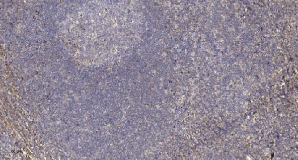
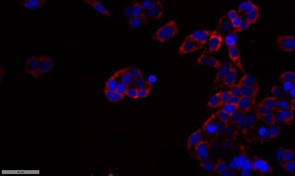
A06354 has IHC data from paraffin-embedded human tonsil and IF data from MCF7 cells (catalog image captions). Its listed reactivity covers human, mouse, and rat (catalog).

Real IHC data Immunohistochemical analysis of paraffin-embedded human tonsil. 1, Antibody was diluted at 1:200 (4° overnight). 2, Tris-EDTA,pH9.0 was used for antigen retrieval. 3,Secondary antibody was diluted at 1:200 (room temperature, 30min).
Anti-DGK- alpha Antibody
Cat # A06354

A06354 is listed for IHC and IF, with human, mouse, and rat reactivity (catalog: applications and reactivity). Its images show IHC on paraffin-embedded human tonsil and IF on MCF7 cells (A06354 image captions).

Which to pick: Choose A06354 for tissue IHC: its own caption documents paraffin-embedded human tonsil with Tris-EDTA retrieval at pH 9; the fixative is unreported (A06354 IHC image caption). For IF/ICC, A06354 is listed for IF and has an IF image of MCF7 cells; it is a rabbit polyclonal antibody (catalog; A06354 IF image caption). For mouse or rat samples, A06354 has stated reactivity, although the supplied IHC image shows human tonsil (catalog: reactivity; A06354 IHC image caption).

Each figure is that product's own IHC / IF validation image from its datasheet.

References

  1. UniProt Consortium. UniProt entry P23743 (DGKA_HUMAN, Diacylglycerol kinase alpha).
  2. Human Protein Atlas. DGKA tissue IHC expression (reliability: Approved).
  3. Human Protein Atlas. DGKA subcellular location (ICC-IF): Mainly localized to vesicles. In addition localized to the nucleoplasm and mitochondria..
  4. Human Protein Atlas. DGKA antibody validation summary (2 antibodies).
  5. High expression of DGKA indicates poor overall survival of nasopharyngeal carcinoma. Discover oncology 2025 — PMC12271027.
  6. Decreased LIPF expression is correlated with DGKA and predicts poor outcome of gastric cancer. Oncology reports 2016 — PMC5022960.
  7. DGKA Provides Platinum Resistance in Ovarian Cancer Through Activation of c-JUN-WEE1 Signaling. Clinical cancer research : an official journal of the American Association for Cancer Research 2020 — PMC7367757.
  8. Multiomics analysis reveals metabolic subtypes and identifies diacylglycerol kinase α (DGKA) as a potential therapeutic target for intrahepatic cholangiocarcinoma. Cancer communications (London, England) 2024 — PMC10876206.
  9. PubMed PMID:2175712 — UniProt-cited evidence.
  10. PubMed PMID:16541075 — UniProt-cited evidence.
  11. PubMed PMID:15489334 — UniProt-cited evidence.