EEA1 / Early endosome antigen 1 · IHC design guide

Design Immunohistochemistry for EEA1

This guide helps plan EEA1 staining in paraffin sections using its variable cytoplasmic tissue profile (HPA tissue IHC). It covers fixation, controls, and interpretation of its expected early endosome association (UniProt).

Evidence assembled Oct 2026 · For research use; verify linked source records and product datasheet before use
Immunohistochemistry protocol sheet for EEA1 (IHC for EEA1): expected localisation Cytoplasmic tissue staining (HPA tissue IHC); early endosome membrane association (UniProt), antibody A02296-3, validated IHC image, and IHC protocol steps
Printable EEA1 IHC protocol sheet — expected localisation Cytoplasmic tissue staining (HPA tissue IHC); early endosome membrane association (UniProt), antibody A02296-3, controls and protocol steps. Open the full EEA1 IHC guide →

EEA1 Immunohistochemistry Experimental Design Guide

Expected localisation, validated protocols, controls and antibodies — the at-a-glance facts below, then the full design guide.

Must know before staining
Expected localisation Cytoplasmic tissue staining (HPA tissue IHC); early endosome membrane association (UniProt)
Staining pattern Variable cytoplasmic staining in most tissues (HPA tissue IHC)
Antigen retrieval EDTA pH 8.0 HIER, heat-mediated (datasheet A02296-3)
Positive control ⓘ Cerebral cortex+4 more · see all
Negative control ⓘ Liver+1 more · see all
Important caveats
Reasons your staining may differ from the expected pattern.
Fixation Keep fixation consistent across sections (standard IHC practice; not target-specific)
Caveat Variable tissue expression can yield weak or absent signal (HPA tissue IHC)
Regulation No expression regulator annotated (UniProt)
Isoform / epitope No isoforms annotated; one 1–1411 chain (UniProt)
Section 1

Recommended EEA1 IHC & IF Protocols

The catalog antibody uses EDTA pH 8.0 retrieval (datasheet A02296-3). Published EEA1 IHC protocols below cover paraffin colonic biopsies (PMC10046606) and pre-embedding brain sections (PMC7315513).

Recommended immunohistochemistry (IHC-P) protocol parameters
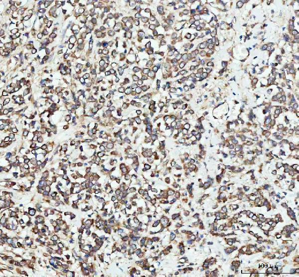
SampleParaffin-embedded human breast cancer tissue; fixative not specified (datasheet A02296-3)
FixationImage fixative and duration unreported (datasheet A02296-3); verify before use.
Sectioning4–5 µm sections on charged slides (standard)
DeparaffinisationXylene, graded ethanol series to water (standard)
Antigen retrievalHeat retrieval: EDTA pH 8.0 (datasheet A02296-3); 20 min, 95–100 °C (standard)
Peroxidase block3% H2O2, 10 min, room temperature (standard)
Blocking10% goat serum (datasheet A02296-3)
Primary antibodyRabbit anti-EEA1, 2-5 μg/ml (datasheet A02296-3)
Primary incubationOvernight at 4 °C (datasheet A02296-3)
DetectionHRP-conjugated secondary, DAB chromogen (datasheet A02296-3)
CounterstainHematoxylin, blue, dehydrate and mount (standard)
Expected resultEEA1-positive staining in neuronal cells of cerebral cortex (HPA tissue IHC: High). HPA tissue profile: Cytoplasmic expression at variable levels in most tissues. No signal in the no-primary control.
💡Decision noteStart with heat-mediated EDTA pH 8.0 for the catalog antibody (datasheet A02296-3); citrate pH 6 is a published alternative for paraffin colonic sections (PMC10046606).
Section 2

What Is the Expected EEA1 Staining Pattern?

EEA1 associates with the early endosome membrane and also localizes to the cytoplasm; it has no transmembrane segment (UniProt Q15075 topology and subcellular location). In paraffin sections, expect cytoplasmic staining in the cell populations HPA reports as positive, including cerebral cortex neurons and esophageal squamous epithelial cells (HPA: High in both). HPA rates its tissue IHC evidence Supported, with medium consistency between staining and RNA expression (HPA: reliability).

What am I looking at on my slide?
Cytoplasmic, sometimes granular staining in the expected cells.This fits HPA's variable cytoplasmic tissue pattern and EEA1's endosome association (HPA: tissue IHC; UniProt Q15075 subcellular location). Discrete vesicles may be hard to resolve with chromogenic IHC; HPA's vesicle assignment comes from ICC-IF (HPA: subcellular).
Predominantly nuclear or cell-surface signal, with little cytoplasmic signal.That distribution does not match the reported cytoplasmic tissue pattern or early endosome location (HPA: tissue IHC; UniProt Q15075 subcellular location). Treat it as suspect and check controls before scoring it as EEA1.
Strong signal in liver cholangiocytes or smooth muscle cells.HPA reports EEA1 as Not detected in those specific cell populations (HPA: liver cholangiocytes; HPA: smooth muscle cells). Consider cross-reactivity or endogenous chromogen activity; the observation alone cannot distinguish them (general IHC practice).
Diffuse stain covers cells and surrounding tissue, obscuring cell boundaries.This is not a reliable basis for assigning the HPA cytoplasmic pattern (HPA: tissue IHC). Background may arise from nonspecific binding or detection chemistry; inspect a matched negative control and review blocking and washes (general IHC practice).
No stain appears in a chosen known-positive cell population.For example, HPA reports High staining in cerebral cortex neurons and fallopian tube glandular cells (HPA: tissue IHC). A blank result warrants a check of tissue identity, antibody and detection controls; it does not establish absence of EEA1 (general IHC practice).
💡Expected EEA1 appearanceCall a result positive when the expected cells show cytoplasmic staining, potentially granular, with signal consistent with their HPA level; predominantly nuclear signal or strong staining in HPA Not detected cell populations is suspect (HPA: tissue IHC; UniProt Q15075 subcellular location).
How each factor affects the staining
Compartment and topologyEEA1 associates with the early endosome membrane, is also cytoplasmic, and has no transmembrane segment (UniProt Q15075). Score the cytoplasmic compartment in IHC; membrane-edge staining alone does not establish the expected pattern (HPA: tissue IHC).
Cell-specific tissue referenceHPA reports High staining in hippocampal neurons, lung alveolar type I cells, placental decidual cells, and round or early spermatids (HPA: tissue IHC). Its Not detected calls apply to liver cholangiocytes and smooth muscle cells, not every cell in those tissues (HPA: tissue IHC).
Strength of IHC evidenceHPA labels the tissue pattern Supported but reports medium consistency between antibody staining and RNA expression (HPA: reliability). Interpret a mismatch with the cited cell-specific pattern cautiously; the evidence does not make every tissue a definitive positive or negative control.
ICC-IF evidence boundaryHPA reports a supported vesicle location in ICC-IF and lists A-431, U-251MG, and U2OS images (HPA: subcellular). This supports a vesicular expectation for IF/ICC; it does not require individually resolved vesicles in chromogenic tissue IHC.
Fixation sensitivityTarget-specific effects of fixation on EEA1 staining are unreported in the supplied UniProt and HPA evidence (UniProt Q15075; HPA: tissue IHC). Evaluate retrieval and processing with run controls as general IHC practice, without attributing a result to an EEA1-specific fixation effect.
Why is my staining missing, weak or wrong?
SituationLikely causeNext action
Expected positive cells stain weakly or remain blank.The cause is undetermined from the tissue pattern alone; HPA reports variable cytoplasmic staining across tissues (HPA: tissue IHC).Confirm that the section contains the intended cell population, then inspect positive controls, retrieval conditions, antibody dilution, and detection reagents (general IHC practice).
Nuclear or cell-surface staining dominates.The compartment conflicts with EEA1's early endosome and cytoplasmic location (UniProt Q15075) and HPA's cytoplasmic tissue pattern (HPA: tissue IHC).Check the antibody and detection controls, compare the same cell population with the expected cytoplasmic pattern, and avoid scoring the discordant signal as positive (general IHC practice).
Strong staining appears in liver cholangiocytes or smooth muscle cells.Both populations are Not detected in HPA tissue IHC (HPA: liver cholangiocytes; HPA: smooth muscle cells). Cross-reactivity or endogenous detection activity is possible (general IHC practice).Inspect a matched control lacking primary antibody and review detection blocking; reassess cell identity before interpreting the stain (general IHC practice).
Diffuse color obscures the cells of interest.Nonspecific binding or background from detection chemistry may obscure a cytoplasmic result (general IHC practice; HPA: cytoplasmic tissue pattern).Compare a matched negative control, then review blocking, washes, antibody dilution, and development time (general IHC practice).
Signal is low in bronchial respiratory epithelium.HPA lists bronchial respiratory epithelial cells as Low, while some other populations are High (HPA: tissue IHC). Weak staining there may agree with the reference pattern.Assess a documented High cell population in the same run before changing conditions; compare like cell types when scoring intensity (HPA: tissue IHC; general IHC practice).
Q: What pattern should IF/ICC show?HPA supports a mainly vesicular ICC-IF location, whereas tissue IHC is described as cytoplasmic (HPA: subcellular; HPA: tissue IHC).A: Look for vesicular cytoplasmic signal in IF/ICC and assess that assay on its own guide page; do not infer an IF/ICC protocol from this IHC result (HPA: subcellular; general assay practice).

Sample controls for EEA1 IHC & IF

🧪Run cerebral cortex first; neuronal cells should stain (HPA: High in neuronal cells). Run smooth muscle as the negative tissue (HPA: Not detected in smooth muscle cells); cells without punctate staining on the cortex slide should remain at background, but HPA does not identify a negative cell type within that slide.
Positive control tissue: Cerebral cortex (Neuronal cells, HPA High)
Negative control tissue: Liver (HPA Not detected)
ICC-IF cell lines (HPA subcellular resource): HPA ICC-IF images show EEA1 in A-431, U-251MG, U2OS, with annotated localisation: Vesicles (supported) (HPA subcellular).
Technical controls: Include a no-primary, secondary-only control; an isotype control using rabbit IgG matched to the primary antibody (caption: rabbit anti-EEA1); and an EEA1-knockout sample or validated peptide-block control. Quench endogenous peroxidase for DAB detection (caption: peroxidase secondary and DAB), and assess neuronal lipofuscin pigment or autofluorescence in cortex (standard textbook IHC/IF practice).
⚠️Feasibility: No target-specific fixation window or fixation effect is reported, and the selected A02296-3 paraffin-section caption leaves the fixative unreported (caption: fixative not stated). Heat retrieval in EDTA at pH 8.0 is a documented IHC starting condition, but the evidence does not establish that retrieval is required (caption: EDTA pH 8.0). Whether frozen sections or IF/ICC are easier is unreported; vesicular IF localization is supported (HPA: Vesicles), while neuronal lipofuscin can complicate punctate signal assessment in cortex (standard textbook IHC/IF practice).

HPA tissue IHC evidence for EEA1

Comprehensive Human Protein Atlas IHC scoring per tissue (reliability: Supported — Medium consistency between antibody staining and RNA expression data.). Rows are taken directly from the HPA tissue chart — click any row's HPA link to view the source.

Positive expression · recommended positive controls

TissueCell typeLevelEvidenceSource
Cerebral cortex Neuronal cells High Protein (IHC) HPA →
Esophagus Squamous epithelial cells High Protein (IHC) HPA →
Fallopian tube Glandular cells High Protein (IHC) HPA →
Gallbladder Glandular cells High Protein (IHC) HPA →
Hippocampus Neuronal cells High Protein (IHC) HPA →

Undetected expression · recommended negative controls

TissueCell typeLevelEvidenceSource
Liver Cholangiocytes Not detected Protein (IHC) HPA →
Smooth muscle Smooth muscle cells Not detected Protein (IHC) HPA →
Section 3

Advanced EEA1 IHC Tips

Troubleshoot EEA1 staining in paraffin sections by checking retrieval, compartmental pattern, controls, and scoring before interpreting signal intensity.

Which retrieval conditions should I start with for EEA1 paraffin-section IHC?
Start with heat-mediated EDTA retrieval at pH 8.0 for this paraffin-section IHC assay (datasheet A02296-3). The selected tissue image used those conditions before incubation with 2 μg/ml antibody overnight at 4°C (datasheet A02296-3). If staining is weak, vary heating duration in small steps while keeping buffer and antibody conditions fixed, and assess tissue integrity alongside signal (standard IHC practice). Include a previously staining section in each comparison so a weak run is distinguishable from a weak specimen (standard IHC practice). Record retrieval conditions before comparing staining intensity across cases (standard IHC practice).
How should I troubleshoot fixation-related loss of EEA1 staining?
Target-specific fixation sensitivity is unknown: the selected paraffin-section caption does not state its fixative (datasheet A02296-3). Do not attribute weak staining to a particular fixative without a controlled comparison using the same antibody and detection conditions (standard IHC practice). First check documented fixation and processing history, then compare affected sections with a consistently processed positive section in the same staining run (standard IHC practice). Keep retrieval at EDTA pH 8.0 while making that comparison (datasheet A02296-3). If processing histories differ, report the difference as a limitation rather than assigning it a target-specific effect (standard IHC practice).
What staining pattern should count as correctly localised EEA1?
Expect cytoplasmic staining associated with early endosomal vesicles, rather than a nuclear or continuous plasma-membrane pattern (UniProt Q15075 localisation; HPA subcellular: vesicles). In chromogenic sections, individual vesicles may merge visually, so assess granular cytoplasmic signal within intact cells at consistent magnification (standard IHC practice; HPA: variable cytoplasmic expression). Compare cell types within the same section before interpreting diffuse brown colour as EEA1 (standard IHC practice). HPA reports high staining in cerebral-cortex neuronal cells and no detected staining in liver cholangiocytes, which can inform control selection when available (HPA tissue IHC). Confirm unexpected patterns with an independent antibody or orthogonal evidence (standard IHC practice).
Could an isoform or modified epitope explain discordant EEA1 staining?
The supplied record annotates 0 isoforms and one chain spanning residues 1–1411 (UniProt Q15075 record). It lists phosphoserines at residues 52 and 70, but does not locate this antibody’s epitope (UniProt Q15075 record; datasheet A02296-3). An epitope-dependent effect therefore remains untested and should not be assigned to either phosphorylation site (standard IHC interpretation). Compare retrieval-matched sections and a second antibody with a documented, distinct epitope if epitope masking is suspected (standard IHC practice). Report concordance by cellular pattern as well as intensity, because intensity alone cannot identify the molecular cause (standard IHC practice).
How can I cross-check EEA1 by IF without misreading tissue autofluorescence?
For a separate IF assay, expect vesicular EEA1 signal within the cytoplasm (HPA subcellular: vesicles; UniProt Q15075 localisation). Multiplex with a marker identifying the expected cell population, such as neuronal cells in cerebral cortex, while checking each channel separately for bleed-through (HPA tissue IHC: high in cerebral-cortex neuronal cells; standard IF practice). Choose a fluorophore channel with low measured autofluorescence in the specimen, often a far-red channel, and include an unstained section (standard IF practice). Permeabilise sufficiently to expose cytoplasmic EEA1, which has no transmembrane segment, without assuming its antibody epitope is mapped (UniProt Q15075 topology; standard IF practice). Keep IF results distinct from the catalog antibody’s chromogenic IHC evidence (datasheet A02296-3).
What should I check when EEA1 DAB staining looks diffuse?
The selected paraffin-section assay used 10% goat-serum blocking, a peroxidase-conjugated secondary, and DAB detection (datasheet A02296-3). For diffuse brown signal, compare a no-primary control and a known staining section, then inspect whether background follows tissue edges, damaged areas, or the entire section (standard IHC practice). Include an endogenous-peroxidase blocking step and verify wash and DAB development conditions, treating these as general workflow controls rather than EEA1-specific evidence (standard IHC practice). Adjust one condition at a time while preserving EDTA pH 8.0 retrieval (datasheet A02296-3; standard IHC practice). Score cellular cytoplasmic signal only after background has been separated from staining (HPA tissue IHC; standard IHC practice).
How should I quantify EEA1 staining across paraffin sections? ⚠ ANSWER MARKED FOR VERIFICATION
Define the cell population and tissue region before scoring, since HPA reports variable cytoplasmic expression across most tissues (HPA tissue IHC; standard IHC practice). For chromogenic IHC, record the percentage of positive cells and staining intensity, or calculate an H-score from 0–300 using intensity categories 0–3 (standard IHC practice). If counting stained profiles, report density per mm² of evaluable tissue and keep the counting rule fixed (standard IHC practice). Normalise cell counts to the number of eligible cells, or area counts to viable tissue area, excluding folds and necrosis (standard IHC practice). Use identical retrieval, imaging, and scoring thresholds across compared sections (standard IHC practice).
How do I distinguish genuine EEA1 positivity from staining artefact?
Genuine EEA1 staining should fit a cytoplasmic, vesicle-associated pattern in intact cells (UniProt Q15075 localisation; HPA subcellular: vesicles). A predominantly nuclear pattern, strong edge-only staining, or signal restricted to necrosis warrants an artefact check before interpretation (standard IHC practice). Compare the suspect region with no-primary controls and with plausible positive cells; HPA reports high staining in fallopian-tube glandular cells and no detected staining in smooth-muscle cells (HPA tissue IHC; standard IHC practice). Residual endogenous-peroxidase activity can produce misleading DAB colour, so review the peroxidase block when diffuse brown signal persists (standard IHC practice). Report uncertain staining separately from scored cellular positivity (standard IHC practice).
Boster reagents

Best EEA1 / Early endosome antigen 1 IHC Antibodies

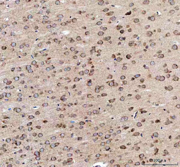
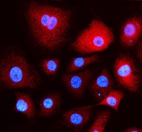
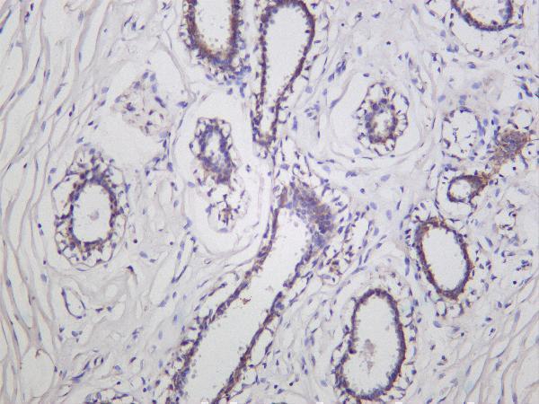
Catalog antibodies have IHC images from human breast cancer, rat brain, and human ductal breast carcinoma, plus IF data in A549 cells (catalog image captions). Human, mouse, and rat reactivity is listed (catalog).

Real IHC data IHC analysis of EEA1 using anti-EEA1 antibody (A02296-3). EEA1 was detected in a paraffin-embedded section of human breast cancer tissue. Heat mediated antigen retrieval was performed in EDTA buffer (pH 8.0, epitope retrieval solution). The tissue section was blocked with 10% goat serum. The tissue section was then incubated with 2 μg/ml rabbit anti-EEA1 Antibody (A02296-3) overnight at 4°C. Peroxidase Conjugated Goat Anti-rabbit IgG was used as secondary antibody and incubated for 30 minutes at 37°C. The tissue section was developed using HRP Conjugated Rabbit IgG Super Vision Assay Kit (Catalog # SV0002) with DAB as the chromogen.
Anti-EEA1 Antibody ®
Cat # A02296-3
Real IHC data Human ductal breast carcinoma was stained with Anti-EEA1 rabbit antibody
Anti-EEA1 Rabbit Monoclonal Antibody
Cat # M02296-3

A02296-3 has paraffin-section IHC images from human breast cancer and rat brain, and an IF image from A549 cells (A02296-3 image captions). M02296-3 has an IHC image from human ductal breast carcinoma; its listed applications include IHC and IF (M02296-3 image caption and catalog applications).

Which to pick: Choose A02296-3 for paraffin-section IHC because its images document staining in human breast cancer and rat brain; the captions do not report the fixative (A02296-3 IHC image captions). Choose A02296-3 for IF/ICC when image-backed cell staining matters, since its IF image shows A549 cells and ICC is listed as an application (A02296-3 IF image caption and catalog applications). For work across species, A02296-3 has human and rat IHC images and listed mouse reactivity; M02296-3 is a monoclonal alternative with listed human, mouse, and rat reactivity, but its supplied IHC image documents human tissue only (catalog reactivity, M02296-3 catalog description, image captions).

Each figure is that product's own IHC / IF validation image from its datasheet.

References

  1. UniProt Consortium. UniProt entry Q15075 (EEA1_HUMAN, Early endosome antigen 1).
  2. Human Protein Atlas. EEA1 tissue IHC expression (reliability: Supported).
  3. Human Protein Atlas. EEA1 subcellular location (ICC-IF): Mainly localized to vesicles..
  4. Human Protein Atlas. EEA1 antibody validation summary (4 antibodies).
  5. Intestinal Barrier in Post-Campylobacter jejuni Irritable Bowel Syndrome. Biomolecules 2023 — PMC10046606.
  6. Heterogeneous intracellular trafficking dynamics of brain-derived neurotrophic factor complexes in the neuronal soma revealed by single quantum dot tracking. PloS one 2014 — PMC3986401.
  7. The Alzheimer susceptibility gene BIN1 induces isoform-dependent neurotoxicity through early endosome defects. Acta neuropathologica communications 2022 — PMC8742943.
  8. Ultrastructural and dynamic studies of the endosomal compartment in Down syndrome. Acta neuropathologica communications 2020 — PMC7315513.
  9. PubMed PMID:7768953 — UniProt-cited evidence.
  10. PubMed PMID:16541075 — UniProt-cited evidence.
  11. PubMed PMID:9697774 — UniProt-cited evidence.