This website uses cookies to ensure you get the best experience on our website.


- Table of Contents
Plan EPB41L2 chromogenic paraffin IHC with colon glandular cells as a positive control and prostate glandular cells as a negative comparator (HPA tissue IHC). This guide covers cytoplasmic and membranous staining (HPA tissue IHC) and the catalog antibody's 1:100–1:300 IHC dilution (datasheet A03718).

Expected localisation, validated protocols, controls and antibodies — the at-a-glance facts below, then the full design guide.
| Expected localisation | Cytoplasmic and membranous tissue staining (HPA tissue IHC) | |
| Staining pattern | Glandular cells show cytoplasmic and membranous staining (HPA tissue IHC) | |
| Antigen retrieval | Tris-EDTA pH 9.0 HIER, heat-mediated (datasheet A03718) | |
| Positive control | Appendix+4 more · see all | |
| Negative control | Adipose tissue+4 more · see all |
| Fixation | Keep fixation consistent across sections. (standard IHC practice; not target-specific) | |
| Caveat | Glandular staining is high or undetected depending on tissue (HPA tissue IHC) | |
| Regulation | No expression regulator reported (UniProt) | |
| Isoform / epitope | 4 isoforms; no transmembrane segment; epitope unreported (UniProt) |
Compare the catalog antibody’s IHC-P protocol (datasheet A03718) with a published liver-section IHC protocol (PMC13226250).
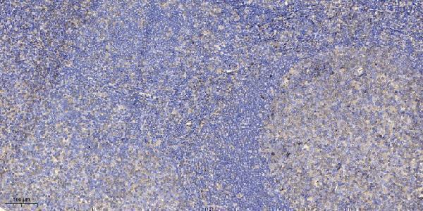
| Sample | Paraffin-embedded human tonsil tissue; fixative not specified (datasheet A03718) |
| Fixation | Image fixative and duration unreported (datasheet A03718); verify before use. |
| Sectioning | 4–5 µm sections on charged slides (standard) |
| Deparaffinisation | Xylene, graded ethanol series to water (standard) |
| Antigen retrieval | Heat retrieval: Tris-EDTA pH 9.0 (datasheet A03718); 20 min, 95–100 °C (standard) |
| Peroxidase block | 3% H2O2, 10 min, room temperature (standard) |
| Blocking | 10% normal serum of the secondary host, 30 min, room temperature (standard) |
| Primary antibody | Rabbit anti-EPB41L2, 1:100-1:300 (datasheet A03718) |
| Primary incubation | Overnight at 4 °C (standard) |
| Detection | HRP-polymer secondary, DAB chromogen 5–10 min (standard) |
| Counterstain | Hematoxylin, blue, dehydrate and mount (standard) |
| Expected result | EPB41L2-positive staining in glandular cells of appendix (HPA tissue IHC: High). HPA tissue profile: Cytoplasmic and membranous expression in several tissues. No signal in the no-primary control. |
EPB41L2 is a cytoskeletal protein found in the cytoplasm, cell cortex, and at the cell membrane; it has no transmembrane segment (UniProt O43491). In tissue IHC, expect cytoplasmic and membranous staining in selected cell populations, including appendix glandular cells and bronchial basal cells (HPA tissue IHC). HPA rates the tissue pattern Supported, with medium consistency between staining and RNA data; external verification is pending (HPA tissue IHC).
| Cytoplasmic or membranous signal is clear in appendix or colon glandular cells, bronchial basal cells, or bone marrow hematopoietic cells. | This matches the reported tissue compartments and cell populations; HPA records High staining in each example (HPA tissue IHC). Compare signal within the identified cell type, since a tissue-level label does not mean every cell on the section must stain. |
| Signal is confined to nuclei, with no convincing cytoplasmic or membranous pattern in tissue IHC. | This diverges from the reported tissue IHC pattern (HPA tissue IHC). Check staining specificity and section interpretation before calling it positive. Nucleoplasmic localization is reported in ICC-IF, so a nuclear signal alone cannot establish an artefact (HPA subcellular ICC-IF). |
| Adipocytes or cardiomyocytes stain as strongly as the selected positive cells. | HPA reports EPB41L2 as Not detected in those cell populations (HPA tissue IHC). Recheck cell identification and controls; cross-reactivity or endogenous detection activity are possible explanations (general IHC practice). A single discordant section does not prove either cause. |
| Color spreads across cell types and tissue spaces, obscuring cell borders and cytoplasm. | The expected cell-associated pattern cannot be scored reliably (HPA tissue IHC). Diffuse deposit may reflect nonspecific primary-antibody binding, insufficient blocking or washing, or detection background (general IHC practice); inspect the negative control before assigning a cause. |
| No signal appears in appendix glandular cells or another HPA High population. | The result conflicts with the reported positive population (HPA tissue IHC). First assess section quality, antibody and detection controls, and the identification of the expected cells (general IHC practice). HPA's Supported rating and pending external verification also limit certainty about any individual specimen (HPA tissue IHC). |
| Tissue and cell selection | EPB41L2 is widely expressed (UniProt O43491), but HPA staining varies by cell population: appendix glandular cells are High, thyroid glandular cells are Low, and adipocytes are Not detected (HPA tissue IHC). Select and score the actual cell population, rather than treating a whole organ as uniformly positive or negative. |
| Compartment and topology | UniProt places EPB41L2 in the cytoskeleton, cell cortex, and at the cell membrane, with no transmembrane segment (UniProt O43491). HPA describes tissue IHC as cytoplasmic and membranous (HPA tissue IHC). Membrane-associated signal therefore fits the record without implying that EPB41L2 spans the membrane. |
| Antibody validation and isoforms | HPA005730 and HPA006642 each have Supported IHC status; their ICC status is Enhanced (HPA antibodies). UniProt lists four EPB41L2 isoforms (UniProt O43491). These facts do not establish which isoforms either antibody detects, so avoid an isoform-specific interpretation without separate epitope evidence. |
| What does IF/ICC add? | HPA ICC-IF places EPB41L2 mainly at the plasma membrane, with additional supported nucleoplasmic and cell-junction localization (HPA subcellular ICC-IF). Use that as compartment context for the separate IF/ICC guide; it does not replace the cytoplasmic and membranous tissue IHC pattern or provide an IHC protocol (HPA tissue IHC; HPA subcellular ICC-IF). |
| Situation | Likely cause | Next action |
|---|---|---|
| A selected HPA High cell population has no visible chromogenic signal. | The expected cell type may be absent from the examined area, or an IHC workflow or detection step may have failed (HPA tissue IHC; general IHC practice). | Confirm the cell population on a counterstained section, then review the catalog antibody's IHC-P instructions and run appropriate positive and detection controls (general IHC practice). Do not infer EPB41L2-specific fixation sensitivity from this result. |
| Signal appears only in nuclei in a tissue section. | The compartment differs from HPA's cytoplasmic and membranous tissue profile, although nucleoplasmic signal is supported in ICC-IF (HPA tissue IHC; HPA subcellular ICC-IF). | Compare with the expected cytoplasmic and membranous cells on the same section and inspect negative controls (HPA tissue IHC; general IHC practice). Record nuclear-only tissue staining as unresolved until independently checked. |
| Cells reported as Not detected show prominent staining. | A mismatch with the HPA cell-level pattern may reflect cell misidentification, cross-reactivity, or endogenous detection activity (HPA tissue IHC; general IHC practice). | Verify morphology and review primary-omission and detection controls (general IHC practice). Compare with a documented High population, such as bronchial basal cells, before accepting the signal as EPB41L2 (HPA tissue IHC). |
| Background masks cell borders or fills tissue spaces. | Nonspecific binding, insufficient blocking or washing, or detection background can obscure a localized IHC pattern (general IHC practice). | Inspect the primary-omission control, blocking and wash steps, and chromogen development (general IHC practice). Score EPB41L2 only where a cell-associated cytoplasmic or membranous pattern can be resolved (HPA tissue IHC). |
| Two cell populations in one section have different staining intensities. | Cell-level variation is plausible: HPA reports High, Low, and Not detected populations across tissues (HPA tissue IHC). | Identify and score each cell population separately, using the relevant HPA entry as a reference (HPA tissue IHC). Do not average a strong focal signal into a whole-tissue score. |
| A membrane-rich IF image seems inconsistent with a cytoplasmic tissue IHC section. | ICC-IF reports mainly plasma-membrane localization, while tissue IHC reports cytoplasmic and membranous expression (HPA subcellular ICC-IF; HPA tissue IHC). | Interpret each preparation against its own HPA localization evidence; use the separate IF/ICC guide for IF decisions (HPA subcellular ICC-IF; HPA tissue IHC). Neither image alone establishes an IHC-specific epitope or fixation effect. |


Comprehensive Human Protein Atlas IHC scoring per tissue (reliability: Supported — Medium consistency between antibody staining and RNA expression data. Pending external verification.). Rows are taken directly from the HPA tissue chart — click any row's HPA link to view the source.
| Tissue | Cell type | Level | Evidence | Source |
|---|---|---|---|---|
| Appendix | Glandular cells | High | Protein (IHC) | HPA → |
| Bone marrow | Hematopoietic cells | High | Protein (IHC) | HPA → |
| Bronchus | Basal cells | High | Protein (IHC) | HPA → |
| Caudate | Glial cells | High | Protein (IHC) | HPA → |
| Cervix | Glandular cells | High | Protein (IHC) | HPA → |
| Tissue | Cell type | Level | Evidence | Source |
|---|---|---|---|---|
| Adipose tissue | Adipocytes | Not detected | Protein (IHC) | HPA → |
| Adrenal gland | Glandular cells | Not detected | Protein (IHC) | HPA → |
| Heart muscle | Cardiomyocytes | Not detected | Protein (IHC) | HPA → |
| Placenta | Decidual cells | Not detected | Protein (IHC) | HPA → |
| Prostate | Glandular cells | Not detected | Protein (IHC) | HPA → |
Use documented staining patterns, the catalog antibody’s paraffin-section conditions, and matched controls to troubleshoot EPB41L2 chromogenic IHC.
Both anti-EPB41L2 antibodies list IHC and human/mouse reactivity (catalog: A03718, A03718-1); only A03718 includes a paraffin-section IHC image (A03718 image caption).
A03718 lists human/mouse reactivity and IHC/IF applications (catalog: A03718 applications/reactivity); its IHC image shows paraffin-embedded human tonsil (A03718 image caption). A03718-1 lists human/mouse/rat reactivity and IHC, with no IHC or IF image supplied (catalog: A03718-1 applications/reactivity/images).
Which to pick: For tissue IHC, choose A03718 when a demonstrated paraffin-section workflow matters: its human tonsil image reports 1:200 antibody dilution and Tris-EDTA pH 9.0 retrieval; the fixative is unreported (A03718 image caption). For IF/ICC, A03718 is the only IF-listed option, though neither an IF image nor ICC validation is supplied (catalog: A03718 applications/images). For rat IHC, A03718-1 lists rat reactivity but has no example image; both antibodies have rabbit hosts and unspecified clones, so clonality does not distinguish them (catalog: A03718 and A03718-1 host/clone; A03718-1 reactivity/applications/images).