This website uses cookies to ensure you get the best experience on our website.


- Table of Contents
ERGIC3 shows broad cytoplasmic tissue staining, with high signal in respiratory epithelium and Purkinje cells (HPA tissue IHC). Use this guide to plan paraffin-section controls and titrate the IHC-validated antibody within 1:100–1:300 (datasheet A09260).

Expected localisation, validated protocols, controls and antibodies — the at-a-glance facts below, then the full design guide.
| Expected localisation | Observed cytoplasmic (HPA tissue IHC); expected ER–Golgi membranes (UniProt) | |
| Staining pattern | Cytoplasmic in most cells; sparing squamous, stromal and muscle cells (HPA tissue IHC) | |
| Antigen retrieval | Tris-EDTA pH 9.0 HIER, heat-mediated (datasheet A09260) | |
| Positive control | Bronchus+4 more · see all | |
| Negative control | Adipose tissue+4 more · see all |
| Fixation | Keep paraffin-section fixation consistent across specimens (standard IHC practice). (selected-SKU IHC image A09260) | |
| Caveat | Low staining/RNA concordance; verify tissue signal (HPA tissue IHC) | |
| Regulation | ERGIC2 needed for stable expression (UniProt) | |
| Isoform / epitope | 3 isoforms; epitope coverage across luminal and cytosolic regions is unknown (UniProt) |
Compare the catalog antibody’s IHC-P protocol (datasheet A09260) with the published ERGIC3 lung cancer IHC protocol (PMC3567939).
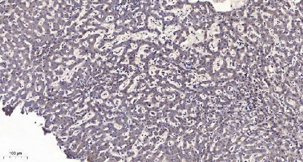
| Sample | Paraffin-embedded human liver cancer tissue; fixative not specified (datasheet A09260) |
| Fixation | Image fixative and duration unreported (datasheet A09260); verify before use. |
| Sectioning | 4–5 µm sections on charged slides (standard) |
| Deparaffinisation | Xylene, graded ethanol series to water (standard) |
| Antigen retrieval | Heat retrieval: Tris-EDTA pH 9.0 (datasheet A09260); 20 min, 95–100 °C (standard) |
| Peroxidase block | 3% H2O2, 10 min, room temperature (standard) |
| Blocking | 10% normal serum of the secondary host, 30 min, room temperature (standard) |
| Primary antibody | Rabbit anti-ERGIC3, 1:100 - 1:300 (datasheet A09260) |
| Primary incubation | Overnight at 4 °C (standard) |
| Detection | HRP-polymer secondary, DAB chromogen 5–10 min (standard) |
| Counterstain | Hematoxylin, blue, dehydrate and mount (standard) |
| Expected result | ERGIC3-positive staining in respiratory epithelial cells of bronchus (HPA tissue IHC: High). HPA tissue profile: Ubiquitous cytoplasmic expression in essentially all cells except in squamous epithelial, stroma cells and myocytes. No signal in the no-primary control. |
ERGIC3 is a membrane protein that cycles between the endoplasmic reticulum and Golgi; its 2 transmembrane segments support an intracellular, cytoplasmic staining pattern in IHC (UniProt Q9Y282 topology and subcellular location). HPA reports widespread cytoplasmic staining, with high signal in selected epithelial, neuronal, trophoblastic and spermatid populations (HPA tissue IHC). Its IHC profile is Approved, but antibody staining has low consistency with RNA expression and awaits external verification (HPA tissue IHC).
| Cytoplasmic staining in bronchial or nasopharyngeal respiratory epithelium, with nearby unstained areas. | This fits a high-signal reference population (HPA: High in respiratory epithelial cells). Assess the intracellular distribution and cell identity together; a brown signal alone does not establish ERGIC3 specificity, especially given the HPA reliability caveat (HPA tissue IHC). |
| Strong, predominantly nuclear or cell-surface staining with little intracellular cytoplasmic signal. | Treat this as a compartment mismatch requiring investigation: ERGIC3 is assigned to ER–Golgi intermediate compartment, cis-Golgi and ER membranes (UniProt Q9Y282 subcellular location). Check the counterstain and whether the apparent signal follows cellular structures before scoring it as positive (general IHC practice). |
| Prominent staining of cardiomyocytes, skeletal myocytes, adipocytes or ovarian stromal cells. | These populations are listed as not detected in the HPA tissue IHC examples (HPA tissue IHC). Reconsider antibody specificity or endogenous detection activity if they dominate the slide; this pattern alone cannot distinguish those causes (general IHC practice). |
| Haze or precipitate across tissue and empty spaces obscures cell boundaries. | A diffuse field-wide signal is difficult to assign to the cytoplasmic pattern reported for ERGIC3 (HPA tissue IHC). Review blocking, washing, chromogen development and detection controls before interpreting cell-specific intensity (general IHC practice). |
| No staining in an adequately preserved bronchial respiratory epithelial control. | HPA reports high staining in this cell population, so absent signal calls for an assay check before interpreting a test section as negative (HPA: High in bronchial respiratory epithelial cells). Confirm that the control contains the intended cells and that primary antibody and detection steps worked (general IHC practice). |
| Subcellular distribution | ERGIC3 occupies ER–Golgi intermediate compartment, cis-Golgi and ER membranes and cycles between ER and Golgi (UniProt Q9Y282 subcellular location). Interpret a localized intracellular pattern within cytoplasm; the record does not specify one invariant punctate shape for paraffin IHC (UniProt Q9Y282 subcellular location). |
| Reference-cell choice | Respiratory epithelial cells, Purkinje cells, cortical neurons, trophoblasts and elongated or late spermatids have high reported staining; endometrial and appendix glandular cells are medium (HPA tissue IHC). Choose a reference with recognizable cell identity and compare like cells, because intensity differs among listed populations (HPA tissue IHC). |
| Negative and low-staining comparators | HPA lists adipocytes, cardiomyocytes, skeletal and smooth myocytes, ovarian stroma and vaginal squamous epithelium as not detected; some other squamous or glandular populations are low (HPA tissue IHC). These are interpretive comparators, subject to the same HPA reliability caveat (HPA tissue IHC). |
| Antibody and molecular scope | HPA assigns IHC Approved to 2 rabbit polyclonal antibodies, HPA015242 and HPA015968, while reporting low agreement between staining and RNA and pending external verification (HPA antibodies; HPA tissue IHC). UniProt lists 3 isoforms, but the supplied record does not establish which isoforms either antibody detects (UniProt Q9Y282 isoforms). |
| IF/ICC Q&A | Can this IHC pattern serve as an IF/ICC acceptance rule? HPA summarizes ERGIC3 as membrane-associated but supplies no main ICC-IF location, imaged cell lines or ICC validation for these antibodies (HPA subcellular; HPA antibodies). Use the separate IF/ICC guide for that application; do not infer an IF pattern from tissue IHC. |
| Situation | Likely cause | Next action |
|---|---|---|
| The expected positive cells are blank. | The chosen control may lack the intended cell population, or an IHC processing or detection step may have failed (general IHC practice). | Confirm cell identity against an HPA high-staining reference, then check section integrity, retrieval, primary antibody application and detection controls (HPA tissue IHC; general IHC practice). The supplied sources give no ERGIC3-specific retrieval setting. |
| Staining is faint in a low-listed tissue. | The selected population may genuinely have low reported staining, such as esophageal squamous cells or liver cholangiocytes (HPA tissue IHC). | Compare with an HPA high-staining cell population processed in the same IHC run before changing assay conditions (HPA tissue IHC; general IHC practice). Avoid calling weak staining a technical failure from that tissue alone. |
| Muscle, adipose or stromal cells stain strongly. | That conflicts with HPA not-detected examples and may reflect off-target binding or endogenous detection activity (HPA tissue IHC; general IHC practice). | Review a no-primary control and the spatial pattern, and compare another HPA-listed cell population if available (general IHC practice; HPA tissue IHC). Interpret persistent signal cautiously because HPA reports low staining–RNA consistency (HPA tissue IHC). |
| Nuclei dominate the apparent positive signal. | Predominantly nuclear staining does not match the annotated ERGIC3 membrane compartments or HPA cytoplasmic tissue profile (UniProt Q9Y282 subcellular location; HPA tissue IHC). | Check the counterstain and detection controls, then score intracellular cytoplasmic signal separately from nuclear color (general IHC practice). Do not use nuclear intensity alone as evidence of ERGIC3. |
| Diffuse brown background prevents cell-level scoring. | Nonspecific reagent binding, insufficient washing or excessive chromogen development can obscure a cellular IHC pattern (general IHC practice). | Inspect a no-primary control and review blocking, washes and development time under the assay's established IHC conditions (general IHC practice). Resume scoring only when cell boundaries and cytoplasmic signal can be distinguished. |
| An IHC-positive tissue gives an unclear IF/ICC result. | Tissue IHC staining and UniProt localization do not establish antibody performance in IF/ICC; HPA lists no ICC validation or imaged cell lines here (HPA antibodies; HPA subcellular; UniProt Q9Y282 subcellular location). | Evaluate IF/ICC under its own guide and controls. Do not transfer the IHC tissue intensity threshold or infer an IF/ICC protocol from these IHC observations (HPA tissue IHC; HPA subcellular; general IF/ICC practice). |


Comprehensive Human Protein Atlas IHC scoring per tissue (reliability: Approved — Low consistency between antibody staining and RNA expression data. Pending external verification.). Rows are taken directly from the HPA tissue chart — click any row's HPA link to view the source.
| Tissue | Cell type | Level | Evidence | Source |
|---|---|---|---|---|
| Bronchus | Respiratory epithelial cells | High | Protein (IHC) | HPA → |
| Cerebellum | Purkinje cells | High | Protein (IHC) | HPA → |
| Cerebral cortex | Neuronal cells | High | Protein (IHC) | HPA → |
| Endometrium | Glandular cells | Medium | Protein (IHC) | HPA → |
| Nasopharynx | Respiratory epithelial cells | High | Protein (IHC) | HPA → |
| Tissue | Cell type | Level | Evidence | Source |
|---|---|---|---|---|
| Adipose tissue | Adipocytes | Not detected | Protein (IHC) | HPA → |
| Heart muscle | Cardiomyocytes | Not detected | Protein (IHC) | HPA → |
| Ovary | Ovarian stroma cells | Not detected | Protein (IHC) | HPA → |
| Skeletal muscle | Myocytes | Not detected | Protein (IHC) | HPA → |
| Smooth muscle | Smooth muscle cells | Not detected | Protein (IHC) | HPA → |
Troubleshoot ERGIC3 staining in paraffin sections by checking retrieval, compartment pattern, controls and cell-specific scoring before interpreting chromogenic signal.
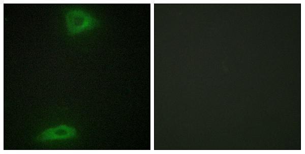
A09260 has human paraffin-section IHC and HepG2-cell IF images (A09260 IHC/IF captions); human and mouse reactivity is listed (A09260 catalog).
A09260 will render with an IHC image of paraffin-embedded human liver cancer (A09260 IHC caption). It is listed for IHC, IF and ICC in human and mouse, with an IF image of HepG2 cells (A09260 catalog; A09260 IF caption).
Which to pick: For tissue IHC, choose A09260: its image caption documents paraffin-embedded human liver cancer, Tris-EDTA retrieval at pH 9.0 and primary antibody at 1:200 overnight at 4°C; the fixative is unreported (A09260 IHC caption). For IF/ICC, A09260 is a rabbit polyclonal listed for both applications, with IF shown in HepG2 cells (A09260 catalog; A09260 IF caption). For cross-species work, A09260 lists human and mouse reactivity, although the supplied IHC image shows human material only (A09260 catalog; A09260 IHC caption).