This website uses cookies to ensure you get the best experience on our website.


- Table of Contents
Plan FCGR3A chromogenic IHC in paraffin sections with the IHC-validated antibody at 2–5 μg/mL (datasheet A01408-1). Assess the cytoplasmic staining reported in lung macrophages, liver Kupffer cells and lymph node cells, while considering the soluble receptor (HPA tissue IHC; UniProt).

Expected localisation, validated protocols, controls and antibodies — the at-a-glance facts below, then the full design guide.
| Expected localisation | Cytoplasmic in tissue (HPA tissue IHC); cell membrane (UniProt) | |
| Staining pattern | Cytoplasmic in lung macrophages, Kupffer and nodal cells (HPA tissue IHC) | |
| Antigen retrieval | EDTA pH 8.0 HIER, heat-mediated (datasheet A01408-1) | |
| Positive control | Lung+4 more · see all | |
| Negative control | Adipose tissue+4 more · see all |
| Fixation | Keep section fixation consistent (standard IHC practice; not target-specific) | |
| Caveat | Soluble receptor may complicate cell assignment (UniProt) | |
| Regulation | Regulation not specified in the record (UniProt) | |
| Isoform / epitope | No isoforms annotated; epitope side affects accessibility (UniProt) |
The catalog antibody’s IHC-P protocol (datasheet A01408-1) is accompanied by three published breast cancer IHC protocols (PMC2987696; PMC2976659; PMC2935495).
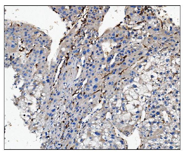
| Sample | Paraffin-embedded human liver cancer tissue; fixative not specified (datasheet A01408-1) |
| Fixation | Image fixative and duration unreported (datasheet A01408-1); verify before use. |
| Sectioning | 4–5 µm sections on charged slides (standard) |
| Deparaffinisation | Xylene, graded ethanol series to water (standard) |
| Antigen retrieval | Heat retrieval: EDTA pH 8.0 (datasheet A01408-1); 20 min, 95–100 °C (standard) |
| Peroxidase block | 3% H2O2, 10 min, room temperature (standard) |
| Blocking | 10% goat serum (datasheet A01408-1) |
| Primary antibody | Rabbit anti-FCGR3A, 2-5μg/ml (datasheet A01408-1) |
| Primary incubation | Overnight at 4 °C (datasheet A01408-1) |
| Detection | Streptavidin-biotin complex (SABC), DAB chromogen (datasheet A01408-1) |
| Counterstain | Hematoxylin, blue, dehydrate and mount (standard) |
| Expected result | FCGR3A-positive staining in macrophages of lung (HPA tissue IHC: High). HPA tissue profile: Cytoplasmic expression in a subset of cells including lung macrophages, Kupffer cells in liver and non-germinal center cells in lymph node. No signal in the no-primary control. |
FCGR3A is a cell-membrane receptor with an extracellular region at residues 17–208 and a transmembrane segment at 209–229; a soluble form also exists (UniProt P08637 topology and subcellular location). In paraffin-section IHC, expect staining in subsets of cells, especially lung macrophages, placental Hofbauer cells and splenic red-pulp cells (HPA: High in each). HPA describes the tissue pattern as cytoplasmic and rates its reliability Supported (HPA: tissue IHC).
| Distinct staining marks a subset of lung macrophages; adjacent respiratory epithelial cells remain unstained. | This distribution fits the observed tissue pattern: lung macrophages are High, while bronchial respiratory epithelial cells are Not detected (HPA: tissue IHC). Record which cells stain rather than assigning one intensity to the entire section (general IHC practice). |
| Positive cells show a cytoplasmic appearance, with or without a discernible cell-edge signal. | HPA describes cytoplasmic staining in selected tissue cells, while UniProt places the intact receptor at the cell membrane and reports a soluble form (HPA: tissue IHC; UniProt P08637 subcellular location). A membrane-only scoring rule would miss the reported IHC appearance. |
| Signal is predominantly nuclear or uniformly fills cells outside the expected positive subsets. | Treat this as a compartment or distribution mismatch requiring a specificity check; nuclear localisation is not reported for FCGR3A here (UniProt P08637 subcellular location; HPA: tissue IHC). Compare a no-primary control and the expected positive-cell pattern (general IHC practice). |
| Strong staining appears in bronchial respiratory epithelium or adipocytes. | Those cell types are Not detected in the supplied HPA tissue observations (HPA: bronchus respiratory epithelial cells; adipose adipocytes). Cross-reactivity or endogenous chromogenic activity are possibilities, not diagnoses; inspect controls before interpreting the signal (general IHC practice). |
| A known-positive section has no convincing positive cells. | Absence in lung macrophages, placental Hofbauer cells or splenic red-pulp cells conflicts with their High HPA observations (HPA: tissue IHC). Check cell identification and the staining run before calling the specimen negative (general IHC practice). |
| Cell selection and comparator tissue | HPA rates lung macrophages, Hofbauer cells and splenic red-pulp cells High, but lymph-node non-germinal-center cells Low (HPA: tissue IHC). Compare like cell types when judging signal. |
| Membrane topology and soluble receptor | The extracellular domain spans residues 17–208, and a soluble receptor also exists (UniProt P08637 topology and subcellular location). Membrane topology alone does not define the appearance of a paraffin-section stain; use HPA's observed cytoplasmic pattern as the IHC reference (HPA: tissue IHC). |
| Antibody evidence | HPA lists IHC as Supported for HPA055431 and CAB032435; ICC status is unlisted for both (HPA: antibody validation). Supported tissue IHC does not establish an IF staining pattern. |
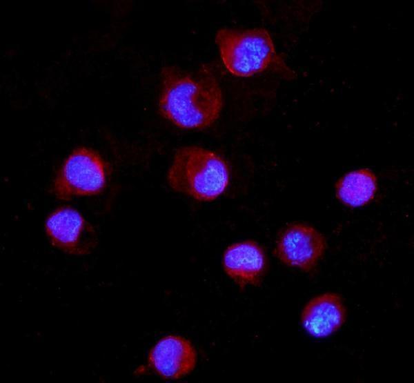
| IF/ICC Q: What should a positive cell look like? | A: Membrane-associated signal is biologically plausible for intact FCGR3A (UniProt P08637 topology). HPA gives no main ICC-IF location or cell-line images, so the supplied record cannot validate a specific IF appearance (HPA: subcellular record). |
| Chromogenic background | Endogenous enzyme activity can contribute signal in chromogenic IHC; assess it with appropriate staining controls and blocking for the chosen detection chemistry (general IHC practice). This is a workflow consideration, not an FCGR3A-specific effect. |
| Situation | Likely cause | Next action |
|---|---|---|
| Lung macrophages show no staining. | A run failure, insufficient detection or difficulty identifying macrophages may explain the discrepancy; HPA reports these cells as High (HPA: lung macrophages). | Check the positive-control section, primary-antibody incubation and detection reagents, then reassess cell identity (general IHC practice). |
| Most cells show a faint, even chromogenic haze. | Nonspecific background or residual detection activity may obscure a subset pattern (general IHC practice); HPA describes expression in a subset of cells (HPA: tissue IHC). | Inspect the no-primary control; review blocking, washing and detection development for the chosen assay (general IHC practice). |
| Bronchial respiratory epithelial cells stain prominently. | This differs from the HPA Not detected observation for that cell type (HPA: bronchus respiratory epithelial cells); specificity or detection background needs assessment. | Compare positive tissue and no-primary controls, then check whether staining follows cell type or a section-wide background pattern (general IHC practice). |
| Signal appears only in nuclei. | A nuclear-only pattern conflicts with the supplied membrane and cytoplasmic descriptions (UniProt P08637 subcellular location; HPA: tissue IHC). | Review staining controls and morphology; withhold an FCGR3A-positive call until the expected cell distribution is demonstrated (general IHC practice). |
| Lymph node looks weaker than lung or spleen. | HPA records Low staining in lymph-node non-germinal-center cells, versus High in lung macrophages and splenic red-pulp cells (HPA: tissue IHC). | Identify the stained cell population and compare it with its own HPA category before changing assay conditions (HPA: tissue IHC; general IHC practice). |
| An IF/ICC result differs from the paraffin-section IHC pattern. | The supplied HPA subcellular record has no main ICC-IF location or cell-line images, and neither listed antibody has an ICC status (HPA: subcellular record; antibody validation). | Treat the IF result as unvalidated by these records; assess its localisation and controls within an IF-specific experiment (UniProt P08637 topology; general IF practice). |


Comprehensive Human Protein Atlas IHC scoring per tissue (reliability: Supported — At least one protein variant secreted, tissue location of RNA and protein might differ and correlation is complex. External characterization data supports antibody staining.). Rows are taken directly from the HPA tissue chart — click any row's HPA link to view the source.
| Tissue | Cell type | Level | Evidence | Source |
|---|---|---|---|---|
| Lung | Macrophages | High | Protein (IHC) | HPA → |
| Placenta | Hofbauer cells | High | Protein (IHC) | HPA → |
| Skin | Fibrohistiocytic cells | Medium | Protein (IHC) | HPA → |
| Spleen | Cells in red pulp | High | Protein (IHC) | HPA → |
| Bone marrow | Hematopoietic cells | Medium | Protein (IHC) | HPA → |
| Tissue | Cell type | Level | Evidence | Source |
|---|---|---|---|---|
| Adipose tissue | Adipocytes | Not detected | Protein (IHC) | HPA → |
| Adrenal gland | Glandular cells | Not detected | Protein (IHC) | HPA → |
| Appendix | Glandular cells | Not detected | Protein (IHC) | HPA → |
| Breast | Adipocytes | Not detected | Protein (IHC) | HPA → |
| Bronchus | Respiratory epithelial cells | Not detected | Protein (IHC) | HPA → |
Troubleshoot FCGR3A staining in paraffin sections by checking retrieval, cell identity, compartment, controls, and scoring against the supplied tissue evidence.
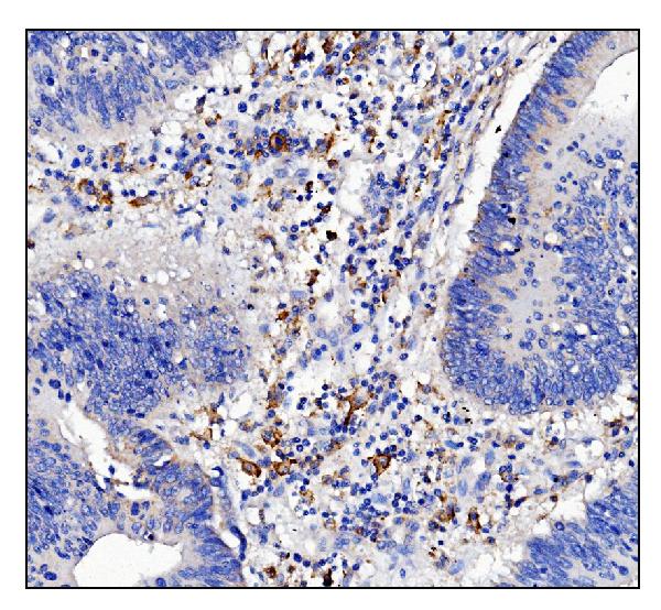
A01408-1 has IHC images from human paraffin sections and an IF image from K562 cells (A01408-1 image captions). M01408-1 lists human, mouse and rat reactivity (catalog).
A01408-1 will render with its own IHC figure from a paraffin-embedded human liver cancer section (A01408-1 IHC image caption). Its catalog also documents IHC in human rectal cancer paraffin sections and IF in K562 cells (A01408-1 image captions).
Which to pick: Choose A01408-1 for tissue IHC: its own figure documents a paraffin-embedded human section, with no fixative reported (A01408-1 IHC image caption). For IF/ICC, A01408-1 has a K562 IF image (A01408-1 IF image caption). For human, mouse or rat IF/ICC, M01408-1 is a rabbit monoclonal option on the application and reactivity lists; IHC is not listed for it (M01408-1 catalog).