This website uses cookies to ensure you get the best experience on our website.
- Table of Contents
6 Citations 6 Q&As

10 Citations 6 Q&As

2 Citations 7 Q&As


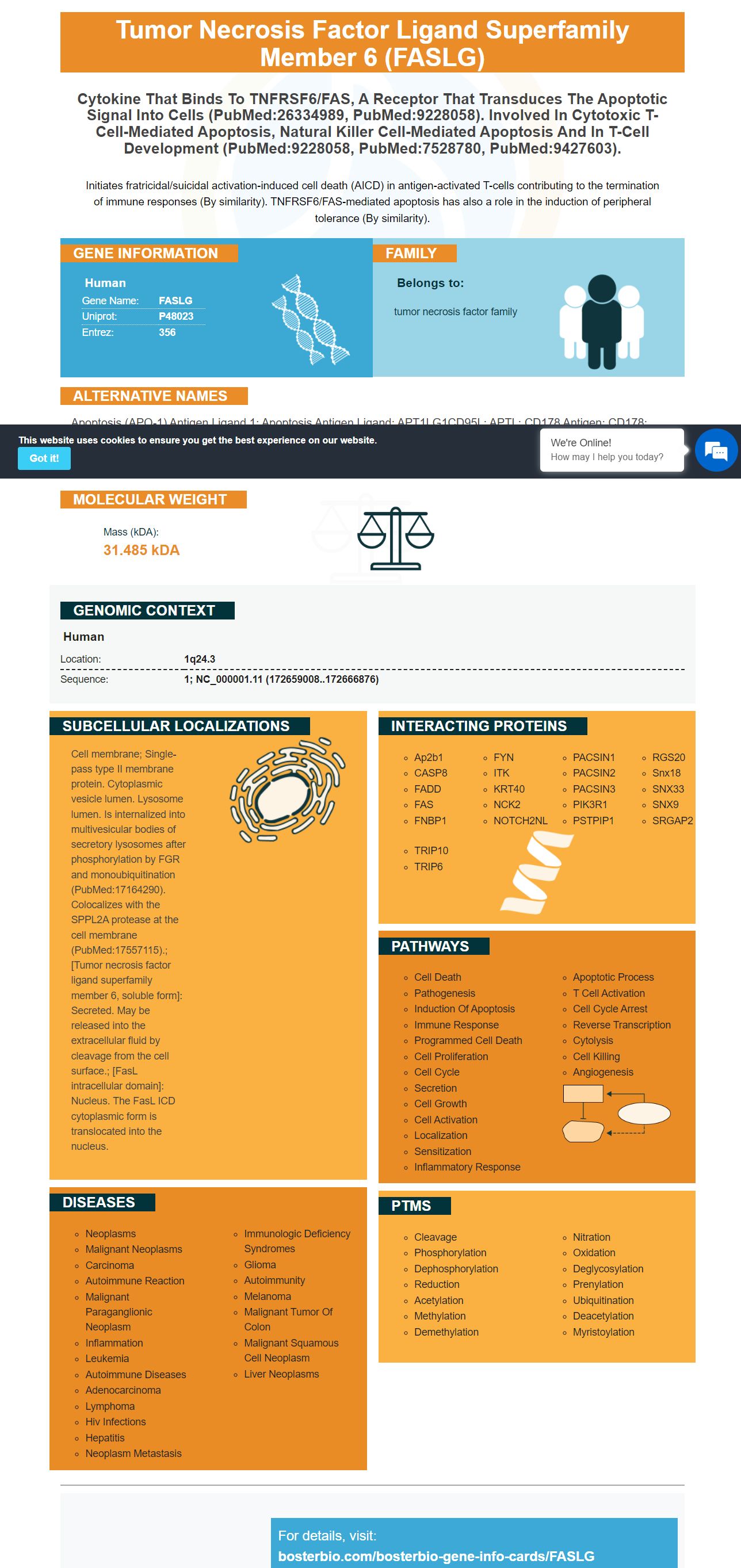
Facts about Tumor necrosis factor ligand superfamily member 6.

Initiates fratricidal/suicidal activation-induced cell death (AICD) in antigen-activated T-cells contributing to the termination of immune responses (By similarity). TNFRSF6/FAS-mediated apoptosis has also a role in the induction of peripheral tolerance (By similarity).
| Human | |
|---|---|
| Gene Name: | FASLG |
| Uniprot: | P48023 |
| Entrez: | 356 |

| Belongs to: |
|---|
| tumor necrosis factor family |

apoptosis (APO-1) antigen ligand 1; Apoptosis antigen ligand; APT1LG1CD95L; APTL; CD178 antigen; CD178; CD95L; CD95-L; Fas antigen ligand; Fas ligand (TNF superfamily, member 6); Fas Ligand; FASLCD95 ligand; FASLG; TNFSF6; TNFSF6FasL; tumor necrosis factor (ligand) superfamily, member 6; tumor necrosis factor ligand superfamily member 6
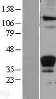
Mass (kDA):
31.485 kDA

| Human | |
|---|---|
| Location: | 1q24.3 |
| Sequence: | 1; NC_000001.11 (172659008..172666876) |
Cell membrane; Single-pass type II membrane protein. Cytoplasmic vesicle lumen. Lysosome lumen. Is internalized into multivesicular bodies of secretory lysosomes after phosphorylation by FGR and monoubiquitination (PubMed:17164290). Colocalizes with the SPPL2A protease at the cell membrane (PubMed:17557115).; [Tumor necrosis factor ligand superfamily member 6, soluble form]: Secreted. May be released into the extracellular fluid by cleavage from the cell surface.; [FasL intracellular domain]: Nucleus. The FasL ICD cytoplasmic form is translocated into the nucleus.


The FASLG marker is a distinctive monoclonal antibody, which has many applications in medicine and biology. It has been used to detect the process of apoptosis. This monoclonal antibody is also capable of detecting the anti-Bax protein, which is a specific marker that detects the apoptosis process. The FASLG marker is applicable to all scientists in the world as well as scientists working on species and their applications.
This monoclonal antibody recognizes Bax (Apoptosis Marker) which is a transcription factor that is linked to apoptosis. Antibodies to this protein could stop the process of apoptosis. These antibodies are made by various companies. Boster Bio, the company that developed the antibody has a variety of types available.
Boster Bio's monoclonal anti-Bax (Apoptosus Marker), reacts to human Bax. It has been tested in a variety of tests and has multiple applications in different research labs. Multiple assays have been used to validate the antibodies. These results are crucial in diagnosing apoptosis.
In a research study using the monoclonal antibody, the N27-2 clone was transfected by mutant hPRA1, the BHRF1, and p-Bax. The OE group had a higher rate of apoptosis than the other two groups. Western blot tests were performed to measure the expression of proteins. Anti-Bax decreased Bcl-2, cleaved-PARP, and caspase-3 expression in OE cells. The pro-apoptotic protein PCNA was not affected.
The active immunization with this antibody boosted the apoptotic marker, CC3, in tumors, but passive immunization did not increase CC3 or Ki67 in tumor cells. The results suggest that both immunotherapy options have an antitumor affect. The immune response is activated when apoptotic cells are triggered by the antibody.
PMID: 7528780 by Alderson M.; Fas ligand mediates activation-induced cell death in human T lymphocytes.
PMID: 7826947 by Takahashi T., et al. Human Fas ligand: gene structure, chromosomal location and species specificity.
*More publications can be found for each product on its corresponding product page