GATA2 / Endothelial transcription factor GATA-2 · IHC design guide

Design Immunohistochemistry for GATA2

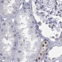
Plan GATA2 paraffin-section IHC around nuclear staining in seminal vesicle glands, placental trophoblasts and prostate (HPA tissue IHC). The IHC-validated antibody has a listed dilution of 1:50 (datasheet: M01415); consider its GATA2/3 designation when scoring staining (datasheet: M01415 title).

Evidence assembled Oct 2026 · For research use; verify linked source records and product datasheet before use
Immunohistochemistry protocol sheet for GATA2 (IHC for GATA2): expected localisation Nuclear staining (HPA tissue IHC), antibody M01415, validated IHC image, and IHC protocol steps
Printable GATA2 IHC protocol sheet — expected localisation Nuclear staining (HPA tissue IHC), antibody M01415, controls and protocol steps. Open the full GATA2 IHC guide →

GATA2 Immunohistochemistry Experimental Design Guide

Expected localisation, validated protocols, controls and antibodies — the at-a-glance facts below, then the full design guide.

Must know before staining
Expected localisation Nuclear staining (HPA tissue IHC)
Staining pattern Nuclear in seminal vesicle glands, trophoblasts and prostate (HPA tissue IHC)
Antigen retrieval Tris-EDTA pH 9.0 HIER, 95–98 °C, 20 min (rule: nuclear antigen)
Positive control ⓘ Duodenum+4 more · see all
Negative control ⓘ Adipose tissue+4 more · see all
Important caveats
Reasons your staining may differ from the expected pattern.
Fixation Keep fixation consistent across sections. (standard IHC practice; not target-specific)
Caveat GATA2/3 reactivity may confound GATA2 scoring (datasheet: M01415 title)
Regulation No specific intensity regulator reported (UniProt)
Isoform / epitope 2 isoforms; epitope coverage is unreported (UniProt)
Section 1

Recommended GATA2 IHC & IF Protocols

The catalog antibody’s IHC-P protocol is paired with one published GATA2 protocol for colorectal cancer specimens (PMC4546112).

Recommended immunohistochemistry (IHC-P) protocol parameters
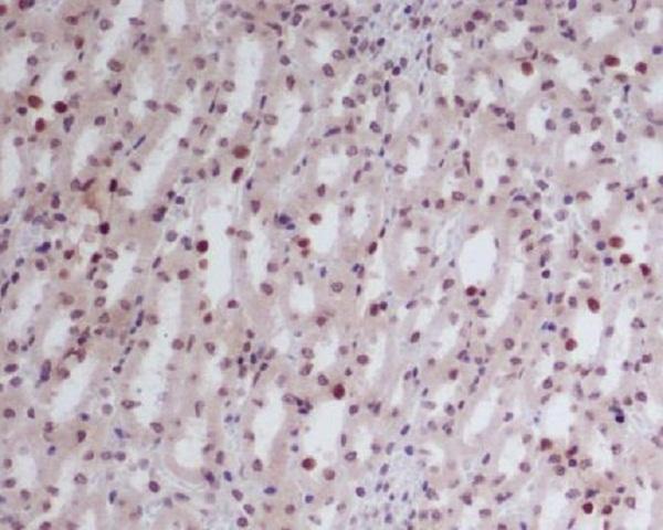
SampleParaffin-embedded mouse kidney tissue; fixative not specified (datasheet M01415)
FixationImage fixative and duration unreported (datasheet M01415); verify before use.
Sectioning4–5 µm sections on charged slides (standard)
DeparaffinisationXylene, graded ethanol series to water (standard)
Antigen retrievalHeat-induced epitope retrieval in Tris-EDTA buffer, pH 9.0, 20 min at 95–98 °C (standard rule: nuclear antigen)
Peroxidase block3% H2O2, 10 min, room temperature (standard)
Blocking10% normal serum of the secondary host, 30 min, room temperature (standard)
Primary antibodyRabbit monoclonal (clone DHA-7) anti-GATA2, 1:50 (datasheet M01415)
Primary incubationOvernight at 4 °C (standard)
DetectionHRP-polymer secondary, DAB chromogen 5–10 min (standard)
CounterstainHematoxylin, blue, dehydrate and mount (standard)
Expected resultGATA2-positive staining in glands of Brunner of duodenum (HPA tissue IHC: High). HPA tissue profile: Nuclear expression in several tissues, mainly in seminal vesicle, placenta and prostate. No signal in the no-primary control.
💡Decision noteStart with Tris-EDTA pH 9.0 retrieval at 95–98 °C for 20 min (page antigen retrieval rule; UniProt P23769: nucleus).
Section 2

What Is the Expected GATA2 Staining Pattern?

GATA2 is a nuclear transcription factor with no transmembrane segment (UniProt P23769: subcellular location; topology). In paraffin-section IHC, expect nuclear staining in selected cells, including placental trophoblastic cells, kidney collecting ducts and seminal-vesicle glandular cells (HPA tissue IHC: High). HPA calls its tissue staining “Enhanced,” while reporting medium consistency between antibody staining and RNA expression (HPA tissue IHC: reliability).

What am I looking at on my slide?
Distinct nuclear staining in placental trophoblastic cells or kidney collecting-duct cells.This fits the expected compartment and an HPA High-staining cell population (UniProt P23769: Nucleus; HPA tissue IHC: Placenta and Kidney, High). Compare nuclei within the same structure; HPA's tissue profile does not imply that every cell in a section must stain.
Strong cytoplasmic or membrane staining dominates, with little nuclear signal.This conflicts with the annotated nuclear location and absence of a transmembrane segment (UniProt P23769: subcellular location; topology). Treat it as an unconfirmed pattern and check control sections, detection background and antibody specificity using general IHC practice.
A cell population reported as not detected shows convincing staining, such as adipocytes.HPA reports GATA2 as Not detected in adipocytes (HPA tissue IHC: Adipose tissue). Check whether signal is truly nuclear and whether the detection control also stains; cross-reactivity or endogenous detection activity are possible general IHC explanations, not established GATA2-specific causes.
Weak color covers nuclei, cytoplasm and surrounding tissue without clear cell boundaries.A diffuse pattern cannot establish the expected nuclear localization (UniProt P23769: Nucleus). In general IHC practice, nonspecific binding or detection background can obscure interpretation; judge the section against a matched control before assigning positive cells.
No nuclear signal appears in an HPA High-staining cell population.An absent result in placental trophoblastic cells or seminal-vesicle glandular cells is unexpected relative to the HPA examples (HPA tissue IHC: Placenta and Seminal vesicle, High). Review section identity and staining controls before concluding that the sample lacks GATA2.
💡Expected GATA2 appearanceCall a positive result when selected HPA High-staining cells show clear nuclear chromogen; predominantly cytoplasmic, membrane or diffuse color does not match the annotated GATA2 location (HPA tissue IHC: High; UniProt P23769: Nucleus; topology).
How each factor affects the staining
Tissue and cell selectionHPA reports High staining in several specific populations, including Brunner glands, collecting ducts and trophoblastic cells, but Low staining in lung type I alveolar cells (HPA tissue IHC). Choose the stated cell population when assessing a section; a whole-tissue label alone does not identify the cells expected to stain.
Subcellular locationUniProt places GATA2 in the nucleus, and HPA ICC-IF supports a nucleoplasmic location (UniProt P23769: subcellular location; HPA subcellular). Score nuclear signal separately from cytoplasmic color in IHC. The ICC-IF finding supports localization; it is not an IF protocol option for this IHC guide.
Antibody validationTwo listed antibodies have Enhanced IHC validation; one also has Supported ICC validation (HPA antibodies: HPA005633 and CAB024847). HPA describes the tissue profile as having medium staining–RNA consistency (HPA tissue IHC: reliability). Use these qualifications when weighing a surprising pattern; they do not certify every cell or specimen.
Isoforms and modified residuesUniProt lists two isoforms and modified residues, including phosphoserine at positions 73 and 192 (UniProt P23769: isoforms; modified residues). The payload gives no antibody epitope or isoform coverage, so it cannot predict an isoform-specific IHC pattern or a modification-dependent staining change.
Why is my staining missing, weak or wrong?
SituationLikely causeNext action
Known HPA High-staining cells show no nuclear signal.The result differs from the reported pattern (HPA tissue IHC: High). The supplied sources do not identify a GATA2-specific failure mechanism.Using general IHC practice, verify tissue and cell identity, the positive and detection controls, and that the catalog antibody was used within its IHC-P instructions. Record any retrieval or dilution changes as assay checks, not established GATA2 requirements.
Color is mainly cytoplasmic or at cell borders.That distribution conflicts with GATA2's nuclear annotation and lack of a transmembrane segment (UniProt P23769: subcellular location; topology).Check nuclear counterstain alignment and compare with controls using general IHC practice. Withhold a positive GATA2 call until discrete nuclear staining is demonstrated in an expected cell population (HPA tissue IHC: High-staining populations).
A reported Not detected population appears strongly positive.For example, adipocytes are Not detected in the HPA tissue profile (HPA tissue IHC: Adipose tissue). Cross-reactivity or endogenous detection activity are possible general IHC causes.Confirm the stained cell type and nuclear localization, then inspect an appropriate detection control using general IHC practice. Report a discordant result as unconfirmed rather than extending the HPA expression pattern.
Background covers both tissue and negative areas.Diffuse color lacks the compartment contrast expected for a nuclear target (UniProt P23769: Nucleus). Its cause cannot be assigned from the GATA2 payload.Use general IHC practice to review blocking, washes and detection controls, then reassess nuclei in an HPA High-staining population (HPA tissue IHC: High). Avoid scoring diffuse color as cellular expression.
Only a few cells stain in a mixed tissue.HPA identifies staining by cell population and reports different levels across populations, including High and Low (HPA tissue IHC: positive and low lists).Identify the relevant cells morphologically and score nuclear staining within that population using general IHC practice. Do not convert a focal pattern into a whole-tissue negative or positive call without cell-level review.
IF/ICC images seem inconsistent with the IHC slide.HPA supports nucleoplasmic localization in ICC-IF, while its tissue observations come from IHC (HPA subcellular; HPA tissue IHC). The payload supplies no paired-sample equivalence.Use the ICC-IF result as a localization check only: look for nuclear IHC signal in the HPA-listed tissue cells (HPA subcellular; HPA tissue IHC). Assess the paraffin section with its own IHC controls using general IHC practice.

Sample controls for GATA2 IHC & IF

🧪Run kidney first: collecting ducts show High GATA2 staining (HPA: Kidney, collecting ducts, High). Use adipose tissue as a negative comparator because adipocytes are Not detected (HPA: Adipose tissue, adipocytes, Not detected); on the kidney slide, cells outside the collecting ducts are internal background comparators, and any nuclear staining in them needs interpretation against the controls rather than an assumption that all are GATA2-negative (HPA: Kidney, collecting ducts, High; UniProt P23769: Nucleus).
Positive control tissue: Duodenum (Glands of Brunner, HPA High)
Negative control tissue: Adipose tissue (HPA Not detected)
ICC-IF cell lines (HPA subcellular resource): HPA ICC-IF images show GATA2 in A-431, U-251MG, U2OS, with annotated localisation: Nucleoplasm (supported) (HPA subcellular).
Technical controls: Include a no-primary (secondary-only) control, a host- and clonality-matched isotype control, and GATA2-knockout material as a biological negative where available (standard IHC/IF control practice). For chromogenic kidney IHC, quench endogenous peroxidase and account for renal endogenous biotin if using avidin–biotin detection; for kidney IF, assess tissue autofluorescence with the no-primary control (standard IHC/IF practice).
⚠️Feasibility: A target-specific fixation window and its effect are unreported, and the selected M01415 paraffin-section mouse-kidney caption does not state a fixative (selected-SKU tissue-IHC caption). Antigen-retrieval dependency is unreported; optimize retrieval for paraffin IHC rather than inferring it from staining patterns (supplied target/application evidence; standard IHC practice). The supplied evidence does not establish whether frozen sections or IF are easier; renal endogenous biotin may complicate avidin–biotin IHC, while kidney autofluorescence may complicate IF (standard IHC/IF practice).

HPA tissue IHC evidence for GATA2

Comprehensive Human Protein Atlas IHC scoring per tissue (reliability: Enhanced — Medium consistency between antibody staining and RNA expression data.). Rows are taken directly from the HPA tissue chart — click any row's HPA link to view the source.

Positive expression · recommended positive controls

TissueCell typeLevelEvidenceSource
Duodenum Glands of Brunner High Protein (IHC) HPA →
Kidney Collecting ducts High Protein (IHC) HPA →
Placenta Trophoblastic cells High Protein (IHC) HPA →
Seminal vesicle Glandular cells High Protein (IHC) HPA →
Testis Round or early spermatids High Protein (IHC) HPA →

Undetected expression · recommended negative controls

TissueCell typeLevelEvidenceSource
Adipose tissue Adipocytes Not detected Protein (IHC) HPA →
Appendix Endocrine cells Not detected Protein (IHC) HPA →
Bone marrow Hematopoietic cells Not detected Protein (IHC) HPA →
Bronchus Basal cells Not detected Protein (IHC) HPA →
Colon Endocrine cells Not detected Protein (IHC) HPA →
Section 3

Advanced GATA2 IHC Tips

Troubleshoot GATA2 staining by checking nuclear localisation, cell identity and controls before comparing signal intensity across sections.

How should I adjust retrieval when GATA2 nuclear staining is weak?
Start with Tris-EDTA pH 9.0 heat-induced retrieval at 95–98 °C for 20 min (page retrieval setting: nuclear antigen rule). Compare a known positive control, such as kidney collecting ducts, on the same staining run before changing conditions (HPA: High in kidney collecting ducts). If nuclei remain weak, test a shorter or longer heating interval on adjacent sections while keeping detection and imaging conditions constant (standard IHC practice). Check for tissue lifting or damaged morphology after heating, and accept a revised condition only when nuclear signal improves without increasing diffuse background (UniProt P23769: nucleus; standard IHC practice).
Could fixation explain weak or uneven GATA2 staining?
Target-specific fixation sensitivity is unknown because no GATA2 fixation comparison is supplied (provided evidence: no target-specific fixation comparison). The selected M01415 image shows a paraffin-embedded mouse kidney section, but its caption does not state the fixative (M01415 caption). Record fixative, fixation duration and section thickness, then compare sections processed consistently with a positive control in the same run (standard IHC practice; HPA: High in kidney collecting ducts). If staining varies across a specimen, inspect morphology and section edges before attributing the pattern to fixation; do not infer a GATA2-specific fixation effect from its nuclear location or modified residues (UniProt P23769: nucleus and modified residues; standard IHC practice).
What localisation should count as a credible GATA2 IHC signal?
Score distinct nuclear staining as the expected GATA2 pattern (UniProt P23769: nucleus; HPA subcellular: supported nucleoplasm). Evaluate each cell type separately because the HPA tissue profile reports nuclear expression across several tissues, including High staining in kidney collecting ducts and placental trophoblastic cells (HPA tissue IHC). Use a nuclear counterstain to verify that chromogen overlaps intact nuclei rather than adjacent cytoplasm, pigment or luminal material (standard IHC practice). If cytoplasmic signal dominates, review retrieval, antibody concentration and detection background against a no-primary control before treating that signal as GATA2 (UniProt P23769: nucleus; standard IHC practice).
Could isoforms or epitope masking change the GATA2 staining pattern?
GATA2 has 2 annotated isoforms, but the supplied evidence does not map this antibody’s epitope to either one (UniProt P23769: isoforms 1 and 2; provided evidence: epitope unspecified). Its 480-residue annotated chain has no signal peptide or transmembrane segment, and reported modifications include phosphoserine at residues 73 and 192 (UniProt P23769). Those features do not establish whether a particular tissue section exposes the antibody’s binding site after retrieval (provided evidence: epitope unspecified; standard IHC practice). If a staining discrepancy persists, obtain epitope information for the catalog antibody and compare matched sections under controlled retrieval conditions before claiming isoform-specific expression (standard IHC practice).
How can I check a GATA2 result with multiplex IF?
On the separate IF/ICC guide, pair GATA2 with a validated endothelial marker when testing endothelial identity, while also checking whether GATA2 signal occupies nuclei (UniProt P23769: endothelial-cell specificity and nucleus). Choose fluorophores after inspecting unstained tissue for autofluorescence, and place the weaker target signal in a channel with lower measured background (standard IF practice). Use permeabilisation appropriate for antibody access to the nucleoplasm, because the expected epitope compartment is nuclear rather than across a plasma membrane (HPA subcellular: supported nucleoplasm; UniProt P23769: no transmembrane segment; standard IF practice). Include single-stain and no-primary controls to distinguish channel bleed-through and background from genuine colocalisation (standard IF practice).
Why is my GATA2 chromogen signal diffuse or widespread?
First compare the section with a no-primary control and inspect whether the chromogen follows intact nuclei (UniProt P23769: nucleus; standard IHC practice). If signal remains in the control, review the peroxidase block, secondary reagent and DAB development, since these are general chromogenic workflow sources of background (standard IHC practice). If background appears only with primary antibody, compare an antibody titration and shorter development time on adjacent sections, keeping the positive control in the same run (standard IHC practice; HPA: High in kidney collecting ducts). Treat diffuse cytoplasmic staining cautiously, particularly when it overwhelms the expected nuclear pattern (UniProt P23769: nucleus; HPA subcellular: supported nucleoplasm).
How should I quantify GATA2 staining across tissue sections? ⚠ ANSWER MARKED FOR VERIFICATION
Define the cell population and count only interpretable nuclei within comparable regions before calculating the percentage of GATA2-positive cells (UniProt P23769: nucleus; standard IHC scoring practice). An H-score can combine nuclear intensity categories with their percentages, while positive nuclei per mm² can describe spatial density (standard IHC scoring practice). Normalise the positive count to all eligible nuclei of the same cell type, or report density with its measured tissue area, and keep threshold and counterstain settings fixed across samples (standard IHC scoring practice). Report regions separately when tissue composition differs, since the HPA profile shows substantial cell-type variation (HPA tissue IHC).
How can I separate true GATA2 staining from artefact?
A plausible positive is signal in intact nuclei of an appropriate cell population, supported by a positive control such as kidney collecting ducts (UniProt P23769: nucleus; HPA: High in kidney collecting ducts). Examine wrong-compartment staining, section edges and necrotic areas for uneven deposition or damaged morphology before scoring them (standard IHC practice). Use a no-primary control to check endogenous enzyme or detection signal, and compare morphology with a counterstained adjacent section when cell identity is uncertain (standard IHC practice). Avoid declaring every unstained cell negative on technical grounds alone: HPA reports variable expression and only Medium consistency between antibody staining and RNA data (HPA tissue IHC: Enhanced reliability).
Boster reagents

Best GATA2 / Endothelial transcription factor GATA-2 IHC Antibodies

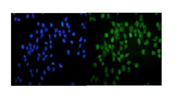
Catalog antibodies list human, mouse and rat reactivity (catalog: reactivity). The figures show IHC in paraffin-embedded mouse kidney (M01415 image caption) and IF in A431 cells (A01415-1 image caption).

Real IHC data Immunohistochemical analysis of paraffin-embedded mouse kidney, using GATA2/3 Antibody.
Anti-GATA2/3 Rabbit Monoclonal Antibody
Cat # M01415
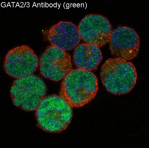
Real IF data IF analysis of GATA2 using anti-GATA2 antibody (A01415-1). GATA2 was detected in immunocytochemical section of A431 cells. Enzyme antigen retrieval was performed using IHC enzyme antigen retrieval reagent (AR0022) for 15 mins. The cells were blocked with 10% goat serum. And then incubated with 5μg/mL rabbit anti-GATA2 Antibody (A01415-1) overnight at 4°C. DyLight®488 Conjugated Goat Anti-Rabbit IgG (BA1127) was used as secondary antibody at 1:100 dilution and incubated for 30 minutes at 37°C. The section was counterstained with DAPI. Visualize using a fluorescence microscope and filter sets appropriate for the label used.
Anti-GATA2 Antibody ®
Cat # A01415-1

M01415 lists IHC and IF applications and shows IHC in paraffin-embedded mouse kidney (catalog: applications; M01415 image caption). A01415-1 lists IF/ICC and shows IF in A431 cells (catalog: applications; A01415-1 image caption).

Which to pick: For tissue IHC, choose M01415: it is a rabbit monoclonal listed for IHC at 1:50, with an image from paraffin-embedded mouse kidney; the fixative is unreported (catalog: host, clone, applications and IHC dilution; M01415 image caption). For IF/ICC, choose A01415-1: its A431 IF image documents staining at 5 μg/mL, while its catalog lists no IHC application (A01415-1 image caption; catalog: applications). Both list human, mouse and rat reactivity for cross-species planning, but the supplied IHC image demonstrates mouse tissue only (catalog: reactivity; M01415 image caption).

Each figure is that product's own IHC / IF validation image from its datasheet.

References

  1. UniProt Consortium. UniProt entry P23769 (GATA2_HUMAN, Endothelial transcription factor GATA-2).
  2. Human Protein Atlas. GATA2 tissue IHC expression (reliability: Enhanced).
  3. Human Protein Atlas. GATA2 subcellular location (ICC-IF): Localized to the nucleoplasm..
  4. Human Protein Atlas. GATA2 antibody validation summary (2 antibodies).
  5. Immunohistochemical Analysis of GATA2 Expression in Endometrium and its Relationship with Hormone Receptor Expression in Benign and Premalignant Endometrial Disorders. Reproductive sciences (Thousand Oaks, Calif.) 2024 — PMC11611599.
  6. Downregulation of endothelial microRNA-200b supports cutaneous wound angiogenesis by desilencing GATA binding protein 2 and vascular endothelial growth factor receptor 2. Arteriosclerosis, thrombosis, and vascular biology 2012 — PMC3399424.
  7. GATA2 rs2335052 Polymorphism Predicts the Survival of Patients with Colorectal Cancer. PloS one 2015 — PMC4546112.
  8. Linking GATA2 to myeloid dysplasia and complex cytogenetics in adult myelodysplastic neoplasm and acute myeloid leukemia. Blood advances 2024 — PMC10787255.
  9. PubMed PMID:1714909 — UniProt-cited evidence.
  10. PubMed PMID:1370462 — UniProt-cited evidence.
  11. PubMed PMID:14702039 — UniProt-cited evidence.