This website uses cookies to ensure you get the best experience on our website.


- Table of Contents
Plan chromogenic IHC for GORASP2 using the reported cytoplasmic and membranous tissue pattern, including strong staining in gastrointestinal endocrine cells (HPA tissue IHC). This guide covers fixation, antibody dilution, controls and scoring, while accounting for the reported mismatch between staining and RNA expression (HPA tissue IHC).

Expected localisation, validated protocols, controls and antibodies — the at-a-glance facts below, then the full design guide.
| Expected localisation | Cytoplasmic and membranous tissue staining (HPA tissue IHC) | |
| Staining pattern | Cytoplasmic/membranous staining in GI endocrine cells (HPA tissue IHC) | |
| Antigen retrieval | Tris-EDTA pH 9.0 HIER, heat-mediated (datasheet A07850-1) | |
| Positive control | Appendix+4 more · see all | |
| Negative control | Caudate+4 more · see all |
| Fixation | Matched tissue-IHC evidence does not establish the fixation claim. Validate the specimen-specific method before use. (selected-SKU IHC image A07850-1) | |
| Caveat | Staining may diverge from RNA abundance (HPA tissue IHC) | |
| Regulation | Intensity regulation unreported (UniProt) | |
| Isoform / epitope | 3 isoforms; confirm epitope coverage (UniProt) |
The catalog antibody’s IHC-P protocol is paired with one published GORASP2 protocol using FFPE sections (PMC6422190).
| Sample | Paraffin-embedded human liver cancer tissue; fixative not specified (datasheet A07850-1) |
| Fixation | Image fixative and duration unreported (datasheet A07850-1); verify before use. |
| Sectioning | 4–5 µm sections on charged slides (standard) |
| Deparaffinisation | Xylene, graded ethanol series to water (standard) |
| Antigen retrieval | Heat retrieval: Tris-EDTA pH 9.0 (datasheet A07850-1); 20 min, 95–100 °C (standard) |
| Peroxidase block | 3% H2O2, 10 min, room temperature (standard) |
| Blocking | 10% normal serum of the secondary host, 30 min, room temperature (standard) |
| Primary antibody | Rabbit anti-GORASP2, 1:100-1:300 (datasheet A07850-1) |
| Primary incubation | Overnight at 4 °C (standard) |
| Detection | HRP-polymer secondary, DAB chromogen 5–10 min (standard) |
| Counterstain | Hematoxylin, blue, dehydrate and mount (standard) |
| Expected result | GORASP2-positive staining in endocrine cells of appendix (HPA tissue IHC: High). HPA tissue profile: Cytoplasmic and membranous expression in several tissues, mainly in the gastrointestinal tract. No signal in the no-primary control. |
GORASP2 is a Golgi-associated protein with no transmembrane segment; ER stress can relocate it to the ER membrane (UniProt Q9H8Y8). In paraffin-section IHC, expect cytoplasmic and membranous staining in selected cells, including gastrointestinal endocrine cells and respiratory epithelial cells (HPA tissue IHC). HPA rates the tissue staining evidence Enhanced, while noting low consistency with RNA expression; paired antibodies support the protein pattern (HPA tissue IHC).
| Localized cytoplasmic staining in endocrine cells of appendix, colon, or duodenum, with little staining in adjacent cells. | This fits the reported High staining in those endocrine cell populations (HPA tissue IHC). A compact, perinuclear distribution is compatible with Golgi localization (UniProt Q9H8Y8; HPA subcellular ICC-IF). Judge the named cell population, since HPA does not assign one intensity to every cell in the section (HPA tissue IHC). |
| Predominantly nuclear, extracellular, or uniform cell-surface staining replaces the expected cytoplasmic pattern. | These compartments do not match the reported Golgi localization or tissue-level cytoplasmic and membranous profile (HPA subcellular ICC-IF; HPA tissue IHC). Treat the result as suspect and review controls and section morphology (general IHC practice). The tissue IHC profile alone cannot resolve every intracellular membrane. |
| Strong staining appears in glomerular cells or liver cholangiocytes. | HPA reports these specific cell populations as Not detected (HPA tissue IHC). Reproducible staining warrants checking antibody specificity and endogenous detection activity (general IHC practice). Do not extend those cell-specific negative observations to all kidney or liver cells (HPA tissue IHC). |
| Color is diffuse across tissue structures, including areas without plausible cellular localization. | A broadly distributed deposit is hard to reconcile with the reported Golgi-centered localization (HPA subcellular ICC-IF). Background can arise from the detection workflow, inadequate blocking, or excess antibody (general IHC practice); the appearance alone does not identify which step is responsible. |
| No staining is visible in a well-preserved appendix, colon, or duodenum endocrine cell population. | Those populations are reported High by HPA, so absent signal calls for a technical review (HPA tissue IHC; general IHC practice). Confirm the cells are present and examine retrieval, antibody use, and detection controls (general IHC practice). HPA tissue levels do not establish a GORASP2-specific fixation sensitivity. |
| Cell selection in the section | HPA reports High staining in respiratory epithelial cells of bronchus and nasopharynx, and glandular cells of endometrium, epididymis, and prostate, alongside the gastrointestinal endocrine populations (HPA tissue IHC). Score the identified cells rather than treating the entire organ as uniformly positive (general IHC practice). |
| Reliability of the tissue pattern | The IHC profile is Enhanced because paired antibodies show similar protein staining, despite low agreement with RNA expression (HPA tissue IHC). This supports the observed pattern but does not make each positive or negative cell assignment an absolute biological rule. |
| Intracellular location | UniProt places GORASP2 at the Golgi and describes it as membrane-associated, with no transmembrane segment (UniProt Q9H8Y8). HPA reports a Golgi main location by ICC-IF (HPA subcellular ICC-IF). A tissue-level description of membranous staining should therefore not automatically be scored as plasma-membrane staining. |
| ER stress | ER stress can relocalize GORASP2 from the Golgi to the ER membrane (UniProt Q9H8Y8). A less compact cytoplasmic distribution may merit biological review when ER stress is part of the experiment; the supplied HPA tissue images do not establish how that change appears in chromogenic paraffin-section IHC. |
| IF/ICC: where should the signal appear? | HPA reports enhanced Golgi localization in ICC-IF images, including U2OS cells with siRNA and scrambled conditions (HPA subcellular ICC-IF). Use that as a compartment cross-check; ICC-IF images do not specify an IHC-P retrieval or staining protocol. |
| Situation | Likely cause | Next action |
|---|---|---|
| Positive-control endocrine cells are blank. | The target cells may be absent from the examined level, or the IHC workflow may have failed (HPA tissue IHC; general IHC practice). | Verify cell identity and preservation, then review antigen retrieval, primary antibody application, and detection controls (general IHC practice). Do not infer a GORASP2-specific fixation effect from a blank section. |
| All structures carry a pale, even chromogen deposit. | Nonlocalized signal can reflect background from blocking, washing, antibody concentration, or detection chemistry (general IHC practice). | Inspect the negative reagent control, wash quality, blocking, and chromogen development; adjust one workflow variable at a time (general IHC practice). |
| Staining appears in glomerular cells or cholangiocytes. | These cells are reported Not detected by HPA; cross-reactivity or endogenous detection activity is possible (HPA tissue IHC; general IHC practice). | Compare a negative reagent control and an appropriate positive-cell control, and confirm the stained cell type before interpreting the result (general IHC practice). |
| The strongest signal is nuclear. | A nuclear-dominant pattern conflicts with the Golgi localization reported by UniProt and HPA (UniProt Q9H8Y8; HPA subcellular ICC-IF). | Recheck counterstain, chromogen distribution, and controls; report the compartment mismatch rather than scoring it as expected GORASP2 staining (general IHC practice). |
| A presumed negative section contains a few stained cells. | HPA negative calls apply to named cells, such as red-pulp cells in spleen, rather than every cell in an organ (HPA tissue IHC). | Identify the stained cells first, then compare that exact cell population with the HPA entry and the negative reagent control (HPA tissue IHC; general IHC practice). |
| Cytoplasmic staining is broad instead of compact and perinuclear. | ER stress can redistribute GORASP2 to the ER membrane, while nonspecific background remains another possibility (UniProt Q9H8Y8; general IHC practice). | Check whether ER stress is relevant to the specimen, then compare cellular boundaries and negative controls; do not assign ER redistribution from chromogenic appearance alone (UniProt Q9H8Y8; general IHC practice). |


Comprehensive Human Protein Atlas IHC scoring per tissue (reliability: Enhanced — Low consistency between antibody staining and RNA expression data. Paired antibodies with high similarity supports the protein expression.). Rows are taken directly from the HPA tissue chart — click any row's HPA link to view the source.
| Tissue | Cell type | Level | Evidence | Source |
|---|---|---|---|---|
| Appendix | Endocrine cells | High | Protein (IHC) | HPA → |
| Bronchus | Respiratory epithelial cells | High | Protein (IHC) | HPA → |
| Colon | Endocrine cells | High | Protein (IHC) | HPA → |
| Duodenum | Endocrine cells | High | Protein (IHC) | HPA → |
| Endometrium | Glandular cells | High | Protein (IHC) | HPA → |
| Tissue | Cell type | Level | Evidence | Source |
|---|---|---|---|---|
| Caudate | Glial cells | Not detected | Protein (IHC) | HPA → |
| Hippocampus | Glial cells | Not detected | Protein (IHC) | HPA → |
| Kidney | Cells in glomeruli | Not detected | Protein (IHC) | HPA → |
| Liver | Cholangiocytes | Not detected | Protein (IHC) | HPA → |
| Skeletal muscle | Myocytes | Not detected | Protein (IHC) | HPA → |
Troubleshoot GORASP2 chromogenic IHC by checking retrieval, tissue preservation, staining pattern, controls, and scoring before interpreting biological differences.
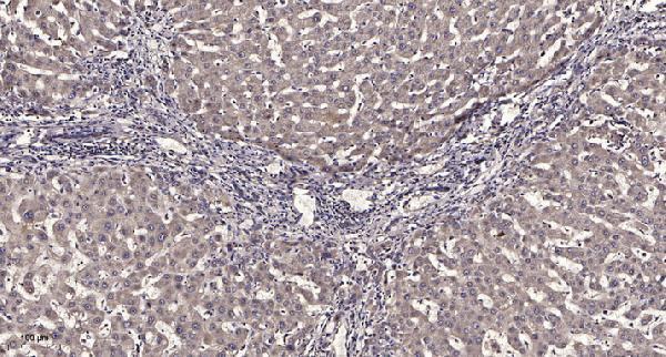
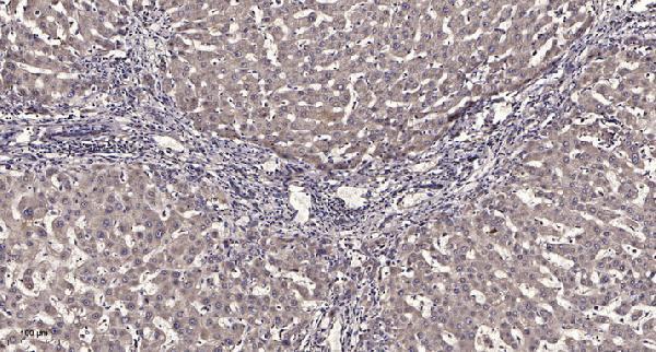
Two anti-GORASP2 antibodies have IHC images from paraffin-embedded human liver cancer (each SKU’s IHC caption); both list IF and human, mouse and rat reactivity (catalog applications and reactivity).
A07850-1 lists IHC and IF for human, mouse and rat, with an IHC image from paraffin-embedded human liver cancer (catalog applications and reactivity; A07850-1 IHC caption). A30705 lists IHC, IF and ICC for human, mouse and rat, with its own IHC image from paraffin-embedded human liver cancer (catalog applications and reactivity; A30705 IHC caption).
Which to pick: For tissue IHC, either SKU has an image from paraffin-embedded human liver cancer; each caption reports 1:200 primary antibody and Tris-EDTA retrieval at pH 9.0, but neither reports the fixative (each SKU’s IHC caption). For IF/ICC, choose A30705 because both applications are listed; A07850-1 lists IF without ICC (catalog applications). For mouse or rat samples, both list reactivity, but the supplied IHC images show human tissue only (catalog reactivity; each SKU’s IHC caption).