GPAT3 / Glycerol-3-phosphate acyltransferase 3 · IHC design guide

Design Immunohistochemistry for GPAT3

Plan GPAT3 paraffin-section IHC around granular cytoplasmic tissue staining (HPA tissue IHC) and its ER membrane location (UniProt). Compare cell types using the catalog antibody's IHC dilution range of 1:100–1:300 (datasheet A07965).

Evidence assembled Oct 2026 · For research use; verify linked source records and product datasheet before use
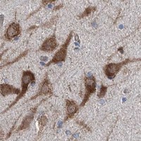
Immunohistochemistry protocol sheet for GPAT3 (IHC for GPAT3): expected localisation Granular cytoplasm (HPA tissue IHC); ER membrane (UniProt), antibody A07965, validated IHC image, and IHC protocol steps
Printable GPAT3 IHC protocol sheet — expected localisation Granular cytoplasm (HPA tissue IHC); ER membrane (UniProt), antibody A07965, controls and protocol steps. Open the full GPAT3 IHC guide →

GPAT3 Immunohistochemistry Experimental Design Guide

Expected localisation, validated protocols, controls and antibodies — the at-a-glance facts below, then the full design guide.

Must know before staining
Expected localisation Granular cytoplasm (HPA tissue IHC); ER membrane (UniProt)
Staining pattern General granular cytoplasmic staining (HPA tissue IHC)
Antigen retrieval Tris-EDTA pH 9.0 HIER, heat-mediated (datasheet A07965)
Positive control ⓘ Appendix+4 more · see all
Negative control ⓘ Adipose tissue+4 more · see all
Important caveats
Reasons your staining may differ from the expected pattern.
Fixation Keep fixation consistent across sections (standard IHC practice; not target-specific)
Caveat Adipocytes undetected (HPA tissue IHC) despite adipose expression (UniProt)
Regulation No staining-linked regulation annotated (UniProt)
Isoform / epitope No isoforms or processing annotated; epitope side unknown (UniProt)
Section 1

Recommended GPAT3 IHC & IF Protocols

The catalog antibody protocol uses Tris-EDTA pH 9.0 heat retrieval (datasheet A07965). The published tissue protocol below uses citrate retrieval (PMC12534568).

Recommended immunohistochemistry (IHC-P) protocol parameters
SampleParaffin-embedded human tonsil tissue; fixative not specified (datasheet A07965)
FixationImage fixative and duration unreported (datasheet A07965); verify before use.
Sectioning4–5 µm sections on charged slides (standard)
DeparaffinisationXylene, graded ethanol series to water (standard)
Antigen retrievalHeat retrieval: Tris-EDTA pH 9.0 (datasheet A07965); 20 min, 95–100 °C (standard)
Peroxidase block3% H2O2, 10 min, room temperature (standard)
Blocking10% normal serum of the secondary host, 30 min, room temperature (standard)
Primary antibodyRabbit anti-GPAT3, 1:100-1:300 (datasheet A07965)
Primary incubationOvernight at 4 °C (standard)
DetectionHRP-polymer secondary, DAB chromogen 5–10 min (standard)
CounterstainHematoxylin, blue, dehydrate and mount (standard)
Expected resultGPAT3-positive staining in glandular cells of appendix (HPA tissue IHC: High). HPA tissue profile: General cytoplasmic expression with a granular pattern. No signal in the no-primary control.
💡Decision noteStart with Tris-EDTA pH 9.0 for the catalog antibody (datasheet A07965); compare citrate retrieval when adapting the published protocol (PMC12534568).
Section 2

What Is the Expected GPAT3 Staining Pattern?

GPAT3 is an endoplasmic reticulum membrane protein with three transmembrane segments (UniProt Q53EU6 topology). In paraffin-section IHC, expect granular cytoplasmic staining in cell populations such as appendix, breast, and colon glandular cells, bronchial basal cells, and selected neurons (HPA: tissue IHC). HPA rates its tissue IHC profile Approved, with external verification pending (HPA: reliability).

What am I looking at on my slide?
Granular cytoplasmic staining in appendix or colon glandular cells.This fits the reported general cytoplasmic, granular pattern and high staining in those cells (HPA: tissue IHC). The membrane-associated signal is compatible with GPAT3's endoplasmic reticulum location (UniProt Q53EU6 subcellular location); chromogenic IHC alone does not prove organelle identity.
Predominantly nuclear staining, with little granular cytoplasmic signal.A dominant nuclear pattern conflicts with GPAT3's annotated endoplasmic reticulum membrane location and HPA's granular cytoplasmic IHC profile (UniProt Q53EU6 subcellular location; HPA: tissue IHC). Check whether the signal persists after appropriate background controls before interpreting it as GPAT3.
Strong staining in adipocytes, cholangiocytes, or skeletal-muscle myocytes.HPA reports GPAT3 as not detected in these specific cell populations (HPA: tissue IHC). Strong signal there warrants a cross-reactivity or detection-background check; it does not establish that every cell in adipose tissue, liver, or skeletal muscle is negative.
Diffuse color across tissue, including cell types expected to be unstained.A uniform haze obscures the cell-specific, granular pattern reported by HPA (HPA: tissue IHC). Assess the no-primary control and endogenous detection activity, then optimize blocking, washes, or detection conditions as general chromogenic IHC practice.
No staining in an intact appendix or colon glandular-cell control.These are reported high-staining populations (HPA: tissue IHC), so a blank result is inconclusive for the specimen being tested. First confirm that the positive control, antibody incubation, and detection reaction worked; HPA's Approved profile is pending external verification (HPA: reliability).
💡Expected GPAT3 appearanceCall a result consistent with GPAT3 when glandular cells in appendix or colon show discernible granular cytoplasmic staining (HPA: High; HPA: general pattern), compatible with an endoplasmic reticulum membrane protein (UniProt Q53EU6); dominant nuclear color or widespread cell-independent haze calls for controls before a positive interpretation.
How each factor affects the staining
Membrane topology and epitope accessGPAT3 has three annotated transmembrane segments (UniProt Q53EU6 topology). Epitope position is not supplied, so no specific retrieval or permeabilisation setting can be inferred; optimize access using the antibody's validated IHC instructions and general IHC controls.
Cell-specific reference patternAppendix and colon glandular cells are high, while adipocytes and skeletal-muscle myocytes are not detected (HPA: tissue IHC). Select and score the named cell population, since a tissue-wide label would conceal these distinctions.
Strength of validationHPA lists the tissue IHC profile and antibody HPA029414 as Approved, with external verification pending for the profile (HPA: reliability; HPA: antibody validation). Treat agreement with the reference pattern as supportive rather than independent proof of specificity.
What should IF/ICC show?A membrane-associated signal is plausible (HPA: subcellular summary; UniProt Q53EU6 subcellular location), but HPA supplies no ICC-IF cell-line images or main location (HPA: subcellular record). This IHC evidence cannot define an IF pattern or an IF protocol.
Why is my staining missing, weak or wrong?
SituationLikely causeNext action
The positive-control glandular cells are blank.The run may have failed; appendix and colon glandular cells are reported high by HPA (HPA: tissue IHC).Confirm that the correct cell population is present. Review the antibody's IHC instructions, retrieval, incubation, and detection steps; repeat with an on-slide positive control (general IHC practice).
Most nuclei stain more strongly than the cytoplasm.This disagrees with the reported granular cytoplasmic profile and endoplasmic reticulum membrane location (HPA: tissue IHC; UniProt Q53EU6).Inspect a no-primary control and compare cytoplasmic signal in an HPA high-staining cell population before assigning target-specific staining (general IHC practice; HPA: tissue IHC).
Adipocytes show strong, widespread color.HPA reports GPAT3 not detected in adipocytes, despite UniProt listing expression in adipose tissue (HPA: tissue IHC; UniProt Q53EU6 tissue specificity).Score adipocytes separately from other cells. Check detection background and antibody specificity with suitable controls; do not treat tissue-level expression as proof of adipocyte staining.
Color spreads across tissue without a granular cellular pattern.The result does not resemble HPA's general granular cytoplasmic profile (HPA: tissue IHC); diffuse chromogen can reflect assay background (general IHC practice).Compare a no-primary slide, check endogenous detection activity, and adjust blocking, washing, or detection conditions as indicated by the controls (general IHC practice).
A weak signal appears only in lung macrophages.HPA lists lung macrophages as Low, whereas appendix and colon glandular cells are High (HPA: tissue IHC).Use a reported high-staining glandular-cell control to assess assay performance. Interpret the macrophage result within its named cell population, without expecting high intensity there (HPA: tissue IHC).
IF/ICC appears to show a precise organelle pattern.HPA summarizes GPAT3 as membrane-associated but lists no main ICC-IF location or cell-line images (HPA: subcellular record).Report the observed IF pattern separately from the paraffin IHC result. Verify organelle identity with an appropriate marker and controls before assigning it to GPAT3 (general IF practice).

Sample controls for GPAT3 IHC & IF

🧪Run appendix first and score its glandular cells for staining (HPA: High in appendix glandular cells). Use skeletal muscle myocytes as a negative tissue comparator (HPA: Not detected in skeletal muscle myocytes); on the appendix slide, assess adjacent non-glandular cells for background staining without assuming they are GPAT3-negative.
Positive control tissue: Appendix (Glandular cells, HPA High)
Negative control tissue: Adipose tissue (HPA Not detected)
ICC-IF cell lines (HPA subcellular resource): HPA carries no ICC-IF cell line for GPAT3; derive a cell-line control from the positive tissue's cell type (Glandular cells) and confirm it by RNA or western blot first.
Technical controls: Include no-primary (secondary-only), host-species-matched isotype control for a monoclonal antibody or nonimmune IgG for a polyclonal antibody, and GPAT3 knockout tissue or peptide competition when a matching immunizing peptide is available. For chromogenic detection in appendix, block endogenous peroxidase and check inflammatory cells for residual signal, which can mimic antibody staining.
⚠️Feasibility: No GPAT3-specific fixation window or fixation effect is reported in the supplied evidence; the selected A07965 paraffin-section caption does not state a fixative (selected-SKU tissue-IHC caption: fixative unreported). The caption uses Tris-EDTA at pH 9.0 for retrieval and primary antibody at 1:200 overnight at 4°C, but does not establish whether retrieval is required under other conditions (selected-SKU tissue-IHC caption). The supplied evidence does not establish that frozen sections or IF are easier; appendix inflammatory cells warrant care when interpreting chromogenic signal.

HPA tissue IHC evidence for GPAT3

Comprehensive Human Protein Atlas IHC scoring per tissue (reliability: Approved — Pending external verification.). Rows are taken directly from the HPA tissue chart — click any row's HPA link to view the source.

Positive expression · recommended positive controls

TissueCell typeLevelEvidenceSource
Appendix Glandular cells High Protein (IHC) HPA →
Breast Glandular cells High Protein (IHC) HPA →
Bronchus Basal cells High Protein (IHC) HPA →
Caudate Neuronal cells High Protein (IHC) HPA →
Cerebellum Cells in molecular layer High Protein (IHC) HPA →

Undetected expression · recommended negative controls

TissueCell typeLevelEvidenceSource
Adipose tissue Adipocytes Not detected Protein (IHC) HPA →
Liver Cholangiocytes Not detected Protein (IHC) HPA →
Lymph node Germinal center cells Not detected Protein (IHC) HPA →
Skeletal muscle Myocytes Not detected Protein (IHC) HPA →
Soft tissue Fibroblasts Not detected Protein (IHC) HPA →
Section 3

Advanced GPAT3 IHC Tips

Troubleshoot GPAT3 staining in paraffin sections by checking retrieval, cell type, intracellular pattern and controls before interpreting chromogenic signal.

Which retrieval condition should I try first for GPAT3 in paraffin sections?
Start with heat-mediated retrieval in Tris-EDTA, pH 9.0 for paraffin sections (datasheet A07965). The A07965 tonsil image used that buffer with primary antibody at 1:200 overnight at 4 °C, but its caption does not report the fixative (datasheet A07965). Keep heating and cooling conditions consistent across sections, then compare signal in the same cell type against a no-primary control (standard IHC practice). If staining remains weak, test an alternative retrieval condition on adjacent sections while holding antibody concentration and detection constant; excessive retrieval can damage morphology or increase background (standard IHC practice).
Could fixation explain weak or uneven GPAT3 staining?
Target-specific fixation sensitivity is unknown: the A07965 paraffin tonsil caption does not state a fixative (datasheet A07965). Record the actual fixative, time in fixative and processing history for each specimen before comparing staining across samples (standard IHC practice). On adjacent sections, hold Tris-EDTA, pH 9.0 retrieval and the 1:200 primary dilution constant while checking whether weak areas coincide with poor tissue preservation (datasheet A07965; standard IHC practice). If preservation varies, optimise processing with matched control tissue; neither GPAT3 membrane topology nor its phosphorylation sites establish a particular fixation effect (UniProt Q53EU6 topology and modified residues).
What intracellular pattern is plausible for GPAT3?
Expect a predominantly cytoplasmic, potentially granular chromogenic pattern: tissue IHC reports general granular cytoplasmic expression, while GPAT3 is assigned to the endoplasmic reticulum membrane (HPA tissue IHC; UniProt Q53EU6 subcellular location). Its 3 annotated transmembrane segments support membrane association, although routine brightfield IHC cannot resolve the membrane precisely (UniProt Q53EU6 topology; standard IHC practice). Compare staining within morphologically intact cells and inspect whether the pattern is reproducible across adjacent sections (standard IHC practice). Strong isolated nuclear staining deserves a control review because it does not match the supplied GPAT3 localisation evidence (UniProt Q53EU6 subcellular location; HPA tissue IHC).
How should epitope placement affect GPAT3 IHC troubleshooting?
Check the catalog antibody's stated immunogen or epitope before attributing absent staining to GPAT3 biology; this payload does not specify its binding site (datasheet A07965). GPAT3 has a 434-residue annotated chain, 3 transmembrane segments at residues 14–34, 137–157 and 161–181, and no annotated isoforms (UniProt Q53EU6). Phosphoserines at residues 68 and 77 are annotated, but their effect on this antibody's staining is unknown (UniProt Q53EU6 modified residues; datasheet A07965). If signal is weak, compare retrieval conditions and an independent epitope reagent on matched sections before proposing epitope masking or an isoform difference (standard IHC practice).
How can I cross-check an IHC pattern using GPAT3 immunofluorescence?
Treat IF/ICC as a separate assay: the supplied A07965 image documents paraffin-section IHC, and the HPA subcellular record lists no ICC/IF cell-line images (datasheet A07965; HPA subcellular). Multiplex GPAT3 with a marker for the cell type being evaluated, such as a validated glandular-cell marker when examining a glandular region, and include single-label controls (HPA tissue IHC; standard IF practice). Choose spectrally separated fluorophores after checking tissue autofluorescence and detector bleed-through, favouring a brighter far-red channel when background warrants it (standard IF practice). Set permeabilisation only after establishing which side of the ER membrane contains the antibody epitope; that orientation is not supplied here (UniProt Q53EU6 topology; datasheet A07965).
How do I distinguish GPAT3 signal from chromogenic background?
Run no-primary and detection-only controls beside the test section to identify secondary-reagent, endogenous-enzyme or chromogen background (standard IHC practice). For peroxidase detection, include a peroxidase block and compare DAB development times across sections; these are general workflow controls, not GPAT3-specific validation (standard IHC practice). The A07965 caption reports a 1:200 primary overnight at 4 °C and a 1:200 secondary for 30 minutes at room temperature (datasheet A07965). If diffuse staining obscures cell boundaries, titrate antibody or shorten chromogen development while retaining the same retrieval and counterstain for comparisons (standard IHC practice).
How should GPAT3 staining be scored across specimens? ⚠ ANSWER MARKED FOR VERIFICATION
Define the cell population and anatomical region before scoring, since HPA reports high staining in several glandular cell populations but no detection in some other listed populations (HPA tissue IHC). For cellular intensity, record the percentage at each intensity level and calculate an H-score; report percentage-positive cells separately when intensity is unreliable (standard IHC practice). If counting discrete positive cells, report density per mm² of evaluable tissue and exclude folds, necrosis and section edges by a prespecified rule (standard IHC practice). Normalise comparisons to the same cell type, section area, staining run and exposure or scanning settings, using matched controls to monitor run variation (standard IHC practice).
When is a GPAT3-positive IHC result convincing?
A convincing result has a repeatable cytoplasmic granular pattern in intact cells, consistent with the reported tissue-IHC pattern and ER-membrane assignment (HPA tissue IHC; UniProt Q53EU6 subcellular location). Cell identity matters: HPA reports high signal in colon glandular cells but no detection in lymph-node germinal-center cells, so score each population separately (HPA tissue IHC). Discount staining concentrated at section edges, folds or necrotic areas, and investigate signal retained in no-primary or peroxidase controls as possible artefact (standard IHC practice). HPA's IHC status is Approved with external verification pending, so corroborate consequential conclusions with independent evidence (HPA tissue IHC).
Boster reagents

Best GPAT3 / Glycerol-3-phosphate acyltransferase 3 IHC Antibodies

Both catalog antibodies have human paraffin-section IHC images (A07965 and A30503 IHC captions), list IF as an application (catalog applications), and list human, mouse and rat reactivity (catalog reactivity).

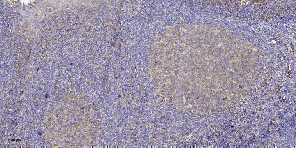
Real IHC data Immunohistochemical analysis of paraffin-embedded human tonsil. 1, Antibody was diluted at 1:200 (4° overnight). 2, Tris-EDTA,pH9.0 was used for antigen retrieval. 3,Secondary antibody was diluted at 1:200 (room temperature, 30min).
Anti-LPAAT- theta GPAT3 Antibody
Cat # A07965
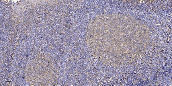
Real IHC data Immunohistochemical analysis of paraffin-embedded human tonsil. 1, Antibody was diluted at 1:200(4° overnight). 2, Tris-EDTA,pH9.0 was used for antigen retrieval. 3,Secondary antibody was diluted at 1:200(room temperature, 30min).
Anti-PLCH GPAT3 Antibody
Cat # A30503

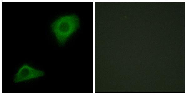
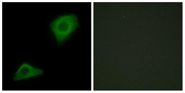
A07965 has an IHC image of paraffin-embedded human tonsil at 1:200 with Tris-EDTA retrieval at pH 9.0 (A07965 IHC caption); IF is listed, but its IF caption identifies a PLCH antibody (A07965 applications and IF caption). A30503 has its own IHC image of paraffin-embedded human tonsil under the same stated conditions (A30503 IHC caption); IF and ICC are listed, but its IF caption also identifies a PLCH antibody (A30503 applications and IF caption).

Which to pick: For tissue IHC, either SKU has its own paraffin-section image; the fixative is unreported in both captions (A07965 and A30503 IHC captions). For ICC, choose A30503 because ICC is listed only for that SKU; both list IF, but the PLCH wording in their IF captions leaves GPAT3-specific IF validation unclear (catalog applications; A07965 and A30503 IF captions). For human, mouse or rat work, both list reactivity and are rabbit polyclonal antibodies, while the supplied tissue images document human tonsil only (catalog reactivity, host and dilution records; A07965 and A30503 IHC captions).

Each figure is that product's own IHC / IF validation image from its datasheet.

References

  1. UniProt Consortium. UniProt entry Q53EU6 (GPAT3_HUMAN, Glycerol-3-phosphate acyltransferase 3).
  2. Human Protein Atlas. GPAT3 tissue IHC expression (reliability: Approved).
  3. Human Protein Atlas. GPAT3 subcellular location (ICC-IF): Membrane.
  4. Human Protein Atlas. GPAT3 antibody validation summary (1 antibodies).
  5. GPAT3 is a potential therapeutic target to overcome sorafenib resistance in hepatocellular carcinoma. Theranostics 2024 — PMC11209725.
  6. CPT2 inhibition enhances selective autophagy and proliferation in colorectal cancer via GPAT4-dependent glycerophospholipid biosynthesis. Communications biology 2025 — PMC12534568.
  7. Integrated Metabolomics and Lipidomics Analysis Reveals the Mechanism Behind the Action of Chiglitazar on the Protection Against Sepsis-Induced Acute Lung Injury. Metabolites 2025 — PMC12112857.
  8. PubMed PMID:17002884 — UniProt-cited evidence.
  9. PubMed PMID:12975309 — UniProt-cited evidence.
  10. PubMed PMID:14702039 — UniProt-cited evidence.