GRIN1 / Glutamate receptor ionotropic, NMDA 1 · IHC design guide

Design Immunohistochemistry for GRIN1

Plan GRIN1 paraffin IHC around synaptic brain staining (HPA tissue IHC) and postsynaptic membrane localisation (UniProt). Use the catalog antibody’s paraffin protocol (datasheet PA1222) and compare matched sections with negative controls (standard IHC practice).

Evidence assembled Oct 2026 · For research use; verify linked source records and product datasheet before use
Immunohistochemistry protocol sheet for GRIN1 (IHC for GRIN1): expected localisation Synaptic brain staining (HPA tissue IHC); postsynaptic membrane (UniProt), antibody PA1222, validated IHC image, and IHC protocol steps
Printable GRIN1 IHC protocol sheet — expected localisation Synaptic brain staining (HPA tissue IHC); postsynaptic membrane (UniProt), antibody PA1222, controls and protocol steps. Open the full GRIN1 IHC guide →

GRIN1 Immunohistochemistry Experimental Design Guide

Expected localisation, validated protocols, controls and antibodies — the at-a-glance facts below, then the full design guide.

Must know before staining
Expected localisation Synaptic brain staining (HPA tissue IHC); postsynaptic membrane (UniProt)
Staining pattern Synaptic staining in cortex; neuronal staining in hippocampus (HPA tissue IHC)
Antigen retrieval EDTA pH 8.0 HIER, heat-mediated (datasheet PA1222)
Positive control ⓘ Cerebellum+2 more · see all
Negative control ⓘ Adipose tissue+4 more · see all
Important caveats
Reasons your staining may differ from the expected pattern.
Fixation Keep fixation consistent across sections (standard IHC practice; not target-specific). Selected-image fixative and duration unreported (datasheet PA1222); verify before use.
Caveat Synaptic targeting depends on GRIN2B (UniProt; by similarity)
Regulation Brain-enriched RNA expression (HPA RNA)
Isoform / epitope 7 isoforms; check extracellular or cytoplasmic epitope coverage (UniProt)
Section 1

Recommended GRIN1 IHC & IF Protocols

The catalog antibody protocol is followed by one published GRIN1 IHC protocol using paraffin sections of glioma and normal brain tissue (PMC8626183).

Recommended immunohistochemistry (IHC-P) protocol parameters
SampleTissue sections; selected-image fixative not specified (standard IHC workflow)
FixationImage fixative and duration unreported (datasheet PA1222); verify before use.
Sectioning4–5 µm sections on charged slides (standard)
DeparaffinisationXylene, graded ethanol series to water (standard)
Antigen retrievalHeat retrieval: EDTA pH 8.0 (datasheet PA1222); 20 min, 95–100 °C (standard)
Peroxidase block3% H2O2, 10 min, room temperature (standard)
Blocking10% goat serum (datasheet PA1222)
Primary antibodyRabbit anti-GRIN1, 0.5-1μg/ml (datasheet PA1222)
Primary incubationOvernight at 4 °C (datasheet PA1222)
DetectionHRP-conjugated secondary, DAB chromogen (datasheet PA1222)
CounterstainHematoxylin, blue, dehydrate and mount (standard)
Expected resultGRIN1-positive staining in cells in granular layer of cerebellum (HPA tissue IHC: Medium). HPA tissue profile: Synaptic expression in brain. No signal in the no-primary control.
💡Decision noteStart with heat-mediated EDTA retrieval at pH 8.0 (datasheet: PA1222); the published protocol reports antigen retrieval without specifying its conditions (PMC8626183).
Section 2

What Is the Expected GRIN1 Staining Pattern?

GRIN1 is a membrane receptor enriched at postsynaptic and synaptic membranes, with three transmembrane segments (UniProt Q05586 topology and subcellular location). In paraffin IHC, expect staining in brain synapses and neuronal populations: cerebral cortex synapses, hippocampal neuronal cells, and cerebellar granular-layer cells show medium staining (HPA tissue IHC). HPA rates the tissue profile Enhanced, while describing only medium consistency between antibody staining and RNA expression (HPA tissue IHC).

What am I looking at on my slide?
Cerebral cortex shows synaptic staining of medium intensity (HPA tissue IHC).This matches the reported brain profile. Interpret signal in relation to synapse-rich regions, since GRIN1 localises to synaptic and postsynaptic membranes (HPA tissue IHC; UniProt Q05586 subcellular location).
Hippocampal neuronal cells or cells in the cerebellar granular layer stain at medium intensity (HPA tissue IHC).Both are reported positive populations. Compare their staining with the cortex on the same run; the HPA tissue profile supports these locations but does not define an exact fraction of positive cells (HPA tissue IHC).
Signal is confined to nuclei, with no corresponding neuronal or synaptic pattern.Treat a nucleus-only pattern as discordant with GRIN1’s membrane localisation; review morphology and controls before scoring it as target staining (UniProt Q05586 subcellular location; standard IHC practice).
Strong staining appears in a cell population reported as not detected, such as adipocytes in adipose tissue (HPA tissue IHC).Consider cross-reactivity or endogenous detection activity. An unexpected positive is a reason to check controls, although one HPA negative observation cannot prove that every specimen of that tissue must be negative (HPA tissue IHC; standard IHC practice).
Staining is diffuse across tissue, or absent from the reported positive brain populations (HPA tissue IHC).Diffuse signal makes compartment-level interpretation unreliable; absent signal in a known-positive control makes a negative test slide inconclusive. Check the staining run before making a biological call (standard IHC practice).
💡Expected GRIN1 appearanceCall a result positive when medium staining follows cerebral cortex synapses or the reported hippocampal neuronal and cerebellar granular-layer populations (HPA tissue IHC), with a membrane-associated pattern consistent with GRIN1 location (UniProt Q05586); isolated nuclear staining or uniform diffuse colour is suspect (standard IHC practice).
How each factor affects the staining
Subcellular topologyGRIN1 has three transmembrane segments and extracellular and cytoplasmic regions (UniProt Q05586 topology). The staining pattern should be interpreted at neuronal or synaptic membranes; the supplied record does not locate the catalog antibody’s epitope.
Tissue evidence and validationCortex synapses, hippocampal neuronal cells, and cerebellar granular-layer cells are medium positives (HPA tissue IHC). The HPA antibody entry rates IHC Enhanced, while the tissue profile reports medium staining–RNA consistency; neither rating guarantees every specimen will match (HPA antibody validation; HPA tissue IHC).
Isoforms and processingSeven isoforms are listed, and the 1–18 signal peptide precedes the 19–938 mature chain (UniProt Q05586 isoforms and processing). Without an antibody epitope, the record cannot establish which isoforms or processed regions the stain detects.
Antigen retrieval and detectionTarget-specific fixation effects are not established by the supplied assay evidence. Verify with a matched IHC source before attributing a result to fixation.
IF/ICC Q: What localisation is supported?A: HPA summarises membrane localisation but lists no main location or ICC-IF image-bearing cell lines (HPA subcellular ICC-IF). UniProt supports synaptic and postsynaptic membranes; this IHC evidence does not supply an IF/ICC protocol (UniProt Q05586 subcellular location; HPA subcellular ICC-IF).
Why is my staining missing, weak or wrong?
SituationLikely causeNext action
Reported positive brain tissue has no visible staining (HPA tissue IHC).The run may have weak retrieval, inadequate antibody or detection conditions, or a failed reagent step (standard IHC practice).Confirm section quality and controls, then optimise retrieval and staining conditions on a reported positive brain section before interpreting test negatives (HPA tissue IHC; standard IHC practice).
All structures, including spaces between cells, appear coloured.Diffuse reagent background or incomplete washing can obscure the expected localised signal (standard IHC practice).Check the primary-omission control, washing, blocking, and detection conditions; score only a pattern separable from background (standard IHC practice).
Nuclei stain strongly without a matching neuronal or synaptic pattern.This is discordant with the reported membrane localisation and may represent nonspecific staining (UniProt Q05586 subcellular location; standard IHC practice).Compare with the counterstain and a primary-omission control, then reassess antibody conditions before assigning a GRIN1-positive score (standard IHC practice).
Unexpected strong colour appears in HPA not-detected cells, such as adipose-tissue adipocytes (HPA tissue IHC).Cross-reactivity or endogenous detection activity is possible; HPA’s not-detected observation is a reference pattern, not a universal exclusion rule (HPA tissue IHC; standard IHC practice).Inspect a primary-omission control and the detection controls, then confirm whether the signal follows the reported neuronal and synaptic pattern in positive tissue (HPA tissue IHC; standard IHC practice).
The positive control works, but a test brain region has less signal than expected.HPA reports medium staining in specific brain cells and synapses, not uniform intensity throughout brain tissue (HPA tissue IHC).Identify the sampled region and cell population, compare like structures, and avoid treating every low-signal field as a failed run (HPA tissue IHC; standard IHC practice).
A negative result is being interpreted from a slide without a known-positive tissue control.Without a positive control in the run, absence of signal cannot distinguish low GRIN1 staining from a staining failure (standard IHC practice).Run a section containing a reported positive population, such as cortex synapses or hippocampal neuronal cells, alongside the test section before reporting a negative result (HPA tissue IHC; standard IHC practice).

Sample controls for GRIN1 IHC & IF

🧪Run hippocampus first and expect neuronal cells to stain (HPA: Medium in hippocampal neuronal cells). Use adipose tissue adipocytes as a negative tissue (HPA: Not detected in adipocytes); on the hippocampal slide, nuclei and non-neuronal regions should remain at background relative to the no-primary control, consistent with GRIN1’s membrane localisation (UniProt Q05586 subcellular location).
Positive control tissue: Cerebellum (Cells in granular layer, HPA Medium)
Negative control tissue: Adipose tissue (HPA Not detected)
ICC-IF cell lines (HPA subcellular resource): HPA carries no ICC-IF cell line for GRIN1; derive a cell-line control from the positive tissue's cell type (Cells in granular layer) and confirm it by RNA or western blot first.
Technical controls: Include a no-primary, secondary-only control and a control matched to the primary antibody’s host species and Ig class: an isotype control for a monoclonal primary or nonimmune IgG for a polyclonal primary (standard IHC practice). Use GRIN1 knockout tissue or antigen-peptide blocking, if available, as a biological specificity control; check endogenous peroxidase in chromogenic IHC and neuronal autofluorescence if interpreting IF (standard IHC/IF practice).
⚠️Feasibility: A target-specific fixation window and fixation effect are unreported in the supplied evidence; the selected PA1222 rat-brain IHC(P) caption does not state a fixative (selected-SKU caption: fixative not stated). Retrieval dependency is unreported, so optimise antigen retrieval on paraffin sections rather than inferring it from staining or topology (supplied evidence; standard IHC practice). The evidence does not establish whether frozen sections or IF are easier; neuronal autofluorescence can complicate IF interpretation (standard IF practice).

HPA tissue IHC evidence for GRIN1

Comprehensive Human Protein Atlas IHC scoring per tissue (reliability: Enhanced — Medium consistency between antibody staining and RNA expression data.). Rows are taken directly from the HPA tissue chart — click any row's HPA link to view the source.

Positive expression · recommended positive controls

TissueCell typeLevelEvidenceSource
Cerebellum Cells in granular layer Medium Protein (IHC) HPA →
Cerebral cortex Synapses Medium Protein (IHC) HPA →
Hippocampus Neuronal cells Medium Protein (IHC) HPA →

Undetected expression · recommended negative controls

TissueCell typeLevelEvidenceSource
Adipose tissue Adipocytes Not detected Protein (IHC) HPA →
Bone marrow Hematopoietic cells Not detected Protein (IHC) HPA →
Breast Adipocytes Not detected Protein (IHC) HPA →
Cervix Glandular cells Not detected Protein (IHC) HPA →
Colon Endothelial cells Not detected Protein (IHC) HPA →
Section 3

Advanced GRIN1 IHC Tips

Troubleshoot GRIN1 staining in paraffin sections by checking retrieval, tissue controls, membrane localisation and detection background before comparing signal across samples.

How should I retrieve GRIN1 in paraffin sections when staining is weak?
Start with heat-mediated retrieval in EDTA at pH 8.0 for the catalog antibody (datasheet PA1222). Use matched sections to compare a 15–20 minute heat exposure with a shorter exposure, then cool for about 20 minutes before staining; these are optimisation starting points, not reported GRIN1-specific conditions (standard IHC practice). If staining remains weak, test an alternative retrieval buffer only after documenting the EDTA result (standard IHC practice). Judge improvement by synaptic or neuronal staining in brain alongside background in a matched negative control (HPA: synaptic expression in brain; standard IHC practice). Excessively harsh retrieval can damage section morphology and make apparent signal difficult to localise (standard IHC practice).
Could fixation explain weak or patchy GRIN1 staining?
The rat brain IHC(P) caption for PA1222 does not state a fixative, so GRIN1-specific fixation sensitivity is unknown here (PA1222 tissue-IHC caption). Record fixative, fixation duration and section thickness for every specimen, and compare adjacent sections processed together before changing antibody concentration (standard IHC practice). For a suspected fixation effect, compare matched samples fixed for approximately 6 and 24 hours under an otherwise identical, locally validated paraffin workflow; those times are troubleshooting conditions, not GRIN1 evidence (standard IHC practice). Keep retrieval at EDTA pH 8.0 during that comparison (datasheet PA1222). Assess preserved neuronal morphology and regional staining together, since damaged tissue can make either condition misleading (HPA: neuronal and synaptic brain staining; standard IHC practice).
Where should credible GRIN1 signal appear in brain sections?
Expect a membrane-associated pattern, including synaptic and postsynaptic compartments, rather than using nuclear staining as the expected pattern (UniProt Q05586: subcellular location). HPA reports synaptic expression in brain, with medium staining at cerebral cortex synapses and in hippocampal neuronal cells (HPA: GRIN1 tissue profile). Compare a 20× regional overview with a higher-power view of neuronal profiles and neuropil, using identical imaging settings across controls (standard IHC practice). A diffuse cytoplasmic component can merit review because GRIN1 can associate with GRIN3A in the endoplasmic reticulum, but that annotation is by similarity and does not validate diffuse staining in a particular section (UniProt Q05586: subcellular location).
How can epitope location or isoforms change the staining pattern?
First obtain the immunogen or mapped epitope for the actual antibody; the supplied PA1222 caption does not identify it (PA1222 tissue-IHC caption). GRIN1 has 7 listed isoforms and extracellular regions at residues 19–559 and 649–811, plus cytoplasmic regions at 581–604, 616–627 and 836–938 (UniProt Q05586: isoforms and topology). A mapped sequence can then be checked against the isoforms before treating missing staining as absent GRIN1 (UniProt Q05586: isoforms; standard IHC practice). If the epitope lies near C-terminal phosphoserines 889–897, consider whether modification could affect antibody recognition, but establish that experimentally rather than assuming a change in abundance (UniProt Q05586: modified residues; standard IHC practice).
How should I adapt GRIN1 localisation checks to multiplex immunofluorescence?
Use IF as a separate assay and multiplex GRIN1 with a validated neuronal or synaptic marker appropriate to the brain region being examined (HPA: neuronal and synaptic brain staining; standard IF practice). Choose a GRIN1 fluorophore in a channel with low measured tissue autofluorescence, and include single-stain controls to assess bleed-through (standard IF practice). Before choosing detergent, map the antibody epitope: intracellular GRIN1 segments at 581–604, 616–627 or 836–938 call for a tested permeabilisation condition, whereas extracellular epitopes can be evaluated with gentler access conditions (UniProt Q05586: topology; standard IF practice). Titrate permeabilisation on paired sections and retain a no-primary control so puncta are judged against background (standard IF practice).
What should I check when DAB obscures GRIN1 staining?
Run a no-primary control through the same secondary antibody and chromogen steps to identify detection-system background (standard IHC practice). For peroxidase-based DAB detection, apply a peroxidase block and inspect whether pigment or endogenous enzyme activity persists; these are general workflow checks, not GRIN1-specific findings (standard IHC practice). Titrate the primary antibody and shorten DAB development in matched sections while holding EDTA retrieval at pH 8.0 constant (datasheet PA1222; standard IHC practice). Compare suspicious staining with the expected brain synaptic pattern and with a tissue compartment reported as undetected, such as adipocytes in adipose tissue (HPA: GRIN1 tissue profile). Interpret that negative reference alongside morphology and its own assay controls (standard IHC practice).
How should GRIN1 staining be scored across brain regions? ⚠ ANSWER MARKED FOR VERIFICATION
Define the anatomical region and scoring compartment before analysis, because HPA describes GRIN1 at synapses in cerebral cortex and in hippocampal neuronal cells (HPA: GRIN1 tissue profile). For neuronal cell staining, report the percentage of positive cells and an intensity-weighted 0–300 H-score; for punctate neuropil signal, report positive area or puncta density per mm² (standard IHC image-analysis practice). Normalise cell counts to eligible neuronal cells and puncta measurements to analysed neuropil area, excluding folds and damaged edges by a preset rule (standard IHC image-analysis practice). Keep retrieval, DAB development, imaging and thresholds matched across samples, and report regions separately rather than pooling unlike compartments (standard IHC practice).
How can I distinguish real GRIN1 staining from artefact?
A credible result should fit the membrane-associated, synaptic localisation of GRIN1 and the reported neuronal or synaptic brain pattern (UniProt Q05586: subcellular location; HPA: GRIN1 tissue profile). Review staining that is exclusively nuclear, confined to cut edges, concentrated in necrotic areas or equally strong in the no-primary control as a possible artefact (standard IHC practice). Check whether pigment or endogenous peroxidase remains after blocking before calling dark DAB deposits positive (standard IHC practice). Use cerebral cortex synapses or hippocampal neuronal cells as contextual positive references, and assess a reported undetected compartment such as adipocytes in adipose tissue as a contextual negative reference (HPA: GRIN1 tissue profile). Confirm borderline calls across at least 2 matched sections with preserved morphology (standard IHC practice).
Boster reagents

Best GRIN1 / Glutamate receptor ionotropic, NMDA 1 IHC Antibodies

PA1222 has IHC images from human and rat brain sections; its catalog also lists mouse reactivity. No IF image is supplied (PA1222 image captions; PA1222 reactivity; PA1222 IF image alts).

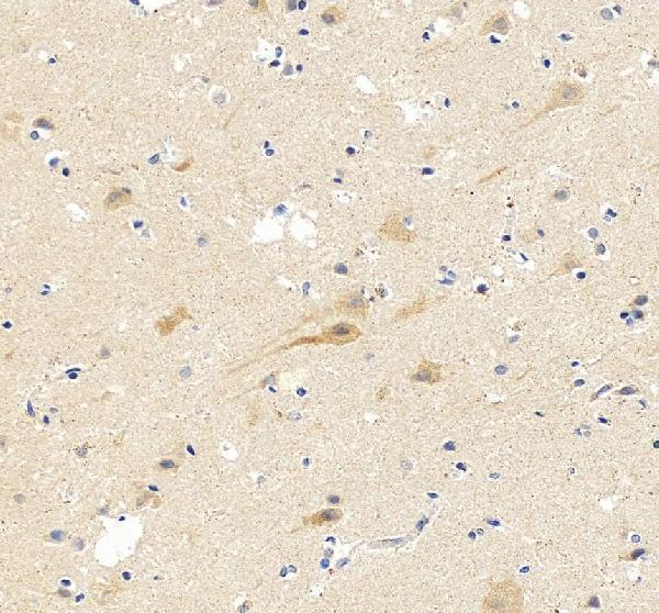
Real IHC data Anti-NMDAR1 antibody, PA1222, IHC(P) IHC(P): Rat Brain Tissue
Anti-NMDAR1/GRIN1 Antibody ®
Cat # PA1222

PA1222 has an IHC(P) image from rat brain tissue and an IHC image from a human brain paraffin section (PA1222 image captions). Its catalog lists IHC and human, mouse, and rat reactivity; the supplied images show human and rat samples (PA1222 applications; PA1222 reactivity; PA1222 image captions).

Which to pick: For tissue IHC, choose PA1222, a rabbit antibody with rat brain IHC(P) and human brain paraffin-section images; the fixative is unreported (PA1222 host; PA1222 image captions). For IF/ICC, this catalog provides no IF-listed SKU or IF image to support a recommendation (PA1222 applications; PA1222 IF image alts). For cross-species IHC, PA1222 lists human, mouse, and rat reactivity, with supplied IHC images for human and rat brain (PA1222 reactivity; PA1222 image captions).

Each figure is that product's own IHC / IF validation image from its datasheet.

References

  1. UniProt Consortium. UniProt entry Q05586 (NMDZ1_HUMAN, Glutamate receptor ionotropic, NMDA 1).
  2. Human Protein Atlas. GRIN1 tissue IHC expression (reliability: Enhanced).
  3. Human Protein Atlas. GRIN1 subcellular location (ICC-IF): Membrane.
  4. Human Protein Atlas. GRIN1 antibody validation summary (1 antibodies).
  5. Dynamic expression patterns of G protein-regulated inducer of neurite outgrowth 1 (GRIN1) and its colocalization with Galphao implicate significant roles of Galphao-GRIN1 signaling in nervous system. Developmental dynamics : an official publication of the American Association of Anatomists 2008 — PMC2631425.
  6. Identification of Hub Gene GRIN1 Correlated with Histological Grade and Prognosis of Glioma by Weighted Gene Coexpression Network Analysis. BioMed research international 2021 — PMC8626183.
  7. Chronic Alcohol Use Induces Molecular Genetic Changes in the Dorsomedial Thalamus of People with Alcohol-Related Disorders. Brain sciences 2021 — PMC8066746.
  8. Glutamic acid promotes hair growth in mice. Scientific reports 2021 — PMC8322389.
  9. PubMed PMID:8406025 — UniProt-cited evidence.
  10. PubMed PMID:7679115 — UniProt-cited evidence.
  11. PubMed PMID:7685113 — UniProt-cited evidence.