This website uses cookies to ensure you get the best experience on our website.


- Table of Contents
Plan paraffin-section HSD17B2 IHC around cytoplasmic and membranous staining in intestinal endocrine cells (HPA tissue IHC). The guide pairs that pattern with a human placenta IHC-P example and a starting antibody concentration of 0.5–1 μg/mL (datasheet: PA1604).

Expected localisation, validated protocols, controls and antibodies — the at-a-glance facts below, then the full design guide.
| Expected localisation | Cytoplasmic and membranous tissue staining (HPA tissue IHC) | |
| Staining pattern | Intestinal endocrine cells: cytoplasmic and membranous signal (HPA tissue IHC) | |
| Antigen retrieval | Citrate pH 6.0 HIER, 95–98 °C, 20 min (rule: cytoplasmic / membrane antigen) | |
| Positive control | Duodenum+1 more · see all | |
| Negative control | Adipose tissue+4 more · see all |
| Fixation | Keep fixation consistent across samples (standard IHC practice; not target-specific) | |
| Caveat | Signal varies by cell type within intestinal tissue (HPA tissue IHC) | |
| Regulation | Stimulus-linked regulation unreported (UniProt) | |
| Isoform / epitope | No annotated isoforms; epitope sidedness unspecified (UniProt) |
The catalog antibody’s IHC-P protocol is supplemented by published HSD17B2 staining protocols for lung, urothelial and endometrial specimens (PMC7170045; PMC5166531; PMC5481366).
| Sample | Tissue sections; selected-image fixative not specified (standard IHC workflow) |
| Fixation | Image fixative and duration unreported (datasheet PA1604); verify before use. |
| Sectioning | 4–5 µm sections on charged slides (standard) |
| Deparaffinisation | Xylene, graded ethanol series to water (standard) |
| Antigen retrieval | Heat-induced epitope retrieval in citrate buffer, pH 6.0, 20 min at 95–98 °C (standard rule: cytoplasmic / membrane antigen) |
| Peroxidase block | 3% H2O2, 10 min, room temperature (standard) |
| Blocking | 10% normal serum of the secondary host, 30 min, room temperature (standard) |
| Primary antibody | Rabbit anti-HSD17B2, 0.5-1μg/ml (datasheet PA1604) |
| Primary incubation | Overnight at 4 °C (standard) |
| Detection | HRP-polymer secondary, DAB chromogen 5–10 min (standard) |
| Counterstain | Hematoxylin, blue, dehydrate and mount (standard) |
| Expected result | HSD17B2-positive staining in endocrine cells of duodenum (HPA tissue IHC: High). HPA tissue profile: Cytoplasmic and membranous expression, most abundant in intestine and placental endothelial cells. No signal in the no-primary control. |
HSD17B2 is an endoplasmic reticulum membrane protein with a transmembrane segment at residues 4–24 (UniProt P37059 topology). In paraffin-section IHC, expect cytoplasmic and membranous staining, especially in intestinal endocrine cells and placental endothelial cells (HPA tissue IHC). HPA rates tissue staining reliability as Enhanced, while reporting medium consistency between staining and RNA expression (HPA tissue IHC).
| Strong cytoplasmic and membranous staining in duodenal or small-intestinal endocrine cells (HPA tissue IHC). | This matches the reported High staining in those cells (HPA tissue IHC). Assess the cell type as well as intensity: a brown signal elsewhere in the section does not establish the expected pattern (general IHC practice). |
| Staining is confined to nuclei, with little cytoplasmic signal. | Nuclear-only staining conflicts with the endoplasmic reticulum membrane location (UniProt P37059) and HPA's cytoplasmic and membranous tissue profile (HPA tissue IHC). Review controls and scoring before calling it HSD17B2. |
| Strong staining appears in adipocytes or adrenal glandular cells. | Those cells are reported as Not detected (HPA tissue IHC). Cross-reactivity or detection background is possible (general IHC practice); the HPA result is a reference pattern, not proof that every specimen must be negative. |
| Brown staining spreads across tissue structures without a discernible cell pattern. | Diffuse signal cannot be assigned confidently to the expected cells or compartment (general IHC practice). Compare a no-primary control and review blocking, washing, and chromogen development (general IHC practice). |
| Duodenal or small-intestinal endocrine cells show no detectable signal. | An absent signal conflicts with their reported High staining (HPA tissue IHC). Check whether the cells are present, then review the IHC-validated antibody and detection controls before interpreting the specimen as negative (general IHC practice). |
| Compartment and topology | HSD17B2 occupies the endoplasmic reticulum membrane and has a transmembrane segment at residues 4–24 (UniProt P37059 topology). Interpret cytoplasmic and membranous IHC together; topology alone does not establish epitope accessibility. |
| Tissue and cell selection | Duodenal and small-intestinal endocrine cells are High; colonic and rectal endocrine cells and hepatocytes are Low (HPA tissue IHC). Match the sampled cell type when comparing stain intensity. |
| Strength of the tissue reference | Two listed antibodies, HPA021826 and HPA050235, have Enhanced IHC validation (HPA antibodies). HPA nevertheless reports medium consistency with RNA expression, so assess localization and controls alongside intensity (HPA tissue IHC; general IHC practice). |
| IF/ICC Q&A: What location should be seen? | An endoplasmic reticulum pattern is the approved ICC-IF location; HPA lists images from Hep-G2 and RT-4 (HPA subcellular). This supports a localization check, not an IF/ICC protocol for this IHC page. |
| Situation | Likely cause | Next action |
|---|---|---|
| No signal in the expected intestinal endocrine cells. | Those cells are High in duodenum and small intestine (HPA tissue IHC); loss of signal may reflect a staining-run problem (general IHC practice). | Confirm that endocrine cells are represented, then review the IHC-validated antibody, detection reagents, and positive control (general IHC practice). |
| The whole section has a brown haze. | Diffuse color can arise from nonspecific binding, endogenous detection activity, or excess chromogen development (general IHC practice). | Compare a no-primary control; review blocking, washing, endogenous-activity control, and development time (general IHC practice). |
| Staining is predominantly nuclear. | That pattern disagrees with the endoplasmic reticulum membrane location (UniProt P37059) and HPA tissue profile (HPA tissue IHC). | Check control sections and reassess whether cytoplasmic and membranous signal is present before scoring nuclei as positive (general IHC practice). |
| Adipocytes or adrenal glandular cells stain strongly. | HPA reports Not detected for those cell types (HPA tissue IHC); cross-reactivity or background is possible (general IHC practice). | Check a no-primary control and compare localization with an independently validated antibody if available (general IHC practice; HPA antibodies). |
| Placental endothelial staining varies across the section. | Target-specific fixation effects are not established by the supplied assay evidence. Verify with a matched IHC source before attributing a result to fixation. | Identify endothelial cells and compare intact regions and run controls before attributing variation to HSD17B2 biology (general IHC practice). |
| Low staining in hepatocytes is mistaken for a failed run. | Hepatocytes are reported as Low, whereas intestinal endocrine cells are High (HPA tissue IHC). | Compare the result with the cell-specific HPA levels and include an intestinal positive control when evaluating assay performance (HPA tissue IHC; general IHC practice). |




Comprehensive Human Protein Atlas IHC scoring per tissue (reliability: Enhanced — Medium consistency between antibody staining and RNA expression data.). Rows are taken directly from the HPA tissue chart — click any row's HPA link to view the source.
| Tissue | Cell type | Level | Evidence | Source |
|---|---|---|---|---|
| Duodenum | Endocrine cells | High | Protein (IHC) | HPA → |
| Small intestine | Endocrine cells | High | Protein (IHC) | HPA → |
| Tissue | Cell type | Level | Evidence | Source |
|---|---|---|---|---|
| Adipose tissue | Adipocytes | Not detected | Protein (IHC) | HPA → |
| Adrenal gland | Glandular cells | Not detected | Protein (IHC) | HPA → |
| Bone marrow | Hematopoietic cells | Not detected | Protein (IHC) | HPA → |
| Breast | Adipocytes | Not detected | Protein (IHC) | HPA → |
| Bronchus | Respiratory epithelial cells | Not detected | Protein (IHC) | HPA → |
Troubleshoot HSD17B2 staining in paraffin sections by checking retrieval, compartment, cell type, controls, and scoring before interpreting chromogenic signal.
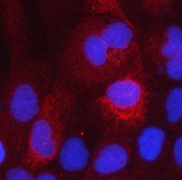
HSD17B2 antibody data cover human placenta paraffin-section IHC (PA1604 image caption) and A431-cell IF (A02506-1 image caption); the catalog also lists mouse reactivity for A02506-1 (catalog reactivity).
PA1604 has a human placenta paraffin-section IHC image and is listed for human IHC (PA1604 image caption; catalog applications and reactivity). A02506-1 has an A431-cell IF image and is listed for human and mouse IF/ICC (A02506-1 image caption; catalog applications and reactivity).
Which to pick: Choose PA1604 for human paraffin-section tissue IHC at 0.5–1 μg/ml; its image caption identifies paraffin sections, but the fixative is unreported (PA1604 image caption; datasheet dilution). Choose A02506-1 for IF/ICC at 5 μg/ml, supported by its A431-cell IF image (A02506-1 image caption; datasheet dilution). For mouse samples, A02506-1 lists mouse reactivity, although its supplied IF image shows A431 cells; clonality is unreported for both antibodies (catalog reactivity; A02506-1 image caption; catalog clone fields).