This website uses cookies to ensure you get the best experience on our website.


- Table of Contents
Plan HSD17B4 paraffin IHC around granular cytoplasmic staining, using hepatocytes as a high-staining reference (HPA tissue IHC). This guide covers fixation consistency, a 0.5–1 μg/mL antibody starting range (datasheet: A02923), chromogenic detection and cell-specific scoring.

Expected localisation, validated protocols, controls and antibodies — the at-a-glance facts below, then the full design guide.
| Expected localisation | Granular cytoplasm (HPA tissue IHC) | |
| Staining pattern | Granular cytoplasm across tissues; high in hepatocytes (HPA tissue IHC) | |
| Antigen retrieval | Citrate pH 6 HIER, heat-mediated (datasheet A02923) | |
| Positive control | Appendix+4 more · see all | |
| Negative control | Adipose tissue+3 more · see all |
| Fixation | Keep fixation consistent across sections (standard IHC practice; not target-specific) | |
| Caveat | Lung macrophages can stain strongly (HPA tissue IHC) | |
| Regulation | Specific expression regulators not reported (UniProt) | |
| Isoform / epitope | 3 isoforms; processed chains warrant epitope mapping (UniProt) |
The catalog antibody’s IHC-P protocol (datasheet A02923) and a published prostate cancer IHC method (PMC3607121) provide starting points.
| Sample | Paraffin-embedded human intestinal cancer tissues; fixative not specified (datasheet A02923) |
| Fixation | Image fixative and duration unreported (datasheet A02923); verify before use. |
| Sectioning | 4–5 µm sections on charged slides (standard) |
| Deparaffinisation | Xylene, graded ethanol series to water (standard) |
| Antigen retrieval | Heat retrieval: Citrate pH 6, 20 min (datasheet A02923) |
| Peroxidase block | 3% H2O2, 10 min, room temperature (standard) |
| Blocking | 10% goat serum (datasheet A02923) |
| Primary antibody | Rabbit anti-HSD17B4, 0.5-1μg/ml (datasheet A02923) |
| Primary incubation | Overnight at 4 °C (datasheet A02923) |
| Detection | Streptavidin-biotin complex (SABC), DAB chromogen (datasheet A02923) |
| Counterstain | Hematoxylin, blue, dehydrate and mount (standard) |
| Expected result | HSD17B4-positive staining in endocrine cells of appendix (HPA tissue IHC: High). HPA tissue profile: Granular cytoplasmic expression in all tissues. No signal in the no-primary control. |
HSD17B4 is a peroxisomal protein with no transmembrane segment (UniProt P51659 topology). In paraffin-section IHC, expect granular cytoplasmic staining, particularly in liver hepatocytes, lung macrophages, and the listed gastrointestinal endocrine cells (HPA: granular cytoplasmic expression; High in those cells). HPA rates the tissue IHC profile Enhanced, citing consistency between antibody staining and RNA expression (HPA: Enhanced reliability).
| Discrete cytoplasmic granules in hepatocytes or lung macrophages. | This matches the reported tissue pattern and cellular distribution (HPA: granular cytoplasmic expression; High in hepatocytes and lung macrophages). The granules are consistent with peroxisomal localisation (UniProt P51659: peroxisome); IHC morphology alone does not identify individual organelles (general IHC practice). |
| Predominantly nuclear or cell-surface staining with little granular cytoplasmic signal. | Treat this as a localisation mismatch: HSD17B4 is peroxisomal and has no transmembrane segment (UniProt P51659 topology; HPA: granular cytoplasmic expression). Review the antibody and detection controls before assigning the signal to HSD17B4 (general IHC practice). |
| Strong staining in adipocytes, ovarian stroma cells, chondrocytes, or vaginal squamous epithelial cells. | These specific cell populations were reported as Not detected (HPA: tissue IHC). Check cell identity and controls; cross-reactivity or endogenous detection activity are possible explanations for unexpected chromogenic signal (general IHC practice). The HPA result does not classify every cell in those tissues as negative. |
| Uniform haze across cells, stroma, and section edges. | A diffuse deposit lacks the reported granular cytoplasmic pattern (HPA: tissue IHC profile). Excess primary antibody, insufficient washing, or endogenous detection activity can produce nonspecific chromogenic background (general IHC practice); a no-primary control helps separate detection background from primary-antibody staining. |
| No granular signal in hepatocytes on a liver section. | Liver hepatocytes are reported High (HPA: liver tissue IHC), so an absent signal calls for a run and specimen check before interpreting a test section as negative. HPA does not establish HSD17B4-specific fixation sensitivity or guarantee staining in every specimen. |
| Tissue and cell selection | Liver hepatocytes, lung macrophages, and appendix, colon, duodenum, or rectum endocrine cells are reported High (HPA: tissue IHC). Choose a known-positive cell population within the section when judging a weak run; HPA staining levels describe those cells, not every cell in the tissue. |
| Compartment and topology | Peroxisomal localisation and the absence of a transmembrane segment support a granular cytoplasmic expectation (UniProt P51659 topology; HPA: tissue IHC and subcellular profile). A chromogenic granule pattern is compatible with that localisation, but does not by itself prove organelle identity (general IHC practice). |
| Antibody evidence | HPA021302, HPA021311, and HPA021479 each have Enhanced IHC status (HPA: antibody validation). That supports the reported pattern at the HPA level; it does not establish how an unspecified catalog antibody performs under different staining conditions. |
| Isoforms, processing, and epitope | UniProt lists three isoforms and chains spanning residues 1–736, 1–311, and 312–736 (UniProt P51659). The supplied record gives no antibody epitope, so it cannot predict which forms a particular reagent detects or explain a staining difference from chain location alone. |
| IF/ICC Q&A: where should signal appear? | Peroxisomal puncta are the expected IF/ICC localisation (HPA: Peroxisomes, enhanced; UniProt P51659: peroxisome). This supports the compartment interpretation of granular IHC, while IF/ICC observations remain a separate application and do not specify an IHC-P staining condition. |
| Antigen retrieval and fixation evidence | Retrieval conditions should be checked against the chosen antibody's IHC-P instructions and controlled empirically (general IHC practice). The supplied UniProt and HPA records give no target-specific fixation effect or retrieval setting; neither a tissue staining level nor a listed modification establishes one. |
| Situation | Likely cause | Next action |
|---|---|---|
| Liver control has no hepatocyte signal. | A failed staining run, unsuitable control section, or ineffective antibody condition is possible (general IHC practice); hepatocytes are reported High (HPA: liver tissue IHC). | Confirm tissue and cell identity, verify the detection controls, and review the chosen antibody's IHC-P instructions before scoring experimental sections (general IHC practice). Do not infer an HSD17B4-specific fixation effect from this result. |
| Signal is mainly nuclear or outlines cell membranes. | That distribution conflicts with the granular cytoplasmic IHC profile and peroxisomal localisation (HPA: tissue IHC; UniProt P51659 topology). | Inspect the no-primary control and compare a known-positive cell population; reassess specificity if the compartment mismatch persists (general IHC practice). |
| Adipocytes or chondrocytes stain strongly. | HPA reports these cell types as Not detected (HPA: adipose tissue adipocytes; soft tissue chondrocytes). Cross-reactivity or endogenous detection activity is possible (general IHC practice). | Confirm cell identity, examine the no-primary control, and seek concordance with an independently validated antibody if available (general IHC practice; HPA: multiple antibodies have Enhanced IHC status). |
| Brown colour is diffuse throughout the section. | Nonspecific primary-antibody binding or detection background can obscure granules (general IHC practice); diffuse colour is unlike the HPA profile (HPA: granular cytoplasmic expression). | Check a no-primary control, then review blocking, primary-antibody dilution, washing, and chromogen development under the chosen IHC-P workflow (general IHC practice). |
| A few weak dots appear in a reported High cell type. | Low contrast can make a granular pattern hard to distinguish from background (general IHC practice); HPA reports High staining in the specified positive cells (HPA: tissue IHC). | Compare the dots with background in adjacent cells and controls, then review the antibody's IHC-P conditions (general IHC practice). Do not score isolated dots as positive solely because the cell type is listed High. |
| Two antibodies give different cell or compartment patterns. | The records list Enhanced IHC status for three HPA antibodies but do not provide their epitopes or explain discordant staining (HPA: antibody validation; UniProt P51659: three isoforms and processed chains). | Compare the same cell populations and controls across runs; prioritise reproducible granular cytoplasmic staining in HPA-reported positive cells (HPA: tissue IHC; general IHC practice). Avoid attributing discordance to a particular isoform without epitope evidence. |
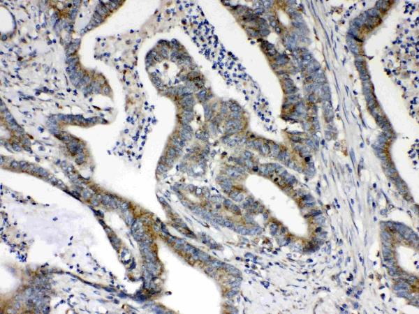
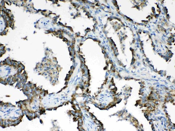


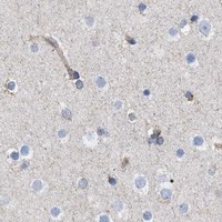
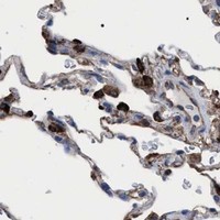

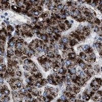


Comprehensive Human Protein Atlas IHC scoring per tissue (reliability: Enhanced — High consistency between antibody staining and RNA expression data.). Rows are taken directly from the HPA tissue chart — click any row's HPA link to view the source.
| Tissue | Cell type | Level | Evidence | Source |
|---|---|---|---|---|
| Appendix | Endocrine cells | High | Protein (IHC) | HPA → |
| Colon | Endocrine cells | High | Protein (IHC) | HPA → |
| Duodenum | Endocrine cells | High | Protein (IHC) | HPA → |
| Epididymis | Glandular cells | High | Protein (IHC) | HPA → |
| Gallbladder | Glandular cells | High | Protein (IHC) | HPA → |
| Tissue | Cell type | Level | Evidence | Source |
|---|---|---|---|---|
| Adipose tissue | Adipocytes | Not detected | Protein (IHC) | HPA → |
| Ovary | Ovarian stroma cells | Not detected | Protein (IHC) | HPA → |
| Soft tissue | Chondrocytes | Not detected | Protein (IHC) | HPA → |
| Vagina | Squamous epithelial cells | Not detected | Protein (IHC) | HPA → |
Troubleshoot HSD17B4 staining in paraffin sections by checking retrieval, signal localisation, controls and cell specific scoring before interpreting chromogenic signal.
Both anti-HSD17B4 antibodies have IHC images from paraffin sections of human cancer tissue (catalog image captions). Both list Human, Mouse, and Rat reactivity (catalog reactivity fields); neither has IF image data (catalog image alts).
A02923 has IHC images from human intestinal, lung, and mammary cancer paraffin sections, and lists Human, Mouse, and Rat reactivity (A02923 image captions; catalog reactivity). PA1727 has IHC images from human lung and intestinal cancer paraffin sections, and lists Human, Mouse, and Rat reactivity (PA1727 image captions; catalog reactivity).
Which to pick: For tissue IHC, choose A02923 if using citrate retrieval at pH 6 with biotin-based DAB detection (A02923 image caption), or PA1727 if using EDTA retrieval at pH 8 with peroxidase-based DAB detection (PA1727 image caption); both captions describe paraffin sections but do not report the fixative (catalog image captions). For IF/ICC, neither SKU lists that application or provides an IF figure (catalog applications; catalog IF image alts). For cross-species IHC, PA1727 explicitly lists Human, Mouse, and Rat for paraffin-section IHC, while A02923's IHC dilution text specifies Human; the IHC figures show human tissue only, and clonality is unreported for both (catalog dilution_raw; catalog image captions; catalog clone fields).