This website uses cookies to ensure you get the best experience on our website.


- Table of Contents
Plan HUWE1 IHC on paraffin sections using the catalog antibody’s 1:50–1:300 IHC range (datasheet A04634-1). Interpret cytoplasmic and nuclear staining (HPA tissue IHC) with consistent fixation and appropriate controls (standard IHC practice).

Expected localisation, validated protocols, controls and antibodies — the at-a-glance facts below, then the full design guide.
| Expected localisation | Cytoplasmic and nuclear tissue staining (HPA tissue IHC) | |
| Staining pattern | Widespread cytoplasmic and nuclear staining (HPA tissue IHC) | |
| Antigen retrieval | Tris-EDTA pH 9.0 HIER, heat-mediated (datasheet A04634-1) | |
| Positive control | Adrenal gland+4 more · see all | |
| Negative control | None in HPA (detected in all 45 tissues); use no-primary + isotype controls |
| Fixation | Keep fixation consistent across sections (standard IHC practice; not target-specific) | |
| Caveat | Nuclear staining may predominate in neuronal cells (UniProt) | |
| Regulation | Staining varies by cell type (HPA tissue IHC) | |
| Isoform / epitope | 3 isoforms; verify epitope coverage across variants (UniProt) |
The catalog antibody protocol uses Tris-EDTA pH 9.0 retrieval (datasheet A04634-1); the published HUWE1 IHC protocol below uses citrate pH 6.0 retrieval (PMC7329634).
| Sample | Paraffin-embedded human brain tumor tissue; fixative not specified (datasheet A04634-1) |
| Fixation | Image fixative and duration unreported (datasheet A04634-1); verify before use. |
| Sectioning | 4–5 µm sections on charged slides (standard) |
| Deparaffinisation | Xylene, graded ethanol series to water (standard) |
| Antigen retrieval | Heat retrieval: Tris-EDTA pH 9.0 (datasheet A04634-1); 20 min, 95–100 °C (standard) |
| Peroxidase block | 3% H2O2, 10 min, room temperature (standard) |
| Blocking | 10% normal serum of the secondary host, 30 min, room temperature (standard) |
| Primary antibody | Rabbit anti-HUWE1, 1:50 recommended; image 1:200 (datasheet A04634-1) |
| Primary incubation | Overnight at 4 °C (standard) |
| Detection | HRP-polymer secondary, DAB chromogen 5–10 min (standard) |
| Counterstain | Hematoxylin, blue, dehydrate and mount (standard) |
| Expected result | HUWE1-positive staining in glandular cells of adrenal gland (HPA tissue IHC: Medium). HPA tissue profile: Ubiquitous cytoplasmic and nuclear expression. No signal in the no-primary control. |
HUWE1 staining is expected in cytoplasm and nuclei across many cell types (HPA tissue IHC: ubiquitous cytoplasmic and nuclear expression). HPA reports medium staining in selected glandular, hematopoietic, basal, and neuronal cells, with low staining in several other cell types (HPA tissue IHC). HUWE1 has no transmembrane segment (UniProt Q7Z6Z7 topology). HPA rates the tissue IHC pattern Approved, with medium consistency between staining and RNA data (HPA tissue IHC).
| Medium cytoplasmic and/or nuclear staining in glandular cells, marrow hematopoietic cells, bronchial basal cells, or brain cells listed as positive (HPA tissue IHC). | This fits the reported tissue pattern (HPA tissue IHC). Compare compartments within the identified cells: UniProt describes HUWE1 as mainly cytoplasmic in most tissues, while nuclear localization in spermatogonia, primary spermatocytes, and neuronal cells is annotated by similarity (UniProt Q7Z6Z7 subcellular location). |
| Staining confined to an unexpected compartment, with no convincing cytoplasmic or nuclear signal in cells expected to stain (HPA tissue IHC). | Treat this as a localisation mismatch, not evidence of a new HUWE1 pattern. HUWE1 is annotated in cytoplasm and nucleus, with mitochondrial recruitment after AMBRA1 interaction (UniProt Q7Z6Z7 subcellular location); the supplied tissue IHC profile does not establish an organelle-specific staining pattern (HPA tissue IHC). |
| Strong staining restricted to cells outside the annotated positive populations, especially when nearby expected cells remain unstained (HPA tissue IHC). | Check cell identification and specificity. The HPA profile is ubiquitous, so absence from its example list alone does not make a cell type wrong; a discordant distribution may reflect cross-reactivity or endogenous chromogen-generating activity (HPA tissue IHC; standard IHC-P practice). |
| Diffuse colour across tissue, extracellular spaces, or the entire section, obscuring cell borders and nuclei. | This is background rather than interpretable cellular localisation (standard IHC-P practice). Assess staining against a no-primary control and tissue morphology before scoring; background cannot be assigned to HUWE1 from the distribution alone (standard IHC-P practice). |
| No convincing staining in a selected HPA medium-staining tissue and cell population (HPA tissue IHC). | Investigate the assay before calling HUWE1 absent. HPA reports medium, rather than uniformly strong, staining in its listed positive populations and rates tissue IHC Approved with medium staining-to-RNA consistency (HPA tissue IHC). Confirm that the relevant cells are present and the detection run worked (standard IHC-P practice). |
| Tissue and cell selection (HPA tissue IHC) | HPA lists medium staining in several glandular populations, marrow hematopoietic cells, bronchial basal cells, and selected brain cells, but low staining in lung alveolar cells, liver cholangiocytes, and other listed populations. Choose a control by its specified cell population, not its organ name alone (HPA tissue IHC). |
| Compartment and topology (UniProt Q7Z6Z7; HPA tissue IHC) | HUWE1 has no transmembrane segment; UniProt places it in cytoplasm and nucleus and notes conditional mitochondrial recruitment. The HPA tissue IHC summary reports cytoplasmic and nuclear expression. Neither source makes membrane-only tissue staining an expected result (UniProt Q7Z6Z7 topology and subcellular location; HPA tissue IHC). |
| Antibody validation (HPA antibodies; HPA tissue IHC) | HPA002548 and CAB022718 are both Approved for IHC; that status should not be described as Enhanced IHC validation. The tissue profile has medium consistency with RNA expression, so interpret an unexpected compartment or cell distribution alongside controls (HPA antibodies; HPA tissue IHC). |
| Isoforms and processing (UniProt Q7Z6Z7) | UniProt lists 3 isoforms, no signal peptide or propeptide, and one annotated chain spanning residues 1–4374. These facts do not identify the antibody epitope or establish isoform-specific IHC staining; avoid assigning a staining difference to an isoform without separate evidence (UniProt Q7Z6Z7). |
| Situation | Likely cause | Next action |
|---|---|---|
| The selected HPA positive cell population is unstained. | The expected cells may be absent from the examined area, or the IHC-P detection run may have failed; an HPA medium designation does not guarantee strong signal in every section (HPA tissue IHC; standard IHC-P practice). | Verify morphology and cell identity, then inspect a concurrently processed positive control and detection controls. Review the catalog antibody's IHC-P instructions for retrieval and dilution rather than inferring HUWE1-specific settings from HPA staining levels (standard IHC-P practice; HPA tissue IHC). |
| Signal appears only in a compartment that conflicts with the expected cellular pattern. | Non-specific staining or misread morphology is possible; HPA tissue IHC reports cytoplasmic and nuclear expression, while UniProt's mitochondrial recruitment is conditional (HPA tissue IHC; UniProt Q7Z6Z7). | Recheck the counterstain and cell boundaries, compare with no-primary staining, and confirm the pattern in an HPA listed positive cell population before assigning subcellular localisation (standard IHC-P practice; HPA tissue IHC). |
| Unexpected cells stain while the nearby expected population does not. | Cross-reactivity, endogenous detection activity, or cell misidentification may explain a discordant distribution (standard IHC-P practice). HPA's ubiquitous profile prevents treating every unlisted cell as intrinsically negative (HPA tissue IHC). | Identify both populations on the counterstained section, check a no-primary control for detection activity, and compare the cellular pattern with the HPA tissue profile (standard IHC-P practice; HPA tissue IHC). |
| Brown colour spreads through the section and obscures cellular detail. | Background from detection chemistry or nonspecific binding can hide a real cellular pattern (standard IHC-P practice). | Check the no-primary control, blocking and wash steps, and whether chromogen development obscured morphology; then reassess cytoplasmic and nuclear staining in identifiable cells (standard IHC-P practice; HPA tissue IHC). |
| A no-primary section still develops chromogenic signal. | The signal does not require primary-antibody binding and may arise from endogenous activity or the detection system (standard IHC-P practice). | Resolve the control staining with the detection system's recommended blocking and control steps before scoring HUWE1. Do not count matching colour in the test section as target-specific without a clean control (standard IHC-P practice). |
| What pattern should an IF/ICC image show? | HPA supports nucleoplasm and cytosol as the main ICC-IF locations; its additional listed locations have separate support levels (HPA subcellular). Tissue IHC appearance and ICC-IF localisation are distinct observations (HPA tissue IHC; HPA subcellular). | For IF/ICC interpretation, compare cellular signal with nucleoplasm and cytosol and consult the separate IF/ICC guide for that application; do not infer an IF/ICC protocol from the tissue IHC pattern (HPA subcellular; HPA tissue IHC). |
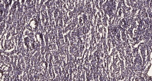
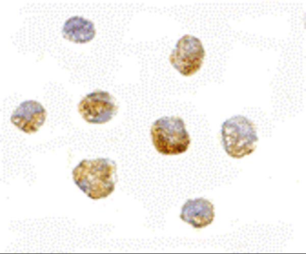
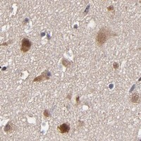
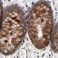

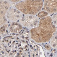


Comprehensive Human Protein Atlas IHC scoring per tissue (reliability: Approved — Medium consistency between antibody staining and RNA expression data.). Rows are taken directly from the HPA tissue chart — click any row's HPA link to view the source.
| Tissue | Cell type | Level | Evidence | Source |
|---|---|---|---|---|
| Adrenal gland | Glandular cells | Medium | Protein (IHC) | HPA → |
| Appendix | Glandular cells | Medium | Protein (IHC) | HPA → |
| Bone marrow | Hematopoietic cells | Medium | Protein (IHC) | HPA → |
| Breast | Glandular cells | Medium | Protein (IHC) | HPA → |
| Bronchus | Basal cells | Medium | Protein (IHC) | HPA → |
| Tissue | Cell type | Level | Evidence | Source |
|---|---|---|---|---|
| None in HPA: HUWE1 is detected in all 45 scored tissues. Use a no-primary (secondary-only) and an isotype control instead. | ||||
Troubleshoot HUWE1 staining in paraffin sections by checking retrieval, compartment patterns, controls, and cell-level scoring before interpreting signal (datasheet A04634-1; HPA tissue IHC).
Two anti-HUWE1 antibodies have image data: paraffin-section IHC in human brain tumor (A04634-1 IHC caption) and IF/ICC in Daudi cells (A04634 image captions). Catalog reactivity includes human and mouse for both, plus rat for A04634 (catalog).
A04634-1 will render with paraffin-section IHC data from human brain tumor; its listed applications are IHC and IF, with human and mouse reactivity (A04634-1 IHC caption; catalog). A04634 will render with Daudi-cell IF/ICC data; its listed applications are IF and ICC, with human, mouse and rat reactivity (A04634 image captions; catalog).
Which to pick: Choose A04634-1 for tissue IHC because its own image documents staining in paraffin-embedded human brain tumor (A04634-1 IHC caption); the fixative is unreported. Choose A04634 for cell IF/ICC because its own images document Daudi-cell staining, including ICC at 5 μg/mL and IF at 20 μg/mL (A04634 image captions). For rat samples, A04634 lists rat reactivity and IF/ICC applications; neither card documents rat staining (catalog; image captions).