IGF2R / Cation-independent mannose-6-phosphate receptor · IHC design guide

Design Immunohistochemistry for IGF2R

Plan chromogenic IGF2R IHC in paraffin sections using type II alveolar or Leydig cells as high-staining references (HPA tissue IHC). Interpret cytoplasmic tissue staining (HPA tissue IHC) alongside the receptor’s Golgi and endosomal membrane localization (UniProt).

Evidence assembled Oct 2026 · For research use; verify linked source records and product datasheet before use
Immunohistochemistry protocol sheet for IGF2R (IHC for IGF2R): expected localisation Cytoplasmic tissue staining; Golgi and endosomal membrane localization (HPA tissue IHC; UniProt), antibody A00951, validated IHC image, and IHC protocol steps
Printable IGF2R IHC protocol sheet — expected localisation Cytoplasmic tissue staining; Golgi and endosomal membrane localization (HPA tissue IHC; UniProt), antibody A00951, controls and protocol steps. Open the full IGF2R IHC guide →

IGF2R Immunohistochemistry Experimental Design Guide

Expected localisation, validated protocols, controls and antibodies — the at-a-glance facts below, then the full design guide.

Must know before staining
Expected localisation Cytoplasmic tissue staining; Golgi and endosomal membrane localization (HPA tissue IHC; UniProt)
Staining pattern Cytoplasmic; high in type II alveolar and Leydig cells (HPA tissue IHC)
Antigen retrieval EDTA pH 8.0 HIER, heat-mediated (datasheet A00951)
Positive control ⓘ Lung+4 more · see all
Negative control ⓘ Adipose tissue+4 more · see all
Important caveats
Reasons your staining may differ from the expected pattern.
Fixation Keep fixation consistent across sections. (standard IHC practice; not target-specific)
Caveat Antibody staining has medium consistency with RNA (HPA tissue IHC)
Regulation Expression regulation unannotated (UniProt)
Isoform / epitope No isoforms listed; lumenal 41–2304 vs cytoplasmic 2328–2491 (UniProt)
Section 1

Recommended IGF2R IHC & IF Protocols

The catalog antibody’s IHC-P protocol (datasheet A00951) is accompanied by published IGF2R IHC methods for canine osteosarcoma (PMC9916217) and human laryngeal tissue (PMC9592118).

Recommended immunohistochemistry (IHC-P) protocol parameters
SampleParaffin-embedded human esophageal squamous carcinoma tissue; fixative not specified (datasheet A00951)
FixationImage fixative and duration unreported (datasheet A00951); verify before use.
Sectioning4–5 µm sections on charged slides (standard)
DeparaffinisationXylene, graded ethanol series to water (standard)
Antigen retrievalHeat retrieval: EDTA pH 8.0 (datasheet A00951); 20 min, 95–100 °C (standard)
Peroxidase block3% H2O2, 10 min, room temperature (standard)
Blocking10% goat serum (datasheet A00951)
Primary antibodyRabbit anti-IGF2R, 2-5μg/ml (datasheet A00951)
Primary incubationOvernight at 4 °C (datasheet A00951)
DetectionHRP-conjugated secondary, DAB chromogen (datasheet A00951)
CounterstainHematoxylin, blue, dehydrate and mount (standard)
Expected resultIGF2R-positive staining in alveolar cells type II of lung (HPA tissue IHC: High). HPA tissue profile: Cytoplasmic expression in most tissues. No signal in the no-primary control.
💡Decision noteStart with EDTA pH 8.0 heat retrieval for the catalog antibody (datasheet A00951). The canine study used Tris-EDTA pH 9 at 97 °C for 20 min (PMC9916217).
Section 2

What Is the Expected IGF2R Staining Pattern?

IGF2R should appear mainly in the cytoplasm, consistent with its Golgi and endosomal membrane localization and single transmembrane segment (HPA tissue IHC; UniProt P11717 topology and subcellular location). Strong staining is reported in lung alveolar type II cells and testis Leydig cells (HPA tissue IHC: High). HPA rates the tissue IHC pattern Supported, with medium consistency between antibody staining and RNA expression (HPA tissue IHC).

What am I looking at on my slide?
Cytoplasmic staining in alveolar type II or Leydig cells, with a concentrated perinuclear component (HPA tissue IHC: High; UniProt P11717 subcellular location).This fits the reported positive cells and Golgi-centered localization. Score the identified cell type and compartment together; intensity alone does not establish specificity (HPA tissue IHC; UniProt P11717 subcellular location; standard IHC practice).
Predominantly nuclear staining, with little cytoplasmic signal in the same cells (HPA tissue IHC; UniProt P11717 subcellular location).A nuclear-dominant pattern conflicts with the reported Golgi and endosomal membrane locations. Treat it as suspect and compare it with a known-positive section and detection controls (UniProt P11717 subcellular location; standard IHC practice).
Strong staining in adipocytes, marrow hematopoietic cells, or squamous epithelium of the esophagus (HPA tissue IHC: Not detected).These reported negative cell populations make prominent signal a reason to check cross-reactivity or endogenous detection activity. An HPA negative observation is a comparison, not proof that every specimen must be negative (HPA tissue IHC; standard IHC practice).
Broad, even color across cells and surrounding tissue, with no discernible cell-specific pattern (standard IHC practice).Diffuse background cannot establish IGF2R localization. Review the no-primary control, blocking, washes, and detection steps before scoring the section (standard IHC practice).
No staining in alveolar type II cells or Leyig cells on an otherwise interpretable section (HPA tissue IHC: High).The absence of signal in a reported strong-positive cell type warrants an assay check. Verify tissue identification, primary antibody conditions, antigen retrieval, and detection before interpreting the specimen as IGF2R-negative (HPA tissue IHC; standard IHC practice).
💡Expected IGF2R appearanceCall positive a cell-associated cytoplasmic signal, potentially concentrated near the Golgi, that is strong in alveolar type II or Leydig cells; isolated nuclear signal or uniform background is suspect (HPA tissue IHC: High; UniProt P11717 subcellular location; standard IHC practice).
How each factor affects the staining
Compartment and epitope position (UniProt P11717 topology).IGF2R has a lumenal region at residues 41–2304, one transmembrane segment at 2305–2327, and a cytoplasmic tail at 2328–2491. Interpret an antibody result in light of its documented epitope, if known; the supplied record gives no epitope for a catalog antibody (UniProt P11717 topology).
Intracellular trafficking (UniProt P11717 function and subcellular location).Golgi and endosomal staining fits receptor recycling. A limited surface-associated component can be biologically plausible, but surface staining alone does not reproduce the reported main localization (UniProt P11717 function and subcellular location).
Choice of comparison cells (HPA tissue IHC).Alveolar type II and Leydig cells are reported High; adipocytes and marrow hematopoietic cells are Not detected. Compare identified cells, not whole-tissue averages; HPA describes cytoplasmic expression in most tissues and low RNA tissue specificity (HPA tissue IHC).
Strength of reference evidence (HPA tissue IHC; HPA antibodies).The tissue profile is Supported with medium staining–RNA consistency, and HPA011332 and CAB009661 each have Supported IHC status. Use the profile as a reference pattern, while retaining control-based assessment of the antibody used in this experiment (HPA tissue IHC; HPA antibodies; standard IHC practice).
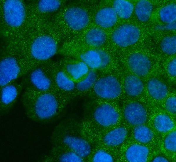
IF/ICC Q&A: Where should IGF2R appear? (HPA subcellular ICC-IF).Mainly at the Golgi apparatus, with additional vesicles; both locations are Supported in HPA ICC-IF. This answers the localization question only; IF/ICC experimental setup belongs on its own guide page (HPA subcellular ICC-IF).
Detection background (standard chromogenic IHC practice).Endogenous enzyme activity can create color independent of primary-antibody binding. Appropriate detection controls help distinguish that activity from a cell-specific cytoplasmic pattern (standard IHC practice).
Why is my staining missing, weak or wrong?
SituationLikely causeNext action
Known-positive alveolar type II or Leydig cells show no signal (HPA tissue IHC: High).The run may have inadequate primary-antibody detection or unsuitable antigen retrieval; the observation alone does not identify which step failed (standard IHC practice).Confirm the cell type and examine a known-positive control, then review the antibody's documented IHC-P conditions, retrieval, dilution, and detection system (HPA tissue IHC; standard IHC practice).
Nuclei stain more strongly than cytoplasm (UniProt P11717 subcellular location).The pattern disagrees with the reported Golgi and endosomal membrane localization; nonspecific staining or detection artifact is possible (UniProt P11717 subcellular location; standard IHC practice).Compare with a no-primary control and a known-positive section; score only a reproducible, cell-associated pattern consistent with the reference localization (standard IHC practice; UniProt P11717 subcellular location).
Reported negative cell populations show strong signal (HPA tissue IHC: Not detected).Cross-reactivity or endogenous detection activity may explain signal in adipocytes, marrow hematopoietic cells, or esophageal squamous cells (HPA tissue IHC; standard IHC practice).Check cell identity and no-primary staining; review blocking and detection controls before assigning IGF2R positivity (standard IHC practice).
Color covers the section without a clear cellular pattern (standard IHC practice).Diffuse background can arise from nonspecific reagent binding or detection activity and cannot be scored as the reported cytoplasmic pattern (standard IHC practice; HPA tissue IHC).Inspect the no-primary control, blocking, washes, and chromogen development; reassess the section only when cells and background are distinguishable (standard IHC practice).
Only a crisp cell-outline signal appears in a reported positive tissue (HPA tissue IHC: High).IGF2R traffics through the cell surface, so some surface signal is plausible, but an outline-only pattern does not match its main Golgi localization (UniProt P11717 function and subcellular location).Look for accompanying cytoplasmic or perinuclear staining in the expected cell type and compare with controls before accepting the outline as specific (HPA tissue IHC; UniProt P11717 subcellular location; standard IHC practice).
Signal differs between tissue sections despite the same antibody (standard IHC practice).Target-specific fixation effects are not established by the supplied assay evidence. Verify with a matched IHC source before attributing a result to fixation.Check section quality, tissue identity, retrieval and detection records, and a same-run positive control before interpreting the difference biologically (standard IHC practice).

Sample controls for IGF2R IHC & IF

🧪Run lung first: alveolar type II cells should stain (HPA: High in lung alveolar type II cells). Use adipose tissue adipocytes as the negative tissue (HPA: Not detected in adipocytes); on the lung slide, compare unstained neighboring cells as a background reference, without treating any particular neighboring cell type as a validated negative (HPA: lung row identifies alveolar type II cells only).
Positive control tissue: Lung (Alveolar cells type II, HPA High)
Negative control tissue: Adipose tissue (HPA Not detected)
ICC-IF cell lines (HPA subcellular resource): HPA ICC-IF images show IGF2R in U-251MG, U2OS, A-431, hTERT-RPE1 (serum starved), with annotated localisation: Golgi apparatus (supported) (HPA subcellular).
Technical controls: Include a no-primary, secondary-only slide and a concentration-matched rabbit IgG isotype control (catalog antibody caption: rabbit primary and anti-rabbit secondary; standard IHC practice). Use IGF2R knockout material, if available, as a biological negative; quench endogenous peroxidase and inspect lung pigment before interpreting DAB signal (standard IHC practice).
⚠️Feasibility: A target-specific fixation window or fixation effect is unreported, and the selected A00951 paraffin-section caption does not state a fixative (selected-SKU tissue-IHC caption: fixative unreported). Heat retrieval in EDTA at pH 8.0 is a documented IHC starting point, though its dependency has not been established for the proposed lung control (selected-SKU tissue-IHC caption: esophageal squamous carcinoma section). Frozen-section or IF ease relative to paraffin IHC is unreported; for IF/ICC, assess permeabilized cells for Golgi and vesicular signal while checking autofluorescence (HPA subcellular: Golgi apparatus and vesicles; standard IF practice).

HPA tissue IHC evidence for IGF2R

Comprehensive Human Protein Atlas IHC scoring per tissue (reliability: Supported — Medium consistency between antibody staining and RNA expression data.). Rows are taken directly from the HPA tissue chart — click any row's HPA link to view the source.

Positive expression · recommended positive controls

TissueCell typeLevelEvidenceSource
Lung Alveolar cells type II High Protein (IHC) HPA →
Testis Leydig cells High Protein (IHC) HPA →
Adrenal gland Glandular cells Medium Protein (IHC) HPA →
Breast Glandular cells Medium Protein (IHC) HPA →
Bronchus Respiratory epithelial cells Medium Protein (IHC) HPA →

Undetected expression · recommended negative controls

TissueCell typeLevelEvidenceSource
Adipose tissue Adipocytes Not detected Protein (IHC) HPA →
Bone marrow Hematopoietic cells Not detected Protein (IHC) HPA →
Caudate Glial cells Not detected Protein (IHC) HPA →
Esophagus Squamous epithelial cells Not detected Protein (IHC) HPA →
Oral mucosa Squamous epithelial cells Not detected Protein (IHC) HPA →
Section 3

Advanced IGF2R IHC Tips

Troubleshoot IGF2R staining in paraffin sections by checking retrieval, compartmental pattern, cell identity, and controls before scoring chromogenic signal.

Which retrieval conditions should I try first for weak IGF2R staining?
Start with heat mediated retrieval in EDTA at pH 8.0 for paraffin sections (datasheet A00951). The selected tissue image used this retrieval before an overnight incubation at 4°C with 2 μg/ml primary antibody (datasheet A00951). If staining remains weak, vary heating duration on matched sections while keeping antibody concentration and detection constant, and include a section processed without primary antibody (standard IHC practice). Assess whether the change improves cytoplasmic staining in expected cells without producing diffuse staining across the section; IGF2R is mainly Golgi associated and also occurs in vesicles (HPA subcellular; UniProt P11717 localisation).
Could fixation explain weak or uneven IGF2R staining?
IGF2R specific sensitivity to fixation is unknown from the supplied evidence; the selected paraffin section caption does not state a fixative (datasheet A00951). Record the specimen's actual fixation method and duration, then compare sections with similar handling before attributing differences to IGF2R abundance (standard IHC practice). For a controlled repeat, hold section thickness, EDTA retrieval at pH 8.0, and primary antibody exposure constant while changing only the documented fixation condition (datasheet A00951; standard IHC practice). Include a known staining control in each run so loss of signal can be distinguished from a failed retrieval or detection step (standard IHC practice).
Where should convincing IGF2R chromogenic staining appear?
Expect predominantly cytoplasmic, often perinuclear staining, consistent with the reported Golgi and vesicular distribution (HPA tissue IHC; HPA subcellular). IGF2R also occupies endosomal membranes, while its steady state distribution is mainly Golgi and is reported as undetectable in lysosomes (UniProt P11717 localisation). Evaluate staining within intact cell boundaries at matched magnification, using morphology and a counterstain to distinguish intracellular signal from deposits over extracellular material (standard IHC practice). Strong staining restricted to nuclei or tissue edges warrants a control review before it is scored as IGF2R, because those patterns do not match the reported compartments (UniProt P11717 localisation; standard IHC practice).
How does epitope position affect IGF2R staining in paraffin sections?
Check the antibody's documented immunogen or epitope before interpreting a change in staining, because the supplied product caption does not specify its binding site (datasheet A00951). IGF2R has a lumenal region at residues 41–2304, one transmembrane segment at 2305–2327, and a cytoplasmic tail at 2328–2491 (UniProt P11717 topology). Its lumenal region has 18 annotated glycosylation sites, so epitope accessibility should be assessed empirically under the stated EDTA retrieval conditions (UniProt P11717 glycosylation; datasheet A00951). No isoforms are listed in the supplied record; that annotation alone does not establish that every possible IGF2R form shares an antibody epitope (UniProt P11717 isoforms; standard interpretation practice).
How should I investigate an IGF2R pattern with immunofluorescence?
Use IF as a companion localisation check for the chromogenic IHC pattern, while treating the paraffin section conditions as evidence for IHC only (datasheet A00951). Multiplex IGF2R with a validated marker for the cell population under study; alveolar type II cells and Leydig cells are reported as high staining examples for choosing a reference population (HPA tissue IHC). Select spectrally separated fluorophores, favoring a far red channel when tissue autofluorescence obscures shorter wavelengths, and inspect single channel controls (standard IF practice). Because the antibody epitope is unspecified, compare mild and stronger permeabilisation if needed: a cytoplasmic tail epitope and a lumenal epitope can differ in accessibility (datasheet A00951; UniProt P11717 topology; standard IF practice).
What should I check when IGF2R DAB staining looks diffuse?
First compare the stained section with a no primary control and inspect whether brown signal follows intact cytoplasm or instead covers edges and damaged areas (standard IHC practice). The selected paraffin tissue image used 10% goat serum blocking, a 2 μg/ml primary antibody incubation overnight at 4°C, and DAB detection (datasheet A00951). Confirm that endogenous peroxidase was blocked, then review wash steps, secondary antibody exposure, and DAB development on matched sections (standard chromogenic IHC practice). Lower primary concentration or shorten chromogen development one variable at a time if the control and tissue morphology indicate excess background (standard IHC practice).
How should I score IGF2R staining across tissue sections? ⚠ ANSWER MARKED FOR VERIFICATION
Define the cell population and region of interest before scoring, then record an H-score or both the percentage of positive cells and staining intensity (standard IHC quantification practice). Normalize comparisons to the number of evaluable cells or the sampled tissue area, and use positive cell density per mm² when cell counts per area are the endpoint (standard IHC quantification practice). Apply the same illumination, counterstain assessment, threshold, and exclusion criteria across sections, excluding folds, necrosis, and edge artefacts (standard IHC quantification practice). Score named cell populations separately because reported IGF2R staining varies among cell types and HPA rates its tissue IHC evidence as supported with medium RNA concordance (HPA tissue IHC).
How can I distinguish genuine IGF2R positivity from an IHC artefact?
Favor reproducible intracellular staining in intact cells, especially a cytoplasmic or perinuclear pattern compatible with Golgi and vesicular localisation (HPA tissue IHC; HPA subcellular). Check cell identity: alveolar type II cells and Leydig cells are reported high, whereas adipocytes and esophageal squamous epithelial cells are reported undetected in the cited normal tissue survey (HPA tissue IHC). The selected image shows staining in human esophageal squamous carcinoma, which should not be equated with the survey's normal esophageal epithelium (datasheet A00951; HPA tissue IHC). Treat nuclear only signal, edge accentuation, necrotic deposits, or staining retained in a no primary control as possible artefacts, and check endogenous peroxidase blocking before assigning positivity (UniProt P11717 localisation; standard IHC practice).
Boster reagents

Best IGF2R / Cation-independent mannose-6-phosphate receptor IHC Antibodies

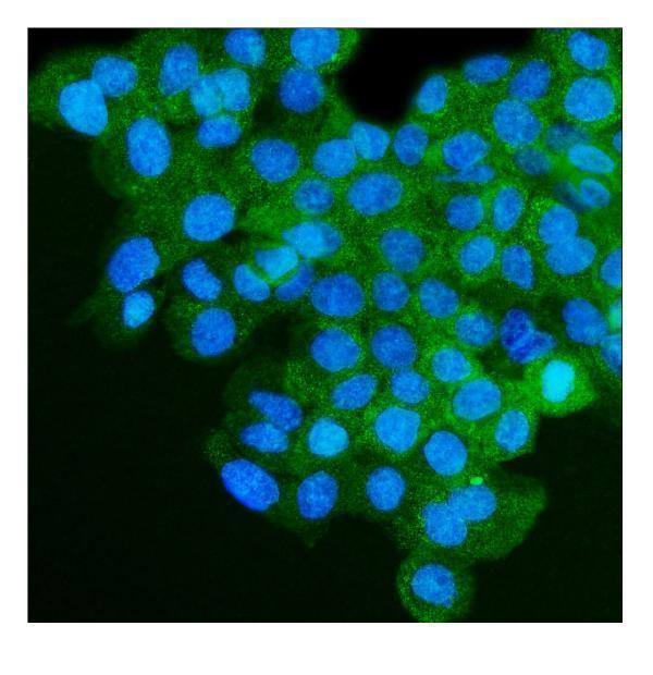
Anti-IGF2R catalog antibodies show human paraffin-section IHC (catalog IHC captions) and A431-cell IF (A00951 and M00951-1 IF captions); listed reactivity includes human, mouse, and rat (catalog reactivity).

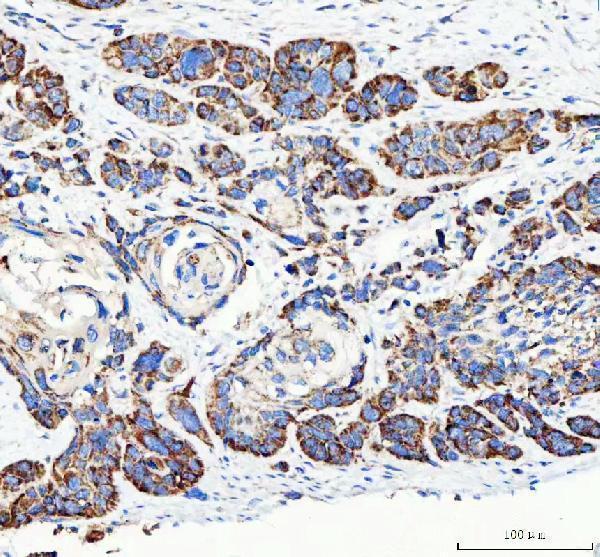
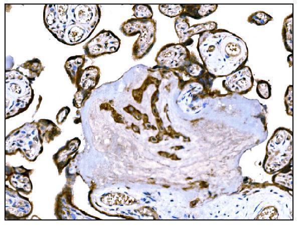
Real IHC data IHC analysis of IGF2R using anti-IGF2R antibody (A00951). IGF2R was detected in a paraffin-embedded section of human esophageal squamous carcinoma tissue. Heat mediated antigen retrieval was performed in EDTA buffer (pH 8.0, epitope retrieval solution). The tissue section was blocked with 10% goat serum. The tissue section was then incubated with 2 μg/ml rabbit anti-IGF2R Antibody (A00951) overnight at 4°C. Peroxidase Conjugated Goat Anti-rabbit IgG was used as secondary antibody and incubated for 30 minutes at 37°C. The tissue section was developed using HRP Conjugated Rabbit IgG Super Vision Assay Kit (Catalog # SV0002) with DAB as the chromogen.
Anti-Mannose 6 Phosphate Receptor (Cation independent)/IGF2R Antibody ®
Cat # A00951
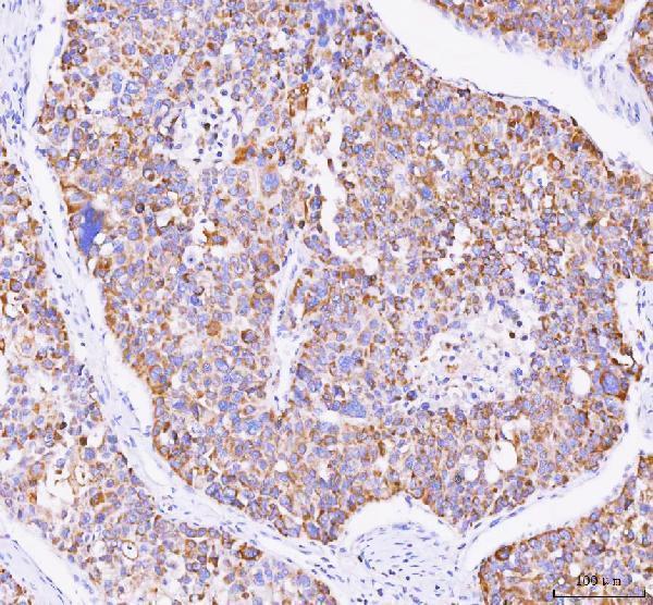
Real IHC data IHC analysis of IGF2R using anti-IGF2R antibody (M00951-2). IGF2R was detected in paraffin-embedded section of human liver cancer tissue. Heat mediated antigen retrieval was performed in EDTA buffer (pH8.0, epitope retrieval solution). The tissue section was blocked with 10% goat serum. The tissue section was then incubated with PBS (Left, blank control) and 2μg/ml mouse anti-IGF2R Antibody (Right) (M00951-2) overnight at 4°C. Biotinylated goat anti-mouse IgG was used as secondary antibody and incubated for 30 minutes at 37°C. The tissue section was developed using Strepavidin-Biotin-Complex (SABC) (Catalog # SA1021) with DAB as the chromogen.
Anti-IGF2R Antibody ® (monoclonal, 6G2)
Cat # M00951-2
Real IHC data IHC analysis of IGF2R using anti-IGF2R antibody (M00951-1). IGF2R was detected in paraffin-embedded section of human colon cancer tissue. Heat mediated antigen retrieval was performed in EDTA buffer (pH8.0, epitope retrieval solution). The tissue section was blocked with 10% goat serum. The tissue section was then incubated with 2μg/ml mouse anti-IGF2R Antibody (M00951-1) overnight at 4°C. Biotinylated goat anti-mouse IgG was used as secondary antibody and incubated for 30 minutes at 37°C. The tissue section was developed using Strepavidin-Biotin-Complex (SABC) (Catalog # SA1021) with DAB as the chromogen.
Anti-IGF2R Antibody ® (monoclonal, 4I11)
Cat # M00951-1
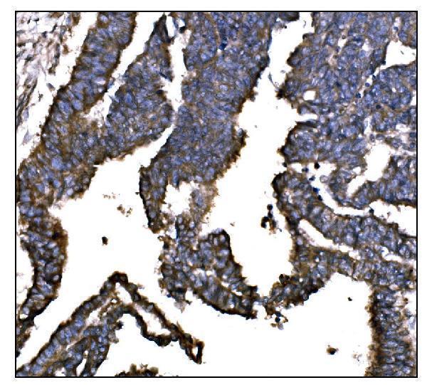
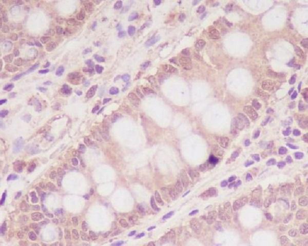
Real IHC data Immunohistochemical analysis of paraffin-embedded human colon, using M6PR/IGF2R Antibody.
Anti-M6PR IGF2R Rabbit Monoclonal Antibody
Cat # M00951

A00951 shows IHC in human esophageal squamous carcinoma, and M00951-2 shows IHC in human liver cancer with a PBS blank control (respective IHC captions). M00951-1 shows IHC in human colon cancer, and M00951 shows IHC in human colon; both figures use paraffin sections (respective IHC captions).

Which to pick: For tissue IHC, consider M00951-2 when a human paraffin-section figure with a PBS blank control is useful; its fixative is unreported (M00951-2 IHC caption). For IF/ICC, choose M00951-1, which lists both applications and has an A431-cell IF image (M00951-1 applications and IF caption). For cross-species work, A00951 lists human, mouse, and rat reactivity plus IHC and IF applications (A00951 catalog); its IHC figure uses a human paraffin section, with fixative unreported (A00951 IHC caption).

Each figure is that product's own IHC / IF validation image from its datasheet.

References

  1. UniProt Consortium. UniProt entry P11717 (MPRI_HUMAN, Cation-independent mannose-6-phosphate receptor).
  2. Human Protein Atlas. IGF2R tissue IHC expression (reliability: Supported).
  3. Human Protein Atlas. IGF2R subcellular location (ICC-IF): Mainly localized to the Golgi apparatus. In addition localized to vesicles..
  4. Human Protein Atlas. IGF2R antibody validation summary (2 antibodies).
  5. Insulin-like growth factors I and II receptors in the breast cancer survival disparity among African-American women. Growth hormone & IGF research : official journal of the Growth Hormone Research Society and the International IGF Research Society 2010 — PMC2875325.
  6. Characterization of IGF2R Molecular Expression in Canine Osteosarcoma as Part of a Novel Comparative Oncology Approach. International journal of molecular sciences 2023 — PMC9916217.
  7. Radioimmunotherapy Targeting IGF2R on Canine-Patient-Derived Osteosarcoma Tumors in Mice and Radiation Dosimetry in Canine and Pediatric Models. Pharmaceuticals (Basel, Switzerland) 2021 — PMC8779382.
  8. Proteomics analysis of cancer tissues identifies IGF2R as a potential therapeutic target in laryngeal carcinoma. Frontiers in endocrinology 2022 — PMC9592118.
  9. PubMed PMID:2957598 — UniProt-cited evidence.
  10. PubMed PMID:2963003 — UniProt-cited evidence.
  11. PubMed PMID:9892739 — UniProt-cited evidence.