IL16 / Pro-interleukin-16 · IHC design guide

Design Immunohistochemistry for IL16

Plan IL16 staining in paraffin sections using cytoplasmic immune-cell staining as the tissue reference (HPA tissue IHC). Compare lymphoid positive tissue with a negative cell population, and check whether the antibody epitope detects the precursor or mature IL16 (HPA tissue IHC; UniProt).

Evidence assembled Oct 2026 · For research use; verify linked source records and product datasheet before use
Immunohistochemistry protocol sheet for IL16 (IHC for IL16): expected localisation Cytoplasm in a subset of immune cells (HPA tissue IHC), antibody PB9238, validated IHC image, and IHC protocol steps
Printable IL16 IHC protocol sheet — expected localisation Cytoplasm in a subset of immune cells (HPA tissue IHC), antibody PB9238, controls and protocol steps. Open the full IL16 IHC guide →

IL16 Immunohistochemistry Experimental Design Guide

Expected localisation, validated protocols, controls and antibodies — the at-a-glance facts below, then the full design guide.

Must know before staining
Expected localisation Cytoplasm in a subset of immune cells (HPA tissue IHC)
Staining pattern Cytoplasmic staining in a subset of immune cells (HPA tissue IHC)
Antigen retrieval Tris-EDTA pH 9.0 HIER, 95–98 °C, 20 min (rule: nuclear antigen)
Positive control ⓘ Appendix+4 more · see all
Negative control ⓘ Adipose tissue+4 more · see all
Important caveats
Reasons your staining may differ from the expected pattern.
Fixation Keep fixation conditions consistent across sections. (standard IHC practice; not target-specific)
Caveat Secreted variants may separate RNA and protein locations (HPA tissue IHC)
Regulation Resting T cells; undetectable during active proliferation (UniProt)
Isoform / epitope 4 isoforms; cleavage yields mature IL16, so check the epitope (UniProt)
Section 1

Recommended IL16 IHC & IF Protocols

The catalog antibody’s IHC-P protocol is accompanied by two published IL16 chromogenic IHC protocols (PMC8805824; PMC4110175 methods).

Recommended immunohistochemistry (IHC-P) protocol parameters
SampleTissue sections; selected-image fixative not specified (standard IHC workflow)
FixationImage fixative and duration unreported (datasheet PB9238); verify before use.
Sectioning4–5 µm sections on charged slides (standard)
DeparaffinisationXylene, graded ethanol series to water (standard)
Antigen retrievalHeat-induced epitope retrieval in Tris-EDTA buffer, pH 9.0, 20 min at 95–98 °C (standard rule: nuclear antigen)
Peroxidase block3% H2O2, 10 min, room temperature (standard)
Blocking10% goat serum (datasheet PB9238)
Primary antibodyRabbit anti-IL16, 0.5-1μg/ml (datasheet PB9238)
Primary incubationOvernight at 4 °C (datasheet PB9238)
DetectionStreptavidin-biotin complex (SABC), DAB chromogen (datasheet PB9238)
CounterstainHematoxylin, blue, dehydrate and mount (standard)
Expected resultIL16-positive staining in germinal center cells of appendix (HPA tissue IHC: High). HPA tissue profile: Cytoplasmic expression in subset of immune cells. No signal in the no-primary control.
💡Decision noteStart with Tris-EDTA pH 9.0 retrieval at 95–98 °C for 20 min (page retrieval rule: nuclear antigen). Neither published excerpt specifies retrieval (PMC8805824; PMC4110175 methods).
Section 2

What Is the Expected IL16 Staining Pattern?

In paraffin-section IHC, expect mainly cytoplasmic IL16 staining in a subset of immune cells, especially in lymphoid tissue (HPA tissue IHC: Enhanced; cytoplasmic expression in a subset of immune cells). IL16 can also localize to the nucleus and be secreted, and it has no transmembrane segment (UniProt Q14005: subcellular location; topology). Interpret tissue staining alongside cell identity and distribution, since secreted protein may appear apart from its source cells (HPA tissue IHC: reliability description).

What am I looking at on my slide?
Subset of cells in appendix germinal centers, lymph-node non-germinal centers, or splenic white pulp stains strongly.This fits the reported high staining in those cell groups (HPA tissue IHC: positive tissues). Score the stained cells and compartment; a strong signal across every cell would differ from HPA’s subset-of-immune-cells profile (HPA tissue IHC: profile).
Staining is confined to an unexpected compartment, such as a sharp membrane-only outline throughout the section.Treat this as a possible artefact and check antibody specificity and detection controls. The tissue IHC profile is cytoplasmic (HPA tissue IHC: profile); membrane localization is uncertain in ICC-IF, while nuclear and cytoplasmic locations are reported (HPA subcellular ICC-IF; UniProt Q14005: subcellular location).
Strong chromogen appears in adipocytes or respiratory epithelial cells instead of the expected immune-cell subset.Consider cross-reactivity or endogenous detection activity and inspect controls. Adipocytes in adipose tissue and respiratory epithelial cells in bronchus were not detected in the reported tissue IHC data (HPA tissue IHC: negative tissues); those findings are cell-specific, not whole-tissue exclusions.
A diffuse haze covers cells and surrounding tissue, obscuring which cells are positive.The slide cannot establish the reported cytoplasmic immune-cell pattern (HPA tissue IHC: profile). Review the negative reagent control and background reduction steps (general IHC practice). Interpret any extracellular signal cautiously because at least one IL16 variant is secreted (HPA tissue IHC: reliability description).
No immune-cell signal appears in appendix, lymph node, or spleen.These are reported high-staining reference sites in the specified cell groups (HPA tissue IHC: positive tissues). Check that the relevant cells are present, then investigate the antibody, retrieval conditions, and detection system (general IHC practice); absence of signal alone does not establish biological absence.
💡Expected IL16 appearanceA convincing positive is distinct cytoplasmic chromogen in a subset of immune cells, potentially high in the specified appendix, lymph-node, or splenic populations (HPA tissue IHC: profile; positive tissues); uniform staining of unrelated cells or diffuse haze calls for control review (general IHC practice).
How each factor affects the staining
Tissue and cell selectionUse the named immune-cell populations when judging a positive: appendix germinal-center cells, lymph-node non-germinal-center cells, and splenic white-pulp cells stain high; bone-marrow hematopoietic and tonsil germinal-center cells stain medium (HPA tissue IHC: positive tissues).
Processing and antibody epitopeUniProt lists a 1–1332 precursor and 1212–1332 interleukin-16 chain (UniProt Q14005: processing). The supplied evidence does not locate the catalog antibody’s epitope, so these records cannot establish which form it detects or predict a form-specific IHC pattern.
Isoforms and secreted proteinFour isoforms are listed (UniProt Q14005: isoforms). At least one protein variant is secreted, so the tissue location of RNA and protein may differ (HPA tissue IHC: reliability description). Avoid assigning a stained cell as the site of synthesis from chromogen alone.
IF/ICC Q: should the same pattern be expected?HPA reports mainly nuclear speckles, with supported cytosol localization and uncertain plasma-membrane localization in ICC-IF (HPA subcellular ICC-IF). Those cell-image observations provide context; they do not replace the cytoplasmic subset-of-immune-cells benchmark for paraffin tissue IHC (HPA tissue IHC: profile).
Why is my staining missing, weak or wrong?
SituationLikely causeNext action
Positive-reference tissue is blank.Relevant cells may be absent from the section, or the IHC workflow may have failed (general IHC practice).Confirm the specified cell population on the counterstain (HPA tissue IHC: positive tissues), then review antibody, retrieval, and detection controls (general IHC practice).
Chromogen marks nearly every cell.This exceeds the reported subset-of-immune-cells pattern and may reflect nonspecific staining (HPA tissue IHC: profile; general IHC practice).Compare with a negative reagent control; review antibody concentration, blocking, and wash conditions (general IHC practice).
The negative reagent control also stains.Background may arise from the detection system or endogenous enzyme activity (general IHC practice).Check detection-only controls and the enzyme-blocking step appropriate to the chromogen system (general IHC practice).
Only an unexpected compartment stains.The result conflicts with the cytoplasmic tissue IHC profile; nuclear localization is also documented, and membrane ICC-IF localization is uncertain (HPA tissue IHC; UniProt Q14005; HPA subcellular ICC-IF).Recheck cell identity and controls, then compare with the reported tissue pattern before calling the signal specific (HPA tissue IHC: profile; general IHC practice).
Signal is diffuse around positive cells.Secreted IL16 can complicate the match between protein signal and the cells that produced it (HPA tissue IHC: reliability description).Score clearly localized cellular staining separately from extracellular haze; use controls to assess nonspecific background (general IHC practice).
An expected negative cell group stains strongly.Cross-reactivity or endogenous detection activity is possible (general IHC practice); HPA reports no detection in the specified adipocytes and respiratory epithelial cells (HPA tissue IHC: negative tissues).Verify cell identity, compare the negative reagent control, and review detection blocking before interpreting the signal as IL16 (general IHC practice).

Sample controls for IL16 IHC & IF

🧪Run appendix first: germinal center cells should stain (HPA: High in appendix germinal center cells). Use adipose tissue as the negative comparator (HPA: Not detected in adipocytes); cells outside the stained germinal centers on the appendix slide should show only background signal, but HPA does not identify a verified internal negative cell type.
Positive control tissue: Appendix (Germinal center cells, HPA High)
Negative control tissue: Adipose tissue (HPA Not detected)
ICC-IF cell lines (HPA subcellular resource): HPA ICC-IF images show IL16 in HEL, SK-MEL-30, U2OS, with annotated localisation: Nuclear speckles (supported) (HPA subcellular).
Technical controls: Include a no-primary, secondary-only control and a concentration-matched isotype control matched to the primary antibody’s host species and clonality (standard IHC practice). Confirm specificity with IL16 knockout material or, where an appropriate immunizing peptide is available, peptide blocking (standard IHC practice). Block endogenous peroxidase in the positive tissue and endogenous biotin if using avidin–biotin detection (standard IHC practice).
⚠️Feasibility: A target-specific fixation window and fixation effect are unreported in the supplied evidence; the selected PB9238 tonsil IHC(P) caption does not report a fixative (product caption: fixative not stated). Retrieval dependence is also unreported, so optimize antigen retrieval for the catalog antibody on paraffin sections (standard IHC practice). The evidence does not establish that frozen sections or IF are easier; endogenous peroxidase in lymphoid tissue can contribute background in chromogenic IHC (standard IHC practice).

HPA tissue IHC evidence for IL16

Comprehensive Human Protein Atlas IHC scoring per tissue (reliability: Enhanced — High consistency between antibody staining and RNA expression data. At least one protein variant secreted, tissue location of RNA and protein might differ and correlation is complex.). Rows are taken directly from the HPA tissue chart — click any row's HPA link to view the source.

Positive expression · recommended positive controls

TissueCell typeLevelEvidenceSource
Appendix Germinal center cells High Protein (IHC) HPA →
Lymph node Non-germinal center cells High Protein (IHC) HPA →
Spleen Cells in white pulp High Protein (IHC) HPA →
Bone marrow Hematopoietic cells Medium Protein (IHC) HPA →
Tonsil Germinal center cells Medium Protein (IHC) HPA →

Undetected expression · recommended negative controls

TissueCell typeLevelEvidenceSource
Adipose tissue Adipocytes Not detected Protein (IHC) HPA →
Adrenal gland Glandular cells Not detected Protein (IHC) HPA →
Breast Adipocytes Not detected Protein (IHC) HPA →
Bronchus Respiratory epithelial cells Not detected Protein (IHC) HPA →
Caudate Glial cells Not detected Protein (IHC) HPA →
Section 3

Advanced IL16 IHC Tips

Troubleshoot IL16 staining in paraffin sections by checking retrieval, cell identity, compartment, and scoring against the available tissue evidence.

What retrieval should I try first if IL16 staining is weak?
Start with Tris-EDTA pH 9.0 heat-induced retrieval at 95–98 °C for 20 minutes (page retrieval setting: nuclear-antigen rule). In chromogenic IHC, compare the retrieved section with a matched no-primary control and a tonsil section, for which this antibody has an IHC(P) image (PB9238 tissue-IHC caption: human tonsil). If staining remains weak, vary retrieval time cautiously while holding antibody dilution and detection constant; excessive heating can damage morphology and make immune-cell scoring unreliable (standard IHC practice). Judge improvement by discrete staining in the expected cell population, since tissue IHC reports cytoplasmic expression in a subset of immune cells (HPA tissue IHC: expression profile).
How should I troubleshoot fixation-related loss of IL16 staining?
Record fixative, fixation duration, processing history, and section age before comparing paraffin sections (standard IHC practice). Target-specific fixation sensitivity for IL16 is unknown from the supplied evidence, and the PB9238 tonsil IHC(P) caption does not state a fixative (PB9238 tissue-IHC caption). If a run is negative, stain a known positive section alongside the study material and keep retrieval and detection conditions identical; a positive control helps distinguish specimen handling from a failed staining run (standard IHC practice). Compare intact areas with preserved cell morphology, and avoid assigning weak staining to fixation alone when retrieval, antibody access, or tissue preservation could also explain it (standard IHC practice).
Should IL16 appear in cytoplasm, nuclei, or extracellular space in tissue sections?
For chromogenic tissue IHC, first look for cytoplasmic staining in a subset of immune cells (HPA tissue IHC: expression profile). IL16 is annotated in cytoplasm, nucleus, and the secreted compartment, while subcellular IF data support nuclear speckles and cytosol (UniProt Q14005: subcellular locations; HPA subcellular: supported locations). These sources describe different assays and compartments, so record nuclear, cytoplasmic, and extracellular signal separately rather than combining them into one positive call (standard IHC scoring practice). Diffuse extracellular colour needs particular caution because secreted protein may lie away from its producing cell, and chromogenic deposits can also obscure cell boundaries (UniProt Q14005: secreted location; standard IHC practice).
Can this antibody distinguish precursor IL16 from the processed cytokine?
Do not assign a positive chromogenic signal to a specific IL16 form until the antibody epitope is established (standard antibody-validation practice). The annotated precursor spans residues 1–1332, whereas the interleukin-16 chain spans 1212–1332; four isoforms are listed (UniProt Q14005: processing and isoforms). An epitope within the retained C-terminal chain could recognize both forms, while an epitope outside that chain would not identify the released chain; the supplied PB9238 caption gives no epitope location (UniProt Q14005: processing; PB9238 tissue-IHC caption). Compare staining across expected compartments and immune-cell populations, and report the readout as antibody-detected IL16 unless epitope mapping supports a narrower claim (standard IHC interpretation practice).
How can IF help resolve ambiguous IL16 staining seen by chromogenic IHC?
On the separate IF/ICC guide, pair IL16 with a marker such as CD4 to identify an expected lymphocyte population, while remembering that IL16 also acts on monocytes and eosinophils (UniProt Q14005: function). Choose a spectrally separated, preferably far-red fluorophore after checking tissue autofluorescence and single-colour controls (standard IF practice). For an intracellular epitope, permeabilise sufficiently to assess cytosol or nuclear speckles; IL16 has no annotated transmembrane segment, but the antibody epitope is not supplied (UniProt Q14005: topology; HPA subcellular: supported locations). Keep extracellular signal distinct in the analysis, since a secreted IL16 form may be displaced from its source cell (UniProt Q14005: secreted location).
What should I check when IL16 chromogenic staining appears widespread?
Run a no-primary control and inspect whether colour follows tissue edges, damaged areas, or vascular contents rather than discrete cells (standard IHC practice). Include an appropriate peroxidase block before DAB detection, then compare blocked and control sections if diffuse brown signal persists (standard chromogenic IHC practice). High signal throughout adipocytes or respiratory epithelial cells would conflict with their reported lack of detection, whereas selected immune-cell staining is expected (HPA tissue IHC: adipocytes and respiratory epithelial cells not detected; expression profile). Titrate the antibody and detection intensity together with positive tissue controls, retaining conditions that preserve cell boundaries and reduce diffuse deposits (standard IHC practice).
How should I quantify IL16-positive cells across paraffin sections? ⚠ ANSWER MARKED FOR VERIFICATION
Define the sampled region and cell population before scoring, because tissue IHC places IL16 in a subset of immune cells (HPA tissue IHC: expression profile). Report the percentage of positive immune cells or their density per mm², and record staining intensity separately; an H-score from 0–300 is useful when intensity varies (standard IHC scoring practice). Normalise counts to the number of eligible cells or sampled tissue area, using the same threshold, section sampling, and exclusion rules across specimens (standard IHC scoring practice). Exclude necrosis and edge staining, and separate cytoplasmic from nuclear calls so a change in compartment does not masquerade as a change in abundance (standard IHC scoring practice).
How do I distinguish genuine IL16 staining from artefact?
Treat discrete cytoplasmic staining in an immune-cell subset as the primary tissue pattern, and compare it with a positive tonsil section from the same staining run (HPA tissue IHC: expression profile; PB9238 tissue-IHC caption: human tonsil). Check whether signal is confined to tissue edges, necrotic regions, or no-primary controls, which would favour a staining artefact (standard IHC practice). Nuclear staining requires separate review: nuclear speckles are supported by subcellular IF evidence, but that pattern does not establish how PB9238 should appear in paraffin tissue (HPA subcellular: nuclear speckles; PB9238 tissue-IHC caption). Investigate diffuse brown deposits with peroxidase controls before attributing them to secreted IL16 (UniProt Q14005: secreted location; standard chromogenic IHC practice).
Boster reagents

Best IL16 / Pro-interleukin-16 IHC Antibodies

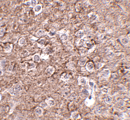
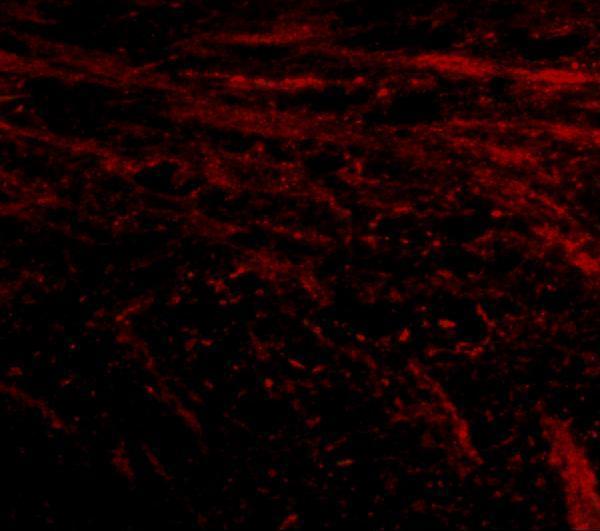
Anti-IL16 antibodies have IHC images from human tonsil and mouse brain (catalog image captions), plus an IF image from mouse brain (A01635-1 image caption).

Real IHC data Anti-IL-16 antibody, PB9238, IHC(P) IHC(P): Human Tonsil Tissue
Anti-IL16 Antibody ®
Cat # PB9238
Real IHC data Immunohistochemistry of IL-16 in mouse brain tissue with IL-16 antibody at 2.5 μg/mL.
Anti-IL-16 Antibody
Cat # A01635-1

PB9238 has a human tonsil IHC(P) image and lists human reactivity (PB9238 image caption; catalog applications and reactivity). A01635-1 has mouse brain IHC and IF images and lists human, mouse, and rat reactivity (A01635-1 image captions; catalog reactivity).

Which to pick: For human paraffin-section IHC, choose PB9238 (PB9238: IHC(P) human tonsil image); its fixative is unreported (PB9238 image caption). For tissue IF, choose A01635-1 (A01635-1: mouse brain IF image); for human ICC, choose PB9238 (PB9238: ICC application and human cord blood immunocytochemistry caption). For work spanning human, mouse, and rat, choose A01635-1 based on its listed reactivity and IHC-P and IF applications (A01635-1 catalog); its mouse brain IHC caption does not report a fixative, and clone type is unreported for both antibodies (catalog payload).

Each figure is that product's own IHC / IF validation image from its datasheet.

References

  1. UniProt Consortium. UniProt entry Q14005 (IL16_HUMAN, Pro-interleukin-16).
  2. Human Protein Atlas. IL16 tissue IHC expression (reliability: Enhanced).
  3. Human Protein Atlas. IL16 subcellular location (ICC-IF): Mainly localized to the nuclear speckles. In addition localized to the plasma membrane and cytosol..
  4. Human Protein Atlas. IL16 antibody validation summary (3 antibodies).
  5. microRNA-128-3p inhibits CD4+ regulatory T cells enrichment by targeting interleukin 16 in gastric cancer. Bioengineered 2022 — PMC8805824.
  6. Cytokine (IL16) and tyrphostin actions on ovarian primordial follicle development. Reproduction (Cambridge, England) 2014 — PMC4110175.
  7. IL16 Regulates Osteoarthritis Progression as a Target Gene of Novel-miR-81. Cartilage 2024 — PMC11368893.
  8. Single-cell hdWGCNA and experimental validation identify an innovative T-cell-associated prognostic model and immune microenvironment in lung adenocarcinoma with lymph node metastasis. BMC cancer 2026 — PMC13041103.
  9. PubMed PMID:12923170 — UniProt-cited evidence.
  10. PubMed PMID:19906316 — UniProt-cited evidence.
  11. PubMed PMID:14702039 — UniProt-cited evidence.