IRAK2 / Interleukin-1 receptor-associated kinase-like 2 · IHC design guide

Design Immunohistochemistry for IRAK2

Plan paraffin-section IRAK2 IHC around cytoplasmic and membranous staining, with kidney tubular cells as a medium-staining reference (HPA tissue IHC). Compare matched sections and interpret intensity cautiously because antibody staining has low consistency with RNA expression (HPA tissue IHC).

Evidence assembled Oct 2026 · For research use; verify linked source records and product datasheet before use
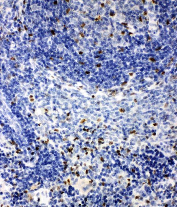
Immunohistochemistry protocol sheet for IRAK2 (IHC for IRAK2): expected localisation Cytoplasmic and membranous tissue staining (HPA tissue IHC), antibody A01559, validated IHC image, and IHC protocol steps
Printable IRAK2 IHC protocol sheet — expected localisation Cytoplasmic and membranous tissue staining (HPA tissue IHC), antibody A01559, controls and protocol steps. Open the full IRAK2 IHC guide →

IRAK2 Immunohistochemistry Experimental Design Guide

Expected localisation, validated protocols, controls and antibodies — the at-a-glance facts below, then the full design guide.

Must know before staining
Expected localisation Cytoplasmic and membranous tissue staining (HPA tissue IHC)
Staining pattern Several cell types show cytoplasmic and membranous staining (HPA tissue IHC)
Antigen retrieval EDTA pH 8.0 HIER, heat-mediated (datasheet A01559)
Positive control ⓘ Adrenal gland+4 more · see all
Negative control ⓘ Cerebellum+4 more · see all
Important caveats
Reasons your staining may differ from the expected pattern.
Fixation Keep fixation consistent across sections. (standard IHC practice; not target-specific)
Caveat Antibody staining has low consistency with RNA data (HPA tissue IHC)
Regulation Expression induction is unreported (UniProt)
Isoform / epitope No annotated isoforms or cleavage; no transmembrane segment (UniProt)
Section 1

Recommended IRAK2 IHC & IF Protocols

The catalog antibody has an IHC-P protocol (datasheet: A01559). The published workflows below describe IRAK2 staining of oral tumor sections and tissue microarrays (PMC8260692; PMC7707239).

Recommended immunohistochemistry (IHC-P) protocol parameters
SampleParaffin-embedded rat liver tissue; fixative not specified (datasheet A01559)
FixationImage fixative and duration unreported (datasheet A01559); verify before use.
Sectioning4–5 µm sections on charged slides (standard)
DeparaffinisationXylene, graded ethanol series to water (standard)
Antigen retrievalHeat retrieval: EDTA pH 8.0 (datasheet A01559); 20 min, 95–100 °C (standard)
Peroxidase block3% H2O2, 10 min, room temperature (standard)
Blocking10% goat serum (datasheet A01559)
Primary antibodyRabbit anti-IRAK2, 0.5-1μg/ml (datasheet A01559)
Primary incubationOvernight at 4 °C (datasheet A01559)
DetectionStreptavidin-biotin complex (SABC), DAB chromogen (datasheet A01559)
CounterstainHematoxylin, blue, dehydrate and mount (standard)
Expected resultIRAK2-positive staining in glandular cells of adrenal gland (HPA tissue IHC: Medium). HPA tissue profile: Cytoplasmic and membranous expression in several tissues. No signal in the no-primary control.
💡Decision noteStart with EDTA pH 8.0 heat retrieval for the catalog antibody (datasheet: A01559). Use each paper’s stated retrieval conditions when reproducing its workflow (PMC8260692; PMC7707239).
Section 2

What Is the Expected IRAK2 Staining Pattern?

For paraffin-section IHC, look for cytoplasmic and membranous IRAK2 staining, particularly in lung macrophages, kidney tubular cells, and bronchial respiratory epithelial cells (HPA tissue IHC). IRAK2 has no transmembrane segment (UniProt O43187 topology), so membranous staining does not establish that it spans the membrane. Treat the pattern as provisional: HPA rates tissue IHC reliability Uncertain because antibody staining and RNA expression have low consistency (HPA tissue IHC).

What am I looking at on my slide?
Moderate cytoplasmic or membranous staining in lung macrophages, kidney tubular cells, or bronchial respiratory epithelium.This resembles the reported IHC pattern and cell-specific Medium staining (HPA tissue IHC). Compare localization and cell identity together; the Uncertain reliability rating means resemblance alone cannot establish antibody specificity (HPA tissue IHC).
Strong nuclear-only staining, with little or no cytoplasmic or membranous staining in the expected cells.Nuclear-only staining conflicts with HPA’s cytoplasmic and membranous tissue profile (HPA tissue IHC). Investigate nonspecific staining and slide-level artefacts before interpreting it as IRAK2; UniProt provides no subcellular annotation that would independently support a nuclear assignment (UniProt O43187).
Prominent staining in pancreatic exocrine glandular cells or cerebellar granular-layer cells.These cell populations were Not detected by HPA tissue IHC (HPA tissue IHC). Consider antibody cross-reactivity or endogenous chromogenic activity as possible explanations (standard IHC practice). HPA’s Uncertain reliability prevents treating either negative population as an absolute exclusion (HPA tissue IHC).
Diffuse color across tissue, including areas without a recognizable cytoplasmic or membranous cell pattern.A diffuse deposit is difficult to assign to IRAK2 because HPA describes a compartmental pattern in several tissues (HPA tissue IHC). Evaluate background from detection chemistry, blocking, washes, and counterstain before scoring cells (standard IHC practice).
No staining in lung macrophages or kidney tubular cells on a technically adequate slide.Those populations showed Medium staining in HPA tissue IHC, so absence warrants a technical check (HPA tissue IHC). It does not prove target absence or antibody failure: HPA labels its IHC evidence Uncertain, and a single section may lack the relevant cells (HPA tissue IHC; standard IHC practice).
💡Expected IRAK2 appearanceA plausible positive is moderate cytoplasmic and membranous chromogenic staining in lung macrophages or kidney tubular cells (HPA tissue IHC); isolated nuclear color or broad, cell-independent deposits call for investigation (HPA tissue IHC; standard IHC practice).
How each factor affects the staining
Compartment and topologyHPA reports cytoplasmic and membranous tissue staining (HPA tissue IHC). UniProt lists no transmembrane segment and no subcellular annotation (UniProt O43187); therefore, do not interpret a membrane-edge signal as proof of membrane insertion.
Cell-specific tissue choiceLung macrophages, kidney tubular cells, and bronchial respiratory epithelial cells showed Medium staining; pancreatic exocrine glandular cells were Not detected (HPA tissue IHC). Choose comparison regions by cell population, since a tissue label alone can hide different cellular patterns.
Strength of the tissue evidenceHPA calls tissue IHC reliability Uncertain because antibody staining and RNA expression show low consistency, pending external verification (HPA tissue IHC). Interpret intensity and positive-versus-negative comparisons as provisional observations rather than validated cutoffs.
Antibody-level validationHPA rates IHC for HPA050520 and CAB013483 Uncertain (HPA antibodies). Agreement with the published tissue pattern can guide review, but the supplied antibody record does not support calling either IHC result independently validated.
IF/ICC Q: Should paraffin-section IHC show vesicles?A: HPA lists vesicles as the approved ICC-IF location and lists HPA050520 ICC as Approved (HPA subcellular; HPA antibodies). This is method-specific evidence; assess paraffin-section IHC against HPA’s cytoplasmic and membranous tissue profile (HPA tissue IHC).
UniProt expression versus HPA stainingUniProt reports expression in pancreas and several other tissues (UniProt O43187 tissue specificity), while HPA reports pancreatic exocrine glandular cells as Not detected by IHC (HPA tissue IHC). Keep tissue-level expression and cell-specific staining distinct when choosing controls.
Why is my staining missing, weak or wrong?
SituationLikely causeNext action
The presumed positive region is blank.The section may lack the relevant cell population, or staining may have failed; HPA positives remain provisional (HPA tissue IHC).Confirm lung macrophages or kidney tubular cells are present, then check the run’s staining controls and detection steps before changing the biological interpretation (HPA tissue IHC; standard IHC practice).
All cells show similar diffuse chromogen.Background from antibody or detection reagents can obscure the compartment pattern (standard IHC practice).Review blocking and washes, inspect a no-primary control, and score only interpretable cell-associated color (standard IHC practice); compare with HPA’s cytoplasmic and membranous profile (HPA tissue IHC).
Color concentrates only in nuclei.The location conflicts with HPA’s reported tissue profile; nonspecific staining or artefact is possible (HPA tissue IHC; standard IHC practice).Inspect controls and morphology, then repeat with the same tissue and carefully compare cellular compartments; avoid calling nuclear-only staining an established IRAK2 pattern (HPA tissue IHC).
Pancreatic exocrine cells stain strongly.HPA reports these cells as Not detected, although UniProt lists expression in pancreas (HPA tissue IHC; UniProt O43187).Check for cross-reactivity and endogenous chromogenic activity with appropriate controls; interpret the discrepancy at the cell level and retain HPA’s Uncertain reliability caveat (standard IHC practice; HPA tissue IHC).
Apparent membrane staining dominates without discernible cytoplasm.HPA allows a membranous component, but UniProt reports no transmembrane segment (HPA tissue IHC; UniProt O43187 topology).Check whether color follows cell boundaries rather than section edges or deposits, and avoid inferring membrane insertion from chromogen distribution alone (standard IHC practice; UniProt O43187 topology).
ICC-IF shows puncta while IHC looks broader.HPA assigns vesicles in ICC-IF but describes cytoplasmic and membranous staining in tissue IHC (HPA subcellular; HPA tissue IHC).Judge each preparation against its own HPA observation; do not require vesicle-like puncta in paraffin-section IHC or use the ICC result to upgrade Uncertain tissue IHC reliability (HPA subcellular; HPA tissue IHC).

Sample controls for IRAK2 IHC & IF

🧪Run colon first and expect glandular cells to stain (HPA: Medium in colon glandular cells); run cerebellum as the negative tissue and expect cells in its granular layer to remain unstained (HPA: Not detected in cerebellum granular layer). On the colon slide, cells without visible signal should show only background chromogen, but do not treat a particular adjacent cell type as IRAK2-negative without validation (standard IHC practice).
Positive control tissue: Adrenal gland (Glandular cells, HPA Medium)
Negative control tissue: Cerebellum (HPA Not detected)
ICC-IF cell lines (HPA subcellular resource): HPA ICC-IF images show IRAK2 in A-431, RT-4, U2OS, with annotated localisation: Vesicles (approved) (HPA subcellular).
Technical controls: Include a no-primary, secondary-only control, a species- and isotype-matched nonimmune rabbit IgG control, and IRAK2-knockout tissue or a validated peptide-block control (hero caption: rabbit primary antibody; standard IHC practice). Block endogenous peroxidase in colon sections and assess endogenous biotin if using the caption’s streptavidin–biotin detection system (hero caption: SABC with DAB; standard IHC practice).
⚠️Feasibility: No target-specific fixation window or fixation effect is reported, and the fixative for the selected A01559 paraffin-section caption is unreported (hero caption: fixative not stated). The caption uses heat retrieval in EDTA at pH 8.0, but does not establish that retrieval is required or that frozen sections are easier (hero caption: EDTA heat retrieval; standard IHC practice). For IF/ICC, HPA reports vesicular localization in A-431, RT-4 and U2OS images; assess tissue autofluorescence separately because those images do not establish an IF fixation method or a colon-specific artefact (HPA: approved vesicle location and listed ICC-IF cell lines; standard IF practice).

HPA tissue IHC evidence for IRAK2

Comprehensive Human Protein Atlas IHC scoring per tissue (reliability: Uncertain — Low consistency between antibody staining and RNA expression data. Pending external verification.). Rows are taken directly from the HPA tissue chart — click any row's HPA link to view the source.

Positive expression · recommended positive controls

TissueCell typeLevelEvidenceSource
Adrenal gland Glandular cells Medium Protein (IHC) HPA →
Bronchus Respiratory epithelial cells Medium Protein (IHC) HPA →
Colon Glandular cells Medium Protein (IHC) HPA →
Duodenum Glandular cells Medium Protein (IHC) HPA →
Gallbladder Glandular cells Medium Protein (IHC) HPA →

Undetected expression · recommended negative controls

TissueCell typeLevelEvidenceSource
Cerebellum Cells in granular layer Not detected Protein (IHC) HPA →
Endometrium Cells in endometrial stroma Not detected Protein (IHC) HPA →
Fallopian tube Glandular cells Not detected Protein (IHC) HPA →
Hippocampus Glial cells Not detected Protein (IHC) HPA →
Ovary Ovarian stroma cells Not detected Protein (IHC) HPA →
Section 3

Advanced IRAK2 IHC Tips

Troubleshoot IRAK2 staining in paraffin sections using the catalog antibody’s IHC example and published tissue observations, while treating staining patterns as provisional.

What retrieval should I use when IRAK2 staining is weak in paraffin sections?
Start with heat-mediated retrieval in EDTA, pH 8.0 (datasheet A01559). The selected rat liver paraffin-section example used that retrieval, followed by 10% goat-serum blocking and 1 µg/mL primary antibody overnight at 4°C (datasheet A01559). If signal remains weak, compare retrieval duration on adjacent sections while keeping antibody concentration and DAB development constant (standard IHC practice). Include a no-primary control and inspect morphology: excessive heating can damage sections or increase diffuse staining (standard IHC practice). Record heating and cooling times so that a useful condition can be repeated (standard IHC practice).
Could fixation explain inconsistent IRAK2 staining between paraffin blocks?
Target-specific fixation sensitivity is unknown: the selected paraffin-section caption does not state a fixative (datasheet A01559). Compare sections from blocks with documented fixation histories under the same EDTA, pH 8.0 retrieval and 1 µg/mL primary-antibody conditions (datasheet A01559; standard IHC practice). Keep section thickness, heating, detection and DAB development consistent, then assess both staining and preserved cell morphology (standard IHC practice). A difference between blocks may reflect fixation, processing or tissue composition, so do not assign a cause from staining alone (standard IHC practice). Use matched controls before changing retrieval or antibody concentration (standard IHC practice).
How should I judge cytoplasmic, membranous or punctate IRAK2 staining?
Assess staining within identified cells before assigning a compartment: tissue IHC describes cytoplasmic and membranous expression, but its reliability is uncertain (HPA tissue IHC). Vesicles are the approved location in cell imaging, while UniProt provides no subcellular annotation and reports no transmembrane segment (HPA subcellular; UniProt O43187 topology). Puncta near a cell boundary can therefore merit review, but chromogenic staining alone may not resolve vesicles from membrane-associated signal (HPA subcellular; standard IHC practice). Compare adjacent cells, the no-primary control and section edges at the same magnification (standard IHC practice). Avoid scoring diffuse extracellular deposit as cellular IRAK2 (standard IHC practice).
Can an epitope choice change which IRAK2 signal my IHC assay detects?
Check the antibody’s documented immunogen or epitope before interpreting a staining difference; the selected caption does not identify either (datasheet A01559). IRAK2 has a death domain at residues 13–94, a protein-kinase domain at 210–489 and a reported phosphoserine at 144 (UniProt O43187). The supplied record lists 0 isoforms, which does not establish what this antibody binds in a section (UniProt O43187; datasheet A01559). Compare the same antibody across matched sections and use an independent antibody with a documented, distinct epitope if available (standard IHC practice). Do not interpret intensity changes as phosphorylation-specific without epitope evidence and an appropriate control (standard IHC practice).
How can IF help investigate ambiguous IRAK2 IHC staining?
Use IF on a separate guide workflow to test whether IRAK2 signal overlaps an independently validated marker for the cell type being examined, such as a macrophage marker in lung (HPA tissue IHC: medium in lung macrophages; standard IF practice). Choose fluorophores after checking tissue autofluorescence and include single-stain and no-primary controls for channel separation (standard IF practice). IRAK2 has no transmembrane segment, and cell imaging assigns it to vesicles; use permeabilisation when testing access to an intracellular epitope (UniProt O43187 topology; HPA subcellular; standard IF practice). The paraffin-section caption supplies IHC conditions, not IF fixation or antibody-performance evidence (datasheet A01559). Interpret colocalisation alongside cell morphology (standard IF practice).
How do I separate IRAK2 signal from chromogenic background?
Run a no-primary control through the same secondary antibody and DAB steps to reveal detection-system background (standard IHC practice). The selected example used biotinylated goat anti-rabbit secondary antibody, a streptavidin-biotin complex and DAB, with 10% goat-serum blocking (datasheet A01559). Apply an appropriate endogenous-peroxidase block and, for this biotin-based system, assess endogenous-biotin background where relevant (standard IHC practice). Compare control and stained sections for edge deposits, uneven chromogen and signal outside intact cells before increasing antibody concentration (standard IHC practice). Shorten DAB development or improve washing when background rises across the section (standard IHC practice).
What should I quantify when comparing IRAK2 IHC across samples? ⚠ ANSWER MARKED FOR VERIFICATION
Define the cell population and compartment before scoring; tissue IHC reports cytoplasmic and membranous patterns with uncertain reliability (HPA tissue IHC). For intact cells in matched regions, record the percentage positive and an H-score based on staining intensity, or count positive cells per mm² when cell density is the question (standard IHC practice). Normalise percentages to the number of evaluable cells of the same type, and density to evaluable tissue area; exclude folds and necrosis consistently (standard IHC practice). Keep retrieval, imaging, DAB development and scoring thresholds fixed across sections (standard IHC practice). Report cell-type and compartment results separately (standard IHC practice).
When is IRAK2 staining convincing rather than artefactual?
Look for reproducible signal in intact, identifiable cells that exceeds the no-primary control and follows a plausible cellular pattern (standard IHC practice). Lung macrophages and kidney tubule cells show medium tissue-IHC staining, while pancreatic exocrine glandular cells are reported as not detected; these observations carry uncertain reliability (HPA tissue IHC). IRAK2 lacks a transmembrane segment, and cell imaging places it in vesicles, so an exclusively sharp surface rim warrants closer review (UniProt O43187 topology; HPA subcellular). Exclude edge staining, necrotic areas and endogenous-enzyme signal with morphology and controls (standard IHC practice). Confirm unexpected cell-type staining independently before calling it biological (standard IHC practice).
Boster reagents

Best IRAK2 / Interleukin-1 receptor-associated kinase-like 2 IHC Antibodies

Anti-IRAK2 antibodies have IHC images from rat liver and spleen, human kidney and lung cancer tissue, plus an IF image from A431 cells (A01559 and PA2236 image captions).

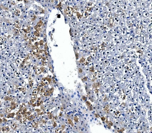
Real IHC data IHC analysis of IRAK2 using anti-IRAK2 antibody (A01559). IRAK2 was detected in paraffin-embedded section of rat liver tissue. Heat mediated antigen retrieval was performed in EDTA buffer (pH8.0, epitope retrieval solution). The tissue section was blocked with 10% goat serum. The tissue section was then incubated with 1μg/ml rabbit anti-IRAK2 Antibody (A01559) overnight at 4°C. Biotinylated goat anti-rabbit IgG was used as secondary antibody and incubated for 30 minutes at 37°C. The tissue section was developed using Strepavidin-Biotin-Complex (SABC) (Catalog # SA1022) with DAB as the chromogen.
Anti-IRAK2 Antibody ®
Cat # A01559
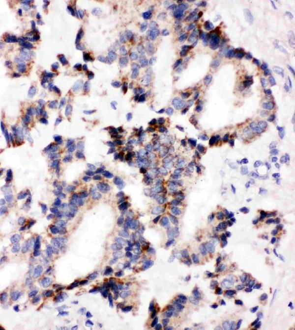
Real IHC data Anti-IRAK2 antibody, PA2236, IHC(P) IHC(P): Human Lung Cancer Tissue
Anti-IRAK2 Antibody ®
Cat # PA2236

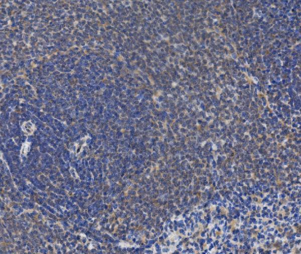
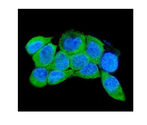
A01559 has paraffin-section IHC images from rat liver, rat spleen and human kidney, plus IF in A431 cells (A01559 image captions). PA2236 has paraffin-section IHC images from human lung cancer tissue and rat spleen (PA2236 image captions).

Which to pick: For tissue IHC, choose the SKU with an image matching your sample: A01559 for the documented liver or kidney examples, or PA2236 for the lung cancer example; both show rat spleen (A01559 and PA2236 IHC image captions). Choose A01559 for IF because it has an A431-cell IF image; PA2236 lists ICC but has no IF image (A01559 IF image caption; PA2236 catalog applications and image list). Both list human, mouse and rat reactivity, so the documented sample and application are the stronger selection criteria; clonality and the fixative used for the paraffin sections are unreported (A01559 and PA2236 catalog reactivity, clone fields and IHC image captions).

Each figure is that product's own IHC / IF validation image from its datasheet.

References

  1. UniProt Consortium. UniProt entry O43187 (IRAK2_HUMAN, Interleukin-1 receptor-associated kinase-like 2).
  2. Human Protein Atlas. IRAK2 tissue IHC expression (reliability: Uncertain).
  3. Human Protein Atlas. IRAK2 subcellular location (ICC-IF): Localized to vesicles..
  4. Human Protein Atlas. IRAK2 antibody validation summary (2 antibodies).
  5. IRAK2, an IL1R/TLR Immune Mediator, Enhances Radiosensitivity via Modulating Caspase 8/3-Mediated Apoptosis in Oral Squamous Cell Carcinoma. Frontiers in oncology 2021 — PMC8260692.
  6. Epigenetic silencing of microRNA-373 to epithelial-mesenchymal transition in non-small cell lung cancer through IRAK2 and LAMP1 axes. Cancer letters 2014 — PMC7707239.
  7. IRAK2, an Immune and Radiation-Response Gene, Correlates with Advanced Disease Features but Predicts Higher Post-Irradiation Local Control in Non-Metastatic and Resected Oral Cancer Patients. International journal of molecular sciences 2023 — PMC10138613.
  8. PubMed PMID:14702039 — UniProt-cited evidence.
  9. PubMed PMID:15489334 — UniProt-cited evidence.
  10. PubMed PMID:9374458 — UniProt-cited evidence.