This website uses cookies to ensure you get the best experience on our website.


- Table of Contents
Plan KAT2B IHC in paraffin sections around the general nuclear staining reported in tissues (HPA tissue IHC). This guide helps select a high-staining control, handle fixation consistently and interpret differentiation-related changes in localisation (UniProt).

Expected localisation, validated protocols, controls and antibodies — the at-a-glance facts below, then the full design guide.
| Expected localisation | General nuclear tissue staining (HPA tissue IHC) | |
| Staining pattern | Nuclear staining; high in placental decidual cells (HPA tissue IHC) | |
| Antigen retrieval | Tris-EDTA pH 9.0 HIER, heat-mediated (datasheet A02403-1) | |
| Positive control | Cerebellum+4 more · see all | |
| Negative control | None in HPA (detected in all 45 tissues); use no-primary + isotype controls |
| Fixation | Matched tissue-IHC evidence does not establish the fixation claim. Validate the specimen-specific method before use. (selected-SKU IHC image A02403-1) | |
| Caveat | Differentiation can shift staining from cytoplasm to nucleus (UniProt) | |
| Regulation | Nuclear signal varies with differentiation (UniProt) | |
| Isoform / epitope | No annotated isoforms; one unprocessed chain (UniProt) |
The catalog antibody’s IHC-P protocol uses Tris-EDTA pH 9.0 retrieval (datasheet A02403-1). Published KAT2B IHC methods provide cervical and nasopharyngeal carcinoma examples (PMC8806817; PMC7719841).
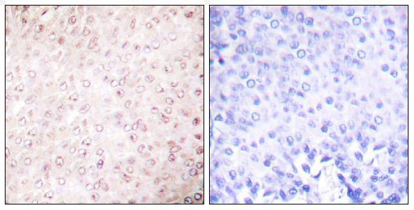
| Sample | Paraffin-embedded human cervical carcinoma tissue; fixative not specified (datasheet A02403-1) |
| Fixation | Image fixative and duration unreported (datasheet A02403-1); verify before use. |
| Sectioning | 4–5 µm sections on charged slides (standard) |
| Deparaffinisation | Xylene, graded ethanol series to water (standard) |
| Antigen retrieval | Heat retrieval: Tris-EDTA pH 9.0 (datasheet A02403-1); 20 min, 95–100 °C (standard) |
| Peroxidase block | 3% H2O2, 10 min, room temperature (standard) |
| Blocking | 10% normal serum of the secondary host, 30 min, room temperature (standard) |
| Primary antibody | Rabbit anti-KAT2B, 1:50 recommended; image 1:200 (datasheet A02403-1) |
| Primary incubation | Overnight at 4 °C (standard) |
| Detection | HRP-polymer secondary, DAB chromogen 5–10 min (standard) |
| Counterstain | Hematoxylin, blue, dehydrate and mount (standard) |
| Expected result | KAT2B-positive staining in cells in molecular layer of cerebellum (HPA tissue IHC: High). HPA tissue profile: General nuclear expression. No signal in the no-primary control. |
KAT2B should stain mainly nuclei in paraffin sections (UniProt Q92831; HPA tissue IHC: general nuclear expression). HPA reports High staining in selected cell populations, including salivary glandular cells, placental decidual cells, and skin melanocytes (HPA tissue IHC). Tissue IHC reliability is Supported, with medium consistency against RNA data (HPA tissue IHC). KAT2B has no transmembrane segment (UniProt Q92831 topology).
| Distinct nuclear chromogen in salivary glandular cells, placental decidual cells, or skin melanocytes (HPA tissue IHC: High). | This fits the reported cell populations and general nuclear pattern (HPA tissue IHC). Compare staining with nuclear morphology and a counterstain when judging individual cells (general IHC practice). |
| Predominantly diffuse cytoplasmic staining with little nuclear signal across a section. | Review specificity and detection background because the reported tissue pattern is generally nuclear (HPA tissue IHC). Cytoplasmic staining alone is not conclusive: localization can vary with differentiation (UniProt Q92831). |
| Strong chromogen in a cell population outside the reported High populations, especially if its nuclei remain unstained. | Consider cross-reactivity or endogenous detection activity and inspect controls (general IHC practice). HPA lists no negative tissue, so an unlisted cell population cannot be classified as KAT2B-negative (HPA tissue IHC). |
| Diffuse color over cells, stroma, and spaces, without clear nuclear boundaries. | This is difficult to score as the general nuclear pattern reported for KAT2B (HPA tissue IHC). Check the no-primary control and detection background before assigning cell-specific expression (general IHC practice). |
| No discernible signal in an HPA High population, such as salivary glandular cells (HPA tissue IHC). | Treat the result as inconclusive until controls and the IHC-validated antibody are checked (general IHC practice). HPA's Supported tissue assessment indicates medium consistency, not guaranteed staining in every section (HPA tissue IHC). |
| Cell and tissue choice | HPA reports High staining in cerebellar molecular-layer cells, decidual cells, salivary and stomach glandular cells, melanocytes, thyroid glandular cells, and urothelial cells; bone-marrow hematopoietic cells are Medium (HPA tissue IHC). Choose a documented High population for interpretation. |
| Compartment | Mainly nuclear localization is supported by UniProt and tissue IHC (UniProt Q92831; HPA tissue IHC). UniProt also records cytoplasm and centrosomes, so a limited nonnuclear component needs cell-context review rather than automatic rejection (UniProt Q92831). |
| Differentiation state | UniProt reports cytoplasmic KAT2B in undifferentiated basal epithelial cells and nuclear KAT2B in differentiated parabasal cells (UniProt Q92831). Assess the layer and cell state before treating a compartment shift as staining failure. |
| Antibody evidence | CAB004526 has an IHC Supported designation; the listed HPA antibodies with ICC Enhanced designations have no IHC designation in this payload (HPA antibody validation). Use the IHC evidence when interpreting paraffin-section chromogen. |
| Target structure | KAT2B is a single listed chain with no signal peptide, propeptide, or transmembrane segment (UniProt Q92831). The supplied record gives no antibody epitope or target-specific fixation effect, so neither epitope access nor fixation sensitivity can be predicted from it. |
| IF/ICC Q: what pattern is expected? | A: Mainly nucleoplasmic signal with additional cytosol signal is reported in cell images (HPA ICC-IF). That observation helps interpret compartment differences; the IF/ICC workflow belongs on its own guide page. |
| Situation | Likely cause | Next action |
|---|---|---|
| No staining in a documented High cell population (HPA tissue IHC). | The section, antibody use, or detection workflow may have failed; absence of color alone cannot identify which step failed (general IHC practice). | Check a documented positive section, the IHC-validated antibody's stated conditions, reagent performance, and the no-primary control before interpreting absence as biological (general IHC practice). |
| Diffuse chromogen obscures nuclei. | Nonspecific primary staining or detection background can reduce contrast (general IHC practice). | Compare a no-primary control, review blocking and washing, and optimize primary-antibody concentration within its stated IHC conditions (general IHC practice). |
| Cytoplasmic color dominates where a general nuclear pattern was expected (HPA tissue IHC). | Cell state may contribute, since KAT2B localization varies with differentiation; technical background is another possibility (UniProt Q92831; general IHC practice). | Check cell identity and layer, then compare nuclear detail and controls. Interpret a compartment shift in that context rather than calling it positive solely from color (UniProt Q92831; general IHC practice). |
| Unexpected cells stain strongly while reported High cells are weak. | Cross-reactivity or endogenous detection activity is possible; HPA provides no negative tissue reference here (general IHC practice; HPA tissue IHC). | Compare the no-primary control and a documented High cell population in the same run; avoid calling an unlisted population negative by definition (general IHC practice; HPA tissue IHC). |
| A low-staining tissue shows faint nuclear color. | Low HPA staining is not an absence claim; liver hepatocytes and several other listed cells have Low staining (HPA tissue IHC). | Score intensity and cellular localization against controls, and report the observation without imposing a positive-versus-negative cutoff unsupported by this payload (HPA tissue IHC; general IHC practice). |
| Color persists in the no-primary control. | Endogenous enzyme activity or detection-reagent background can produce chromogen independently of primary-antibody binding (general chromogenic IHC practice). | Address the detection system's background according to its instructions and repeat the control before attributing sample color to KAT2B (general chromogenic IHC practice). |
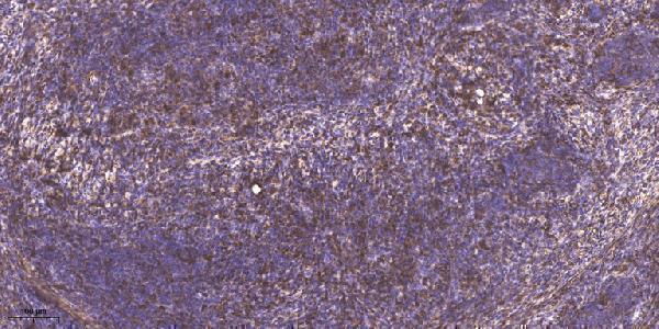


Comprehensive Human Protein Atlas IHC scoring per tissue (reliability: Supported — Medium consistency between antibody staining and RNA expression data.). Rows are taken directly from the HPA tissue chart — click any row's HPA link to view the source.
| Tissue | Cell type | Level | Evidence | Source |
|---|---|---|---|---|
| Cerebellum | Cells in molecular layer | High | Protein (IHC) | HPA → |
| Placenta | Decidual cells | High | Protein (IHC) | HPA → |
| Salivary gland | Glandular cells | High | Protein (IHC) | HPA → |
| Skin | Melanocytes | High | Protein (IHC) | HPA → |
| Stomach | Glandular cells | High | Protein (IHC) | HPA → |
| Tissue | Cell type | Level | Evidence | Source |
|---|---|---|---|---|
| None in HPA: KAT2B is detected in all 45 scored tissues. Use a no-primary (secondary-only) and an isotype control instead. | ||||
Troubleshoot KAT2B staining in paraffin sections by checking retrieval, nuclear localisation, cell type and controls before interpreting chromogenic signal.
Catalog antibodies cover IHC in human paraffin carcinoma sections (A02403 and A02403-1 image captions), with human, mouse, and rat reactivity across the range (catalog). IF is listed, but no IF figure is supplied (catalog).
A02403-1 has an IHC image from paraffin-embedded human cervical carcinoma and lists human and mouse reactivity (A02403-1 image caption; catalog). A02403 has a paraffin-embedded human breast carcinoma IHC caption that names p300/CBP, while A02403-3 lists human IHC, IF, and ICC without an image (A02403 image caption; A02403-3 catalog).
Which to pick: For tissue IHC targeting acetyl-K428, choose A02403-1: its own paraffin-section caption reports Tris-EDTA pH 9.0 retrieval and 1:200 antibody overnight at 4 °C; the fixative is unreported (A02403-1 catalog; image caption). For human IF/ICC, A02403-3 lists both applications at 5 μg/ml, but supplies no IF image (A02403-3 catalog). For cross-species IHC or IF, A02403 lists human, mouse, and rat reactivity and both applications; its breast carcinoma caption names p300/CBP, so verify target identity before use, and note that the fixative is unreported (A02403 catalog; image caption).