This website uses cookies to ensure you get the best experience on our website.


- Table of Contents
Plan chromogenic KCNJ5 IHC in paraffin sections using adrenal glandular cells as a strong positive reference (HPA tissue IHC). This guide covers fixation, antibody use, and interpretation of observed cytoplasmic staining alongside the protein’s membrane topology (HPA tissue IHC; UniProt).

Expected localisation, validated protocols, controls and antibodies — the at-a-glance facts below, then the full design guide.
| Expected localisation | Cytoplasmic staining (HPA tissue IHC); membrane protein (UniProt) | |
| Staining pattern | Glandular cell cytoplasm, strongest in adrenal gland (HPA tissue IHC) | |
| Antigen retrieval | Tris-EDTA pH 9.0 HIER, heat-mediated (datasheet A02160) | |
| Positive control | Adrenal gland+4 more · see all | |
| Negative control | Bone marrow+4 more · see all |
| Fixation | Keep fixation consistent across sections (standard IHC practice; not target-specific) | |
| Caveat | Antibody staining has low consistency with RNA data (HPA tissue IHC) | |
| Regulation | Adrenal and pancreas RNA enrichment (HPA tissue RNA) | |
| Isoform / epitope | No annotated isoforms; epitope side affects access (UniProt) |
The catalog antibody’s IHC-P protocol (datasheet A02160) is paired with published KCNJ5 protocols for FFPE adrenal tissue (PMC6739168; PMC12185499; PMC11787464).
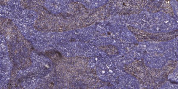
| Sample | Paraffin-embedded human cervical carcinoma tissue; fixative not specified (datasheet A02160) |
| Fixation | Image fixative and duration unreported (datasheet A02160); verify before use. |
| Sectioning | 4–5 µm sections on charged slides (standard) |
| Deparaffinisation | Xylene, graded ethanol series to water (standard) |
| Antigen retrieval | Heat retrieval: Tris-EDTA pH 9.0 (datasheet A02160); 20 min, 95–100 °C (standard) |
| Peroxidase block | 3% H2O2, 10 min, room temperature (standard) |
| Blocking | 10% normal serum of the secondary host, 30 min, room temperature (standard) |
| Primary antibody | Rabbit anti-KCNJ5, 1:50 recommended; image 1:200 (datasheet A02160) |
| Primary incubation | Overnight at 4 °C (standard) |
| Detection | HRP-polymer secondary, DAB chromogen 5–10 min (standard) |
| Counterstain | Hematoxylin, blue, dehydrate and mount (standard) |
| Expected result | KCNJ5-positive staining in glandular cells of adrenal gland (HPA tissue IHC: High). HPA tissue profile: Cytoplasmic expression in several tissues with strongest expression in adrenal gland. No signal in the no-primary control. |
KCNJ5 (GIRK4) is a membrane protein with two transmembrane segments (UniProt P48544 topology). In paraffin-section IHC, HPA describes predominantly cytoplasmic staining, strongest in adrenal glandular cells and also high in pancreatic exocrine glandular cells (HPA tissue IHC). Treat these as observed staining patterns, not proof of precise membrane localisation: HPA rates the tissue IHC evidence Approved but reports low consistency with RNA expression (HPA tissue IHC).
| Strong staining in adrenal glandular cells, with cytoplasmic signal and possible membrane enrichment (HPA tissue IHC; UniProt P48544 topology). | This fits the strongest reported tissue pattern (HPA: High in adrenal glandular cells). Record cytoplasmic and membrane-associated signal separately; the channel topology supports membrane localisation, while the HPA IHC description calls the observed staining cytoplasmic (UniProt P48544 topology; HPA tissue IHC). |
| Predominantly nuclear staining, with little signal in the expected cytoplasmic or membrane-associated area. | A nuclear-only pattern conflicts with KCNJ5 membrane topology and the reported cytoplasmic IHC profile (UniProt P48544 topology; HPA tissue IHC). Check the negative control and repeat staining before assigning it to KCNJ5 (general IHC practice). |
| Strong staining in squamous epithelial cells of esophagus or oral mucosa (HPA: Not detected in those cells). | That distribution conflicts with the reported tissue pattern and may indicate nonspecific antibody binding or endogenous detection activity; it does not establish either cause by itself (HPA tissue IHC; general IHC practice). Compare a no-primary control and the glandular-cell pattern (general IHC practice). |
| Broad, fairly uniform chromogen across tissue and extracellular spaces, obscuring cell boundaries. | This is more consistent with staining background than an interpretable cell-specific pattern (general IHC practice). It prevents comparison with HPA's glandular-cell staining levels; assess the no-primary control before scoring cells (HPA tissue IHC; general IHC practice). |
| No detectable staining in well-preserved adrenal glandular cells (HPA: High in adrenal glandular cells). | A negative result in this expected positive tissue is inconclusive until the run controls and antibody performance are checked (HPA tissue IHC; general IHC practice). HPA's Approved rating comes with low staining-to-RNA consistency, so it is not a guarantee that every section will stain (HPA tissue IHC). |
| Observed compartment versus channel topology | HPA describes cytoplasmic tissue IHC, whereas UniProt assigns KCNJ5 to membranes with two transmembrane segments; score the observed cytoplasmic pattern without claiming that chromogenic IHC resolves the channel's exact membrane boundary (HPA tissue IHC; UniProt P48544 topology). |
| Tissue and cell context | Adrenal glandular cells and pancreatic exocrine glandular cells are High; appendix, breast, cervix, colon, duodenum and endometrial glandular cells are Medium (HPA tissue IHC). Compare like cell populations when judging intensity (general IHC practice). |
| Epitope location, if supplied for the chosen antibody | UniProt places residues 1–86 and 186–419 on the cytoplasmic side and residues 112–135 and 155–163 extracellularly (UniProt P48544 topology). The antibody epitope is unspecified here, so topology alone cannot predict retrieval requirements or staining strength. |
| Strength of tissue evidence | All three listed antibodies have Approved IHC status; the tissue profile also reports low consistency between staining and RNA expression (HPA antibodies; HPA tissue IHC). Use the reported distribution as a comparison, then interpret unexpected staining with controls rather than treating it as definitive expression. |
| IF/ICC: should the same appearance be expected? | HPA summarises subcellular location as Membrane but lists no ICC-IF image cell lines or ICC validation for the three antibodies (HPA subcellular; HPA antibodies). IF/ICC localisation therefore cannot be verified from these payloads; consult its separate guide for that application. |
| Situation | Likely cause | Next action |
|---|---|---|
| Adrenal glandular cells are unstained (HPA: High). | Target-specific fixation effects are not established by the supplied assay evidence. Verify with a matched IHC source before attributing a result to fixation. | Check positive and no-primary controls, reagent delivery and detection steps; document the result as unresolved if controls work (general IHC practice). |
| Pancreatic exocrine glandular cells stain weakly or unevenly (HPA: High). | Field selection or uneven section staining can confound comparison with the reported High category (HPA tissue IHC; general IHC practice). | Inspect multiple intact exocrine fields and compare their background and counterstain before scoring intensity (general IHC practice). |
| Nuclei carry the dominant chromogenic signal. | A nuclear-only distribution is discordant with the reported cytoplasmic IHC profile and membrane topology (HPA tissue IHC; UniProt P48544 topology). | Check the no-primary control and antibody staining conditions; withhold a KCNJ5-positive call unless the expected cellular pattern appears (general IHC practice). |
| HPA-negative squamous cells stain strongly. | Nonspecific binding or endogenous detection activity is possible; esophageal and oral squamous epithelial cells are reported Not detected (HPA tissue IHC; general IHC practice). | Use no-primary and detection controls to localise background, then reassess the glandular-cell pattern under the same run conditions (general IHC practice). |
| Diffuse chromogen makes individual cells hard to score. | Excess background can obscure the cell-specific distribution needed for interpretation (general IHC practice). | Review control sections and the detection and wash steps; score only fields where cells and subcellular signal remain distinguishable (general IHC practice). |
| An IF image appears membrane-localised, but the IHC section looks cytoplasmic. | Those descriptions come from different HPA summaries; the supplied record has no ICC-IF image cell lines for direct comparison (HPA tissue IHC; HPA subcellular). | Report each application and its controls separately; do not use the IF summary to overrule an interpretable paraffin-section IHC result (HPA tissue IHC; HPA subcellular; general IHC practice). |


Comprehensive Human Protein Atlas IHC scoring per tissue (reliability: Approved — Low consistency between antibody staining and RNA expression data.). Rows are taken directly from the HPA tissue chart — click any row's HPA link to view the source.
| Tissue | Cell type | Level | Evidence | Source |
|---|---|---|---|---|
| Adrenal gland | Glandular cells | High | Protein (IHC) | HPA → |
| Pancreas | Exocrine glandular cells | High | Protein (IHC) | HPA → |
| Appendix | Glandular cells | Medium | Protein (IHC) | HPA → |
| Breast | Glandular cells | Medium | Protein (IHC) | HPA → |
| Cervix | Glandular cells | Medium | Protein (IHC) | HPA → |
| Tissue | Cell type | Level | Evidence | Source |
|---|---|---|---|---|
| Bone marrow | Hematopoietic cells | Not detected | Protein (IHC) | HPA → |
| Esophagus | Squamous epithelial cells | Not detected | Protein (IHC) | HPA → |
| Oral mucosa | Squamous epithelial cells | Not detected | Protein (IHC) | HPA → |
| Skeletal muscle | Myocytes | Not detected | Protein (IHC) | HPA → |
| Vagina | Squamous epithelial cells | Not detected | Protein (IHC) | HPA → |
Troubleshoot KCNJ5 chromogenic IHC in paraffin sections using the catalog antibody’s tissue image, protein topology, and tissue staining patterns as distinct evidence sources.
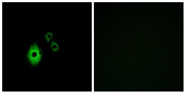
A02160 has a paraffin-section IHC image of human cervical carcinoma and an IF image of A549 cells (catalog image captions). Listed reactivity covers Human, Mouse, and Rat (catalog reactivity).
A02160 will render with its IHC figure of paraffin-embedded human cervical carcinoma (A02160 IHC image caption). The catalog also supplies an A549-cell IF image, although IF/ICC is absent from the listed applications (catalog IF image caption; catalog applications).
Which to pick: Choose A02160 for tissue IHC: it is a rabbit polyclonal with IHC listed, and its own figure used Tris–EDTA at pH 9.0 for antigen retrieval on paraffin-embedded sections (catalog host, dilution_raw, applications; A02160 IHC image caption). For IF/ICC, A02160 has an A549-cell image, but its application list does not include IF/ICC, so confirm suitability before use (catalog IF image caption; catalog applications). A02160 lists Human, Mouse, and Rat reactivity for cross-species planning; the fixative used for its IHC figure is unreported (catalog reactivity; A02160 IHC image caption).