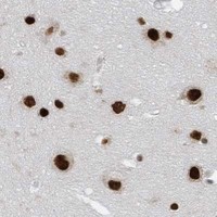
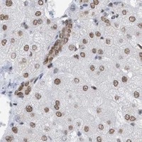
KHSRP / Far upstream element-binding protein 2 · IHC design guide

Design Immunohistochemistry for KHSRP

Plan chromogenic KHSRP IHC on paraffin sections using 2–5 μg/ml of the IHC-validated antibody (datasheet A02770-2). Score nuclear staining with high-staining bone marrow hematopoietic cells and lower-staining heart cardiomyocytes as tissue references (HPA tissue IHC).

Evidence assembled Oct 2026 · For research use; verify linked source records and product datasheet before use
Immunohistochemistry protocol sheet for KHSRP (IHC for KHSRP): expected localisation Nuclear staining in tissue (HPA tissue IHC), antibody A02770-2, validated IHC image, and IHC protocol steps
Printable KHSRP IHC protocol sheet — expected localisation Nuclear staining in tissue (HPA tissue IHC), antibody A02770-2, controls and protocol steps. Open the full KHSRP IHC guide →

KHSRP Immunohistochemistry Experimental Design Guide

Expected localisation, validated protocols, controls and antibodies — the at-a-glance facts below, then the full design guide.

Must know before staining
Expected localisation Nuclear staining in tissue (HPA tissue IHC)
Staining pattern Widespread nuclear staining across tissue cell types (HPA tissue IHC)
Antigen retrieval EDTA pH 8.0 HIER, heat-mediated (datasheet A02770-2)
Positive control ⓘ Adipose tissue+4 more · see all
Negative control ⓘ None in HPA (detected in all 45 tissues); use no-primary + isotype controls
Important caveats
Reasons your staining may differ from the expected pattern.
Fixation Matched tissue-IHC evidence does not establish the fixation claim. Validate the specimen-specific method before use. (selected-SKU IHC image A02770-2)
Caveat Ubiquitous nuclear staining limits cell-specific contrast (HPA tissue IHC)
Regulation Specific expression regulators unreported (UniProt)
Isoform / epitope No annotated isoforms or isoform-specific epitope map (UniProt)
Section 1

Recommended KHSRP IHC & IF Protocols

The catalog antibody protocol is paired with one published KHSRP tissue microarray IHC protocol (PMC11467790 methods).

Recommended immunohistochemistry (IHC-P) protocol parameters
SampleParaffin-embedded human gallbladder adenocarcinoma tissue; fixative not specified (datasheet A02770-2)
FixationImage fixative and duration unreported (datasheet A02770-2); verify before use.
Sectioning4–5 µm sections on charged slides (standard)
DeparaffinisationXylene, graded ethanol series to water (standard)
Antigen retrievalHeat retrieval: EDTA pH 8.0 (datasheet A02770-2); 20 min, 95–100 °C (standard)
Peroxidase block3% H2O2, 10 min, room temperature (standard)
Blocking10% goat serum (datasheet A02770-2)
Primary antibodyRabbit anti-KHSRP, 2-5 μg/ml (datasheet A02770-2)
Primary incubationOvernight at 4 °C (datasheet A02770-2)
DetectionStreptavidin-biotin complex (SABC), DAB chromogen (datasheet A02770-2)
CounterstainHematoxylin, blue, dehydrate and mount (standard)
Expected resultKHSRP-positive staining in adipocytes of adipose tissue (HPA tissue IHC: High). HPA tissue profile: Ubiquitous nuclear expression. No signal in the no-primary control.
💡Decision noteStart with heat-mediated EDTA pH 8.0 retrieval (datasheet A02770-2); the published tissue microarray method does not specify retrieval (PMC11467790 methods).
Section 2

What Is the Expected KHSRP Staining Pattern?

KHSRP is primarily nuclear, with nucleoplasmic localisation in HPA ICC-IF; UniProt also reports cytoplasmic localisation, including a small proportion in neuronal cell bodies and dendrites (HPA: nucleoplasm; UniProt Q92945: subcellular location). Expect nuclear staining across many cell types: HPA describes ubiquitous nuclear expression and rates its tissue IHC profile Supported, citing consistency with RNA expression (HPA: tissue IHC profile and reliability). KHSRP has no transmembrane segment (UniProt Q92945: topology).

What am I looking at on my slide?
Distinct nuclear chromogen in adipocytes, glandular cells or hematopoietic cells.This fits the reported high staining in adipose tissue, adrenal gland and bone marrow (HPA: High in these cell types). Judge localisation within identifiable cells, because pooled tissue colour cannot establish which cells carry the signal (standard IHC interpretation).
Strong signal confined to cell borders or extracellular material, with nuclei unstained.Treat this as a compartment mismatch: KHSRP is nuclear and cytoplasmic, and lacks a transmembrane segment (UniProt Q92945: location and topology; HPA: nucleoplasm). Review morphology and detection controls before assigning the signal to KHSRP (standard IHC practice).
Intense staining in a cell population inconsistent with the expected local pattern.Compare the cell identities with HPA's tissue examples before calling it positive; HPA records high signal in specific cells, such as colon endothelial cells (HPA: High in colon endothelial cells). Cross-reactivity or endogenous detection activity is a possible explanation for an unexpected pattern, not a diagnosis from appearance alone (standard IHC practice).
Weak brown haze covers nuclei, cytoplasm and surrounding tissue without cell boundaries.This does not establish the reported nuclear distribution (HPA: ubiquitous nuclear expression). Diffuse staining can reflect background from detection or blocking conditions; assess a matched negative control and whether chromogen tracks identifiable cells (standard IHC practice).
No nuclear signal in an HPA high-staining tissue.A negative result in adipose tissue adipocytes, adrenal gland glandular cells or bone marrow hematopoietic cells warrants a run-level check (HPA: High in those cells). Check tissue preservation, antibody conditions and detection controls; HPA's tissue profile does not establish KHSRP-specific fixation sensitivity (standard IHC practice; HPA: tissue IHC scope).
💡Expected KHSRP appearanceCall a section positive when identifiable cells show clear nuclear staining, potentially strong in HPA high-staining cell populations, while isolated membrane or diffuse extracellular colour is suspect (HPA: ubiquitous nuclear expression and listed High cells; UniProt Q92945: topology; standard IHC interpretation).
How each factor affects the staining
Tissue and cell selectionHPA reports low tissue specificity and ubiquitous nuclear expression, with High staining in several listed cell types but Low staining in heart cardiomyocytes (HPA: tissue IHC profile). Select controls by the reported cell population, not by the tissue name alone (standard IHC practice).
Compartment and topologyNuclear localisation is the main slide expectation (HPA: ubiquitous nuclear expression); UniProt also lists cytoplasm and notes a small cytoplasmic fraction in neuronal cell bodies and dendrites (UniProt Q92945: subcellular location). No transmembrane segment or signal peptide supports treating dominant cell-surface staining cautiously (UniProt Q92945: topology and processing).
Antibody evidenceHPA rates tissue IHC Supported overall and lists HPA034739 as IHC Supported; HPA056518 has no listed IHC validation status (HPA: tissue reliability and antibody validation). These ratings support comparison with the reported pattern but do not validate an unlisted antibody or every specimen (HPA: antibody validation scope).
IF/ICC comparisonQ: Where should an IF/ICC signal appear? A: Predominantly in the nucleoplasm; HPA calls this location Enhanced and lists images for A-431, U-251MG, U2OS and NIH 3T3 (HPA: subcellular ICC-IF). Use that localisation as context for the IHC readout, while recognising that the assays and preparations differ (standard assay interpretation).
Antigen retrievalRetrieval settings can affect an IHC run in general; optimise against tissue controls under the chosen assay conditions (standard IHC practice). The supplied HPA and UniProt records give no KHSRP-specific retrieval condition or fixation-sensitivity result (HPA: tissue IHC scope; UniProt Q92945: record scope).
Why is my staining missing, weak or wrong?
SituationLikely causeNext action
Nuclear stain is absent in a listed high-staining cell population.A failed run or unsuitable assay conditions are possibilities; the supplied sources do not identify a KHSRP-specific fixation effect (standard IHC practice; HPA: tissue IHC scope).Confirm the expected cells are present, examine a run control, and review the antibody, retrieval and detection steps under the assay's instructions (standard IHC practice; HPA: listed High cells).
Signal is mainly membranous while nearby nuclei are blank.The pattern conflicts with reported nuclear localisation and absence of a transmembrane segment (HPA: tissue IHC profile; UniProt Q92945: topology).Recheck cell boundaries and compare with a negative detection control; withhold a positive call until nuclear staining is supported (standard IHC practice).
A neuronal section shows both nuclear and limited cytoplasmic colour.UniProt reports a small cytoplasmic proportion in neuronal cell bodies and dendrites (UniProt Q92945: subcellular location).Assess whether the nuclear pattern remains clear and whether cytoplasmic colour follows neuronal morphology; avoid rejecting a limited cytoplasmic component solely for its location (UniProt Q92945: location; standard IHC interpretation).
Brown colour appears broadly outside identifiable cells.Diffuse detection background or endogenous activity is possible; colour alone cannot establish KHSRP localisation (standard IHC practice).Review matched negative and endogenous-activity controls, blocking and chromogen development, then score only cell-associated staining with interpretable morphology (standard IHC practice).
Heart cardiomyocytes stain weakly.Low staining in heart cardiomyocytes is reported by HPA, so faint signal alone does not show assay failure (HPA: Low in heart cardiomyocytes).Compare nuclear localisation and a listed high-staining control before judging performance; avoid using this low-staining population as the sole positive control (HPA: tissue IHC levels; standard IHC practice).
Two antibodies give different tissue patterns.Their supplied IHC evidence differs: HPA034739 is IHC Supported, while HPA056518 has no listed IHC status (HPA: antibody validation).Compare localisation, cell identity and run controls for each reagent; do not transfer one antibody's IHC validation status to the other (HPA: antibody validation; standard IHC practice).

Sample controls for KHSRP IHC & IF

🧪Run adipose tissue first and assess staining in adipocytes (HPA: High in adipocytes). HPA detects KHSRP in all 45 scored tissues, so there is no supported negative tissue; use no-primary and isotype controls, and treat any unstained cells on the positive slide as background comparators rather than a validated KHSRP-negative population (HPA: no negative rows; detected in all 45 scored tissues).
Positive control tissue: Adipose tissue (Adipocytes, HPA High)
Negative control tissue: None in HPA: KHSRP is detected in all 45 scored tissues. Use a no-primary (secondary-only) and an isotype control instead.
ICC-IF cell lines (HPA subcellular resource): HPA ICC-IF images show KHSRP in A-431, U-251MG, U2OS, NIH 3T3, with annotated localisation: Nucleoplasm (enhanced) (HPA subcellular).
Technical controls: Include a no-primary, secondary-only slide and a concentration-matched rabbit IgG isotype control for the rabbit catalog antibody; a KHSRP-knockout specimen can test biological specificity (selected-SKU caption: rabbit anti-KHSRP; standard IHC practice). Quench endogenous peroxidase and check for endogenous biotin because the caption uses a biotinylated secondary, streptavidin–biotin detection and DAB (selected-SKU tissue-IHC caption; standard IHC practice).
⚠️Feasibility: A target-specific fixation window or fixation effect is unreported, and the selected-SKU paraffin-section caption does not state a fixative (selected-SKU tissue-IHC caption). The reported IHC example used heat retrieval in EDTA at pH 8.0 and 2 μg/ml primary antibody overnight at 4°C; it does not establish that retrieval is required under other conditions (selected-SKU tissue-IHC caption). The supplied evidence does not establish whether frozen sections or IF are easier; for adipose tissue, the large lipid space leaves a thin rim of cytoplasm, so assess staining in the cell rim and nucleus rather than the empty space (HPA: High in adipocytes; standard tissue morphology).

HPA tissue IHC evidence for KHSRP

Comprehensive Human Protein Atlas IHC scoring per tissue (reliability: Supported — High consistency between antibody staining and RNA expression data.). Rows are taken directly from the HPA tissue chart — click any row's HPA link to view the source.

Positive expression · recommended positive controls

TissueCell typeLevelEvidenceSource
Adipose tissue Adipocytes High Protein (IHC) HPA →
Adrenal gland Glandular cells High Protein (IHC) HPA →
Bone marrow Hematopoietic cells High Protein (IHC) HPA →
Breast Adipocytes High Protein (IHC) HPA →
Caudate Neuronal cells High Protein (IHC) HPA →

Undetected expression · recommended negative controls

TissueCell typeLevelEvidenceSource
None in HPA: KHSRP is detected in all 45 scored tissues. Use a no-primary (secondary-only) and an isotype control instead.
Section 3

Advanced KHSRP IHC Tips

Use the catalog antibody’s paraffin-section example to establish retrieval and detection conditions, then assess KHSRP staining by compartment and cell type.

What retrieval should I try first when KHSRP staining is weak?
Start with heat-mediated retrieval in EDTA at pH 8.0 for paraffin sections (datasheet A02770-2). The catalog antibody detected KHSRP in a paraffin-embedded human gallbladder adenocarcinoma section after this retrieval, followed by 2 μg/ml primary antibody overnight at 4°C (datasheet A02770-2). If nuclear staining remains weak, compare a modest increase in retrieval time with an adjacent section while keeping antibody concentration and detection conditions fixed (standard IHC practice). Check tissue morphology and nuclear counterstain after heating, since excessive retrieval can damage sections and make apparent staining difficult to score (standard IHC practice).
Could fixation explain weak or uneven KHSRP staining?
The selected tissue example identifies a paraffin-embedded section but does not state its fixative, so target-specific fixation sensitivity is unknown (datasheet A02770-2). Record the fixative, fixation duration, tissue thickness and processing history for each specimen before comparing KHSRP signal across sections (standard IHC practice). On adjacent sections, hold EDTA retrieval at pH 8.0 and the primary concentration at 2 μg/ml while assessing whether weak staining tracks with specimen handling (datasheet A02770-2; standard IHC practice). Evaluate nuclear morphology and an appropriate control stain alongside KHSRP; neither the reported tissue pattern nor KHSRP modifications establishes how a particular fixative affects this antibody (HPA: ubiquitous nuclear expression; UniProt Q92945: modified residues).
Where should convincing KHSRP signal appear in a paraffin section?
Prioritise nuclear staining when reviewing chromogenic KHSRP sections: tissue profiling describes ubiquitous nuclear expression, and cell imaging places the protein in the nucleoplasm (HPA: tissue IHC profile; HPA: nucleoplasm, enhanced). UniProt also lists cytoplasmic localisation, including a small proportion in neuronal cell bodies and dendrites, so limited cytoplasmic signal can be biologically plausible in context (UniProt Q92945: subcellular location). Compare the DAB pattern with the nuclear counterstain and score nuclei separately from cytoplasm rather than combining both compartments into one intensity value (standard IHC practice). Diffuse extracellular or sharply peripheral staining warrants a technical check before it is called KHSRP localisation (standard IHC practice).
How should epitope location affect KHSRP troubleshooting?
The supplied record lists 0 annotated isoforms and four KH domains at residues 144–208, 233–299, 322–386 and 424–491 (UniProt Q92945: isoforms and domains). It does not identify this antibody’s epitope, so do not assign a weak IHC result to a particular KH domain or modified residue (UniProt Q92945: record; datasheet A02770-2: tissue-IHC caption). KHSRP has no transmembrane segment, while its annotated modifications include acetylation, methylation and phosphorylation; these annotations alone do not establish epitope accessibility after processing (UniProt Q92945: topology and modified residues). If sections disagree, compare retrieval and tissue handling on adjacent sections before making an epitope-masking claim (standard IHC practice).
How can IF help check a disputed chromogenic KHSRP pattern?
Use IF/ICC as a follow-up localisation check, keeping its antibody validation separate from the catalog antibody’s paraffin-section IHC example (datasheet A02770-2: tissue-IHC caption). Multiplex KHSRP with a marker identifying the cell population under review, then compare nuclear overlap with the nucleoplasmic pattern reported by cell imaging (HPA: nucleoplasm, enhanced; standard IF practice). Choose a fluorophore channel after inspecting unstained tissue for autofluorescence, and include single-label controls when interpreting overlap (standard IF practice). Because KHSRP has no transmembrane segment and is principally nuclear, use permeabilisation appropriate for intracellular and nuclear access; optimise it for the IF antibody rather than treating the IHC retrieval conditions as an IF protocol (UniProt Q92945: topology and location; standard IF practice).
What should I check when DAB obscures nuclear KHSRP staining?
The selected example used a biotinylated secondary antibody, a streptavidin–biotin detection complex and DAB, so assess background against that detection chain (datasheet A02770-2). Include a section without primary antibody to reveal signal from secondary reagents, tissue components or the chromogenic reaction (standard IHC practice). Apply an endogenous peroxidase block and check whether biotin-based detection contributes unwanted signal in the specimen; these are general IHC controls, not demonstrated KHSRP-specific requirements (standard IHC practice). If background persists, compare a shorter DAB development or a primary-antibody titration with a matched control while preserving interpretable nuclear contrast (standard IHC practice; HPA: ubiquitous nuclear expression).
How should I quantify KHSRP when many cells stain? ⚠ ANSWER MARKED FOR VERIFICATION
Define the scored cell population and tissue region before measuring signal, because KHSRP tissue IHC is described as ubiquitously nuclear (HPA: tissue IHC profile). For nuclear DAB, report the percentage of positive nuclei and an H-score from 0–300, calculated as the sum of percentages at intensity grades 0–3 multiplied by their grades (standard IHC scoring practice). If cell counts matter, report positive nuclei per mm² of viable tissue and normalise comparisons to the same cell population and analysed area (standard IHC practice). Keep cytoplasmic signal separate, and apply one counterstain, threshold and scoring rule across sections (UniProt Q92945: cytoplasmic localisation; standard IHC practice).
How do I distinguish true KHSRP staining from section artefact?
A credible result shows interpretable nuclear staining within identifiable cells, consistent with the supported, ubiquitous nuclear tissue profile and enhanced nucleoplasmic cell-imaging pattern (HPA: tissue IHC profile and reliability; HPA: nucleoplasm, enhanced). Assess cytoplasmic staining in context, since UniProt reports a small cytoplasmic fraction in neuronal cell bodies and dendrites (UniProt Q92945: subcellular location). Exclude section edges and necrotic areas from scoring when their staining lacks intact cellular morphology, and compare suspicious signal with a no-primary control (standard IHC practice). If the control also develops DAB, investigate endogenous peroxidase or the biotin-based detection chain before interpreting that signal as KHSRP (standard IHC practice; datasheet A02770-2: detection method).
Boster reagents

Best KHSRP / Far upstream element-binding protein 2 IHC Antibodies

KHSRP antibodies have real IHC data from human paraffin sections (A02770-2 image caption) and IF/ICC data from human cells (M02770-3 image caption).

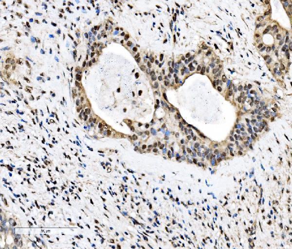
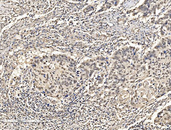
Real IHC data IHC analysis of KHSRP using anti-KHSRP antibody (A02770-2). KHSRP was detected in a paraffin-embedded section of human gallbladder adenocarcinoma tissue. Heat mediated antigen retrieval was performed in EDTA buffer (pH 8.0, epitope retrieval solution). The tissue section was blocked with 10% goat serum. The tissue section was then incubated with 2 μg/ml rabbit anti-KHSRP Antibody (A02770-2) overnight at 4°C. Biotinylated goat anti-rabbit IgG was used as secondary antibody and incubated for 30 minutes at 37°C. The tissue section was developed using Strepavidin-Biotin-Complex (SABC) (Catalog # SA1022) with DAB as the chromogen.
Anti-KHSRP Antibody ®
Cat # A02770-2
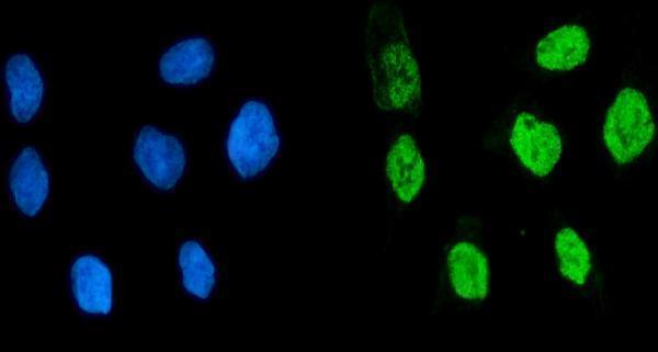
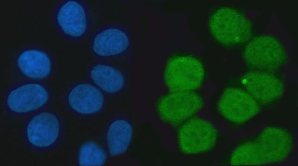
Real IF data IF analysis of KHSRP using anti-KHSRP antibody (M02770-3). KHSRP was detected in an immunocytochemical section of MCF-7 cells. Enzyme antigen retrieval was performed using IHC enzyme antigen retrieval reagent (AR0022) for 15 mins. The cells were blocked with 10% goat serum. And then incubated with 5 μg/mL mouse anti-KHSRP Antibody (M02770-3) overnight at 4°C. DyLight®488 Conjugated Goat Anti-Mouse IgG (BA1126) was used as secondary antibody at 1:100 dilution and incubated for 30 minutes at 37°C. The section was counterstained with DAPI. Visualize using a fluorescence microscope and filter sets appropriate for the label used.
Anti-KHSRP Antibody ® (monoclonal, 4F10D2)
Cat # M02770-3

A02770-2 will render with IHC data from human gallbladder adenocarcinoma paraffin sections and listed Human/Rat reactivity (A02770-2 image caption; catalog: reactivity). M02770-3 will render with IF/ICC data from MCF-7 cells and listed Human/Mouse/Rat reactivity (M02770-3 image caption; catalog: reactivity).

Which to pick: Choose A02770-2 for chromogenic tissue IHC: its own caption documents paraffin sections, EDTA retrieval, and DAB detection; the fixative is unreported (A02770-2 image caption). Choose mouse monoclonal M02770-3 for IF/ICC, supported by its own cell image, with no IHC application listed (M02770-3 image caption; catalog: applications, clone 4F10D2). For cross-species planning, M02770-3 lists Human/Mouse/Rat for IF/ICC, while IHC-listed A02770-2 lists Human/Rat; the pictured examples for both are human (catalog: applications, reactivity; A02770-2 and M02770-3 image captions).

Each figure is that product's own IHC / IF validation image from its datasheet.

References

  1. UniProt Consortium. UniProt entry Q92945 (FUBP2_HUMAN, Far upstream element-binding protein 2).
  2. Human Protein Atlas. KHSRP tissue IHC expression (reliability: Supported).
  3. Human Protein Atlas. KHSRP subcellular location (ICC-IF): Localized to the nucleoplasm..
  4. Human Protein Atlas. KHSRP antibody validation summary (2 antibodies).
  5. Acetylated KHSRP impairs DNA-damage-response-related mRNA decay and facilitates prostate cancer tumorigenesis. Molecular oncology 2024 — PMC11467790.
  6. KHSRP promotes cancer stem cell maintenance, tumorigenesis, and suppresses anti-tumor immunity in gastric cancer. Oncology research 2025 — PMC11753988.
  7. RNA-binding protein KHSRP promotes tumor growth and metastasis in non-small cell lung cancer. Journal of experimental & clinical cancer research : CR 2019 — PMC6882349.
  8. Tumour-derived small extracellular vesicles suppress CD8+ T cell immune function by inhibiting SLC6A8-mediated creatine import in NPM1-mutated acute myeloid leukaemia. Journal of extracellular vesicles 2021 — PMC8607980.
  9. PubMed PMID:9136930 — UniProt-cited evidence.
  10. PubMed PMID:15057824 — UniProt-cited evidence.
  11. PubMed PMID:15489334 — UniProt-cited evidence.