This website uses cookies to ensure you get the best experience on our website.


- Table of Contents
Plan KIF5A paraffin IHC using cerebral cortex or hippocampus neuronal cells as positive controls (HPA tissue IHC). This guide covers cytoplasmic staining in neuronal subsets and the possibility of off-target signal (HPA tissue IHC).

Expected localisation, validated protocols, controls and antibodies — the at-a-glance facts below, then the full design guide.
| Expected localisation | Neuronal cytoplasm (HPA tissue IHC); perinuclear enrichment expected (UniProt) | |
| Staining pattern | Cytoplasmic staining in subsets of neuronal cells (HPA tissue IHC) | |
| Antigen retrieval | Citrate pH 6.0 HIER, 95–98 °C, 20 min (rule: cytoplasmic / membrane antigen) | |
| Positive control | Cerebral cortex+2 more · see all | |
| Negative control | Adipose tissue+4 more · see all |
| Fixation | Keep fixation consistent across sections. (standard IHC practice; not target-specific) | |
| Caveat | Presumed off-target staining can complicate scoring (HPA tissue IHC) | |
| Regulation | Enriched in neuronal subsets (UniProt) | |
| Isoform / epitope | No isoforms recorded; no extracellular segment (UniProt) |
The catalog antibody protocol is followed by published KIF5A IHC methods for bladder, liver and canine nervous tissue (PMC6636440; PMC9853384; PMC4864272).
| Sample | Tissue sections; selected-image fixative not specified (standard IHC workflow) |
| Fixation | Image fixative and duration unreported (datasheet A03635); verify before use. |
| Sectioning | 4–5 µm sections on charged slides (standard) |
| Deparaffinisation | Xylene, graded ethanol series to water (standard) |
| Antigen retrieval | Heat-induced epitope retrieval in citrate buffer, pH 6.0, 20 min at 95–98 °C (standard rule: cytoplasmic / membrane antigen) |
| Peroxidase block | 3% H2O2, 10 min, room temperature (standard) |
| Blocking | 10% normal serum of the secondary host, 30 min, room temperature (standard) |
| Primary antibody | Rabbit anti-KIF5A, 5 μg/mL (datasheet A03635) |
| Primary incubation | Overnight at 4 °C (standard) |
| Detection | HRP-polymer secondary, DAB chromogen 5–10 min (standard) |
| Counterstain | Hematoxylin, blue, dehydrate and mount (standard) |
| Expected result | KIF5A-positive staining in neuronal cells of cerebral cortex (HPA tissue IHC: Medium). HPA tissue profile: Cytoplasmic expression mainly in subsets of neuronal cells. No signal in the no-primary control. |
KIF5A should appear chiefly in the cytoplasm of subsets of neurons, with concentration around the cell body and perinuclear region (HPA tissue IHC; UniProt Q12840). It has no transmembrane segment (UniProt Q12840 topology). HPA rates the tissue IHC pattern Enhanced, while reporting medium consistency with RNA expression and presumed off-target staining that was disregarded (HPA tissue IHC).
| Cytoplasmic staining in subsets of cerebral cortex or hippocampal neurons. | This matches the reported neuronal pattern; both tissues show medium staining in neuronal cells (HPA tissue IHC). Assess the stained cells and their cytoplasmic distribution together: UniProt places KIF5A in the neuronal cell body, particularly around the nucleus (UniProt Q12840). |
| Predominantly nuclear staining in a paraffin section, with little neuronal cytoplasmic signal. | Treat this as an IHC pattern mismatch and investigate nonspecific staining or detection artefact before scoring it as KIF5A (HPA tissue IHC; UniProt Q12840). HPA separately reports approved nucleoplasmic and cytosolic ICC-IF localisation; that ICC-IF observation does not establish a nuclear IHC tissue pattern (HPA subcellular ICC-IF). |
| Strong staining across cell types reported as negative in the same tissue. | Check cell identity before calling the tissue positive. For example, HPA reports adipocytes in adipose tissue and hematopoietic cells in bone marrow as not detected (HPA tissue IHC). Unexpected staining may reflect cross-reactivity or endogenous detection activity; compare it with a negative reagent control (general IHC practice). |
| Diffuse chromogen over cells and surrounding tissue, obscuring cytoplasmic borders. | Background that lacks the reported subset-of-neurons pattern cannot establish KIF5A localisation (HPA tissue IHC). Review reagent controls, blocking, washes, antibody concentration and development time as general IHC troubleshooting steps; HPA reports presumed off-target binding in its tissue assessment (HPA tissue IHC; general IHC practice). |
| No neuronal signal in cerebral cortex or hippocampus. | First confirm that the section contains identifiable neuronal cells and that detection worked: HPA reports medium neuronal staining in both tissues (HPA tissue IHC). A blank result is inconclusive until the IHC workflow and antibody performance are checked (general IHC practice); it does not by itself overturn the reported tissue pattern. |
| Choice of positive and comparison tissues | Cerebral cortex and hippocampus provide reported medium neuronal staining; cerebellar Purkinje cells and caudate neurons are reported low (HPA tissue IHC). A weak result in a low-level tissue is less decisive than a blank result in a reported medium-level neuronal population (HPA tissue IHC; general IHC practice). |
| Antibody validation and specificity | HPA004469 is rated IHC Enhanced, meaning its pattern was supported by independent antibody staining or orthogonal data (HPA antibodies). The tissue assessment also notes presumed off-target binding, so confirm cell type and compartment and review controls before accepting unexpected staining (HPA tissue IHC; general IHC practice). |
| Cellular distribution and topology | KIF5A is a cytoplasmic, cytoskeleton-associated motor concentrated in neuronal perikarya, especially perinuclearly, and lacks a transmembrane segment (UniProt Q12840). Use this distribution to interpret tissue staining; the record supplies no basis for requiring a membrane-only IHC pattern (UniProt Q12840). |
| Antigen retrieval in paraffin sections | Target-specific fixation effects are not established by the supplied assay evidence. Verify with a matched IHC source before attributing a result to fixation. |
| Q: Does ICC-IF localisation define the IHC result? | A: No. HPA approves nucleoplasmic and cytosolic ICC-IF localisation, with additional locations reported, while tissue IHC mainly shows cytoplasmic staining in neuronal subsets (HPA subcellular ICC-IF; HPA tissue IHC). Interpret an IF image in its own assay context; no IF protocol is specified here. |
| Situation | Likely cause | Next action |
|---|---|---|
| Neurons are unstained in a cerebral cortex or hippocampus section. | The expected medium neuronal signal may be missed because of a workflow or antibody performance problem (HPA tissue IHC; general IHC practice). | Verify neuronal morphology, check the positive control and detection reagents, then review the antibody's validated IHC conditions and retrieval settings (general IHC practice). |
| Only nuclei stain in a paraffin section. | The pattern conflicts with the mainly cytoplasmic neuronal tissue pattern; nuclear ICC-IF localisation does not validate nuclear-only IHC (HPA tissue IHC; HPA subcellular ICC-IF). | Compare with a reagent control, inspect counterstain and chromogen localisation, and reassess specificity before scoring cells as positive (general IHC practice). |
| Adipocytes in adipose tissue or marrow hematopoietic cells stain strongly. | These cell populations are listed as not detected; HPA also notes presumed off-target binding in its tissue assessment (HPA tissue IHC). | Confirm cell identity and compare staining with a negative reagent control; investigate antibody specificity and endogenous detection activity (general IHC practice). |
| Background covers most of the section. | Nonselective chromogen deposition can obscure the reported neuronal cytoplasmic pattern (HPA tissue IHC; general IHC practice). | Review blocking, washes, reagent concentration and chromogen development; use a control lacking primary antibody to assess detection background (general IHC practice). |
| Cerebellar Purkinje cells appear faint or absent. | HPA reports low staining in Purkinje cells, so this tissue offers a less decisive positive comparison than cortex or hippocampus (HPA tissue IHC). | Compare with reported medium-level neuronal staining in cerebral cortex or hippocampus before judging assay performance (HPA tissue IHC; general IHC practice). |
| Enterocytes stain despite the chiefly neuronal summary. | HPA reports medium duodenal enterocyte staining as well as mainly neuronal cytoplasmic expression; the summary does not make every non-neuronal signal false (HPA tissue IHC). | Check tissue and cell identity, localisation and controls. Record the enterocyte result separately from the neuronal pattern instead of dismissing it solely by cell type (HPA tissue IHC; general IHC practice). |
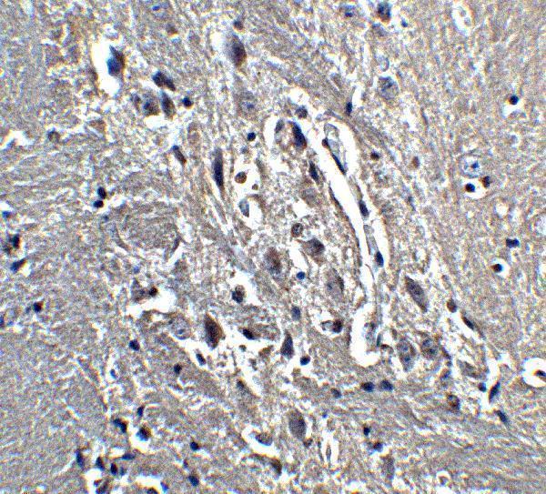
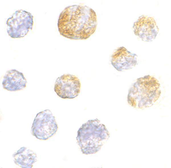

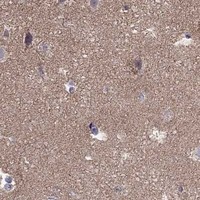


Comprehensive Human Protein Atlas IHC scoring per tissue (reliability: Enhanced — Medium consistency between antibody staining and RNA expression data. Presumed off target binding observed and disregarded.). Rows are taken directly from the HPA tissue chart — click any row's HPA link to view the source.
| Tissue | Cell type | Level | Evidence | Source |
|---|---|---|---|---|
| Cerebral cortex | Neuronal cells | Medium | Protein (IHC) | HPA → |
| Duodenum | Enterocytes | Medium | Protein (IHC) | HPA → |
| Hippocampus | Neuronal cells | Medium | Protein (IHC) | HPA → |
| Tissue | Cell type | Level | Evidence | Source |
|---|---|---|---|---|
| Adipose tissue | Adipocytes | Not detected | Protein (IHC) | HPA → |
| Appendix | Germinal center cells | Not detected | Protein (IHC) | HPA → |
| Bone marrow | Hematopoietic cells | Not detected | Protein (IHC) | HPA → |
| Breast | Adipocytes | Not detected | Protein (IHC) | HPA → |
| Bronchus | Respiratory epithelial cells | Not detected | Protein (IHC) | HPA → |
Troubleshoot KIF5A chromogenic IHC in paraffin sections using neuronal staining patterns, controlled processing changes, and compartment aware scoring.
The catalog antibody has a mouse brain IHC image and K562 ICC and IF images (catalog image captions). Human, mouse, and rat reactivity is listed; KIF5A isoform specificity is unreported (catalog: reactivity and title).
A03635 will render with its mouse brain IHC image at 5 μg/mL (A03635 IHC image caption). Its ICC and IF images show K562 cells, and an IF image shows mouse brain tissue (A03635 IF image captions).
Which to pick: Choose A03635 for tissue IHC because its own image shows staining in mouse brain; section processing and fixative are unreported (A03635 IHC image caption). Choose the same SKU for IF/ICC based on its K562 and mouse brain images (A03635 IF image captions). It is a rabbit antibody listed for human, mouse, and rat, but the captions do not demonstrate IHC in all three species or establish KIF5A isoform specificity (catalog: host, reactivity, title; A03635 image captions).