LAMC1 / Laminin subunit gamma-1 · IHC design guide

Design Immunohistochemistry for LAMC1

Plan paraffin-section LAMC1 IHC around basement membrane and smooth muscle staining (HPA tissue IHC). The catalog antibody PA1581 has a stated IHC dilution of 0.5–1 μg/ml (datasheet).

Evidence assembled Oct 2026 · For research use; verify linked source records and product datasheet before use
Immunohistochemistry protocol sheet for LAMC1 (IHC for LAMC1): expected localisation Basement membrane and extracellular matrix (UniProt), antibody PA1581, validated IHC image, and IHC protocol steps
Printable LAMC1 IHC protocol sheet — expected localisation Basement membrane and extracellular matrix (UniProt), antibody PA1581, controls and protocol steps. Open the full LAMC1 IHC guide →

LAMC1 Immunohistochemistry Experimental Design Guide

Expected localisation, validated protocols, controls and antibodies — the at-a-glance facts below, then the full design guide.

Must know before staining
Expected localisation Basement membrane and extracellular matrix (UniProt)
Staining pattern Basement membrane and smooth muscle staining (HPA tissue IHC)
Antigen retrieval Citrate pH 6.0 HIER, 95–98 °C, 20 min (rule: cytoplasmic / membrane antigen)
Positive control ⓘ Placenta+4 more · see all
Negative control ⓘ Adrenal gland+4 more · see all
Important caveats
Reasons your staining may differ from the expected pattern.
Fixation Keep fixation consistent across sections. (standard IHC practice; not target-specific)
Caveat Secreted LAMC1 may stain away from RNA-positive cells (HPA tissue IHC)
Regulation No expression regulator specified (UniProt)
Isoform / epitope No listed isoforms; signal peptide 1–33 is removed (UniProt)
Section 1

Recommended LAMC1 IHC & IF Protocols

The catalog antibody protocol (datasheet) is accompanied by a published IHC protocol for endometrial specimens (PMC7044014 methods).

Recommended immunohistochemistry (IHC-P) protocol parameters
SampleTissue sections; selected-image fixative not specified (standard IHC workflow)
FixationImage fixative and duration unreported (datasheet PA1581); verify before use.
Sectioning4–5 µm sections on charged slides (standard)
DeparaffinisationXylene, graded ethanol series to water (standard)
Antigen retrievalHeat-induced epitope retrieval in citrate buffer, pH 6.0, 20 min at 95–98 °C (standard rule: cytoplasmic / membrane antigen)
Peroxidase block3% H2O2, 10 min, room temperature (standard)
Blocking10% normal serum of the secondary host, 30 min, room temperature (standard)
Primary antibodyRabbit anti-LAMC1, 0.5-1μg/ml (datasheet PA1581)
Primary incubationOvernight at 4 °C (standard)
DetectionHRP-polymer secondary, DAB chromogen 5–10 min (standard)
CounterstainHematoxylin, blue, dehydrate and mount (standard)
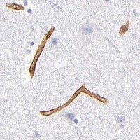
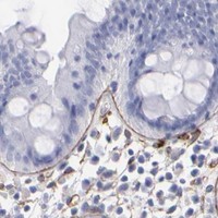
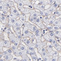
Expected resultLAMC1-positive staining in cytotrophoblasts of placenta (HPA tissue IHC: High). HPA tissue profile: Expression in basement membranes and smooth muscle. No signal in the no-primary control.
💡Decision noteStart with citrate pH 6.0 retrieval at 95–98 °C for 20 min (page retrieval); the published protocol used 95 °C for 20 min (PMC7044014 methods).
Section 2

What Is the Expected LAMC1 Staining Pattern?

LAMC1 is a secreted laminin subunit concentrated in extracellular matrix and basement membranes, with no transmembrane segment (UniProt P11047). In paraffin-section IHC, expect staining that outlines basement membranes and may accompany smooth muscle; HPA reports high staining in placental cytotrophoblasts and medium staining in several other cell populations (HPA: tissue IHC). HPA rates the tissue pattern Enhanced, while noting that secreted protein can complicate agreement between cellular staining and RNA expression (HPA: reliability description).

What am I looking at on my slide?
Continuous staining outlines tissue structures, with strong signal around placental cytotrophoblasts.This fits a basement-membrane protein and the reported high placental cytotrophoblast staining (UniProt P11047; HPA: High in cytotrophoblasts). Assess the outline and surrounding matrix as well as the cells named in HPA's scoring table; a secreted protein need not remain where its RNA was made (HPA: reliability description).
Most signal is sharply nuclear, with little basement-membrane staining.A predominantly nuclear IHC pattern conflicts with the reported extracellular and basement-membrane location (UniProt P11047). Treat it as suspect until a no-primary control and an independently validated antibody support it; HPA's IHC validation does not establish nuclear localisation (HPA: antibody IHC Enhanced).
Strong staining appears in cells scored as not detected, such as bone-marrow hematopoietic cells.HPA reports no detected staining in that specific cell population, so strong cellular signal merits investigation (HPA: Not detected in hematopoietic cells). Compare nearby basement membranes before assigning it to the cells; cross-reactivity or endogenous detection activity are possible general IHC causes.
Brown signal coats most of the section without a discernible structural outline.Diffuse staining makes the expected basement-membrane pattern difficult to judge (UniProt P11047; HPA: basement membranes and smooth muscle). General IHC causes include nonspecific antibody binding, insufficient washing, or excess chromogen development; a no-primary control helps separate detection background from primary-antibody staining.
A placental section has no discernible LAMC1 signal.Absence in a reported high-staining population raises a technical concern, although an individual section and protocol need their own validation (HPA: High in cytotrophoblasts). Check that the section contains the relevant cells, then review antibody performance and detection controls before interpreting a biological loss.
💡Expected LAMC1 appearanceCall the IHC result positive when basement-membrane or associated smooth-muscle staining is discernible, with strong placental cytotrophoblast-associated signal in an appropriate control (UniProt P11047; HPA: basement membranes and smooth muscle; HPA: High in cytotrophoblasts); isolated strong nuclear staining is suspect (UniProt P11047).
How each factor affects the staining
Extracellular topologyLAMC1 is secreted and lacks a transmembrane segment (UniProt P11047). Read a linear extracellular outline as biologically plausible; do not require uniform staining throughout a cell to call the section positive.
Tissue and cell contextHPA records high staining in placental cytotrophoblasts and medium staining in kidney glomerular cells and smooth-muscle cells (HPA: tissue IHC). Its not-detected entries describe named cell populations, not every structure in those tissues (HPA: tissue IHC).
Validation and interpretationFour listed antibodies have Enhanced IHC status (HPA: antibody validation). HPA also notes medium consistency between staining and RNA data because secreted variants can appear away from their site of production (HPA: reliability description). Judge the slide by its structures and controls.
Processing and modificationsUniProt lists a signal peptide at residues 1–33 and a mature chain at 34–1609, plus 14 glycosylation sites (UniProt P11047). Epitope location may matter when comparing antibodies, but these annotations alone do not predict antigen-retrieval needs or fixation sensitivity.
IF/ICC Q&A: Should intracellular fluorescence match IHC?HPA lists plasma-membrane and endoplasmic-reticulum ICC/IF locations as uncertain (HPA: subcellular). Interpret those images cautiously alongside the established secreted, basement-membrane location; ICC/IF localisation does not replace the expected paraffin-section IHC pattern (UniProt P11047).
Why is my staining missing, weak or wrong?
SituationLikely causeNext action
No signal in a placental positive controlThe expected high-staining cytotrophoblast population may be absent from the sampled area, or the IHC detection run may have failed (HPA: High in cytotrophoblasts).Confirm the cells on the counterstained section; check the catalog antibody's IHC-P instructions, detection reagents, and a known-working control. Treat retrieval adjustments as general assay optimisation, not a documented LAMC1 fixation effect.
Weak outlines despite visible tissue architectureThe antibody or chromogenic detection conditions may give inadequate contrast; HPA reports a basement-membrane pattern but supplies no target-specific fixation sensitivity (HPA: tissue IHC).Compare with a reported higher-staining area, then review the antibody's stated IHC-P dilution and detection workflow. Optimise general IHC conditions against a positive control without inferring a specific LAMC1 retrieval requirement.
Strong nuclear staining dominatesNuclear localisation is inconsistent with secreted, extracellular LAMC1 (UniProt P11047); nonspecific staining or detection background is possible.Inspect a no-primary control, verify that the antibody was validated for IHC, and compare the same structures with another validated antibody if available (HPA: antibody IHC Enhanced).
Strong signal in a cell population scored not detectedThe signal may belong to adjacent matrix, or arise from cross-reactivity or endogenous detection activity; HPA's negative calls are cell-specific (HPA: tissue IHC).Review the cell boundaries and surrounding basement membrane, then run no-primary and appropriate detection controls before scoring those cells as positive.
Uniform brown haze obscures structural stainingNonspecific binding, incomplete washing, or excessive chromogen development are general IHC possibilities; the expected pattern is structural (HPA: basement membranes and smooth muscle).Compare the no-primary section, inspect blank spaces and section edges, then adjust general blocking, washing, or chromogen development as the control result indicates.
A negative comparator appears wholly positiveA not-detected HPA entry applies to its named cells; extracellular basement membrane may still be present in the tissue (HPA: tissue IHC; UniProt P11047).Score the named cells separately from extracellular outlines. If the cells themselves stain, investigate detection background and antibody specificity before changing the interpretation.

Sample controls for LAMC1 IHC & IF

🧪Run placenta first and look for staining associated with cytotrophoblasts (High; HPA: placental cytotrophoblasts), then use bone marrow hematopoietic cells as the negative tissue (Not detected; HPA: bone marrow hematopoietic cells). On the placental slide, use cells outside the scored cytotrophoblast compartment to assess background, while allowing for extracellular basement-membrane staining rather than requiring every cell to stain (HPA: placental cytotrophoblasts; UniProt P11047 localization).
Positive control tissue: Placenta (Cytotrophoblasts, HPA High)
Negative control tissue: Adrenal gland (HPA Not detected)
ICC-IF cell lines (HPA subcellular resource): HPA ICC-IF images show LAMC1 in A-431, U-251MG, U2OS, ASC52telo, with annotated localisation: Endoplasmic reticulum (uncertain), Plasma membrane (uncertain) (HPA subcellular).
Technical controls: Include a no-primary secondary-only control, an immunoglobulin control matched to the primary antibody’s host species and monoclonal or polyclonal format, and a knockout specimen or validated immunizing-peptide block as a biological specificity control (standard IHC practice). For placental chromogenic IHC, quench endogenous peroxidase and block endogenous biotin if using avidin–biotin detection (standard IHC practice).
⚠️Feasibility: A target-specific fixation window and fixation effect are unreported in the supplied evidence; the selected PA1581 IHC(P) caption reports no fixative (PA1581 caption). Retrieval dependence is also unreported, so compare retrieved and unretrieved paraffin sections with the same controls; the supplied evidence does not establish that frozen sections or IF are easier for LAMC1 (standard IHC practice; supplied evidence). In placenta, blood-associated endogenous peroxidase can create chromogenic background, and extracellular basement-membrane deposits can complicate assignment of signal to individual cells (standard IHC practice; UniProt P11047 localization).

HPA tissue IHC evidence for LAMC1

Comprehensive Human Protein Atlas IHC scoring per tissue (reliability: Enhanced — Medium consistency between antibody staining and RNA expression data. At least one protein variant secreted, tissue location of RNA and protein might differ and correlation is complex. Antibody staining in cells/structures not annotated, view images.). Rows are taken directly from the HPA tissue chart — click any row's HPA link to view the source.

Positive expression · recommended positive controls

TissueCell typeLevelEvidenceSource
Placenta Cytotrophoblasts High Protein (IHC) HPA →
Breast Adipocytes Medium Protein (IHC) HPA →
Heart muscle Cardiomyocytes Medium Protein (IHC) HPA →
Kidney Cells in glomeruli Medium Protein (IHC) HPA →
Lung Alveolar cells Medium Protein (IHC) HPA →

Undetected expression · recommended negative controls

TissueCell typeLevelEvidenceSource
Adrenal gland Glandular cells Not detected Protein (IHC) HPA →
Bone marrow Hematopoietic cells Not detected Protein (IHC) HPA →
Cerebellum Cells in granular layer Not detected Protein (IHC) HPA →
Duodenum Glandular cells Not detected Protein (IHC) HPA →
Epididymis Glandular cells Not detected Protein (IHC) HPA →
Section 3

Advanced LAMC1 IHC Tips

Troubleshoot LAMC1 staining by checking basement membrane localisation, processing conditions and controls before comparing signal across sections.

How should I adjust retrieval when basement membrane staining is weak?
Start with citrate buffer at pH 6.0, heated to 95–98 °C for 20 min (page retrieval rule). Allow sections to cool in buffer, then compare retrieved and untreated serial sections using the same chromogenic detection conditions (standard IHC practice). LAMC1 is a secreted basement membrane component with multiple glycosylation sites, so judge retrieval by continuous extracellular outlines rather than a general increase in cellular colour (UniProt P11047 localisation and glycosylation). If signal remains weak, optimise retrieval time in small increments while checking morphology and background; treat any alternative buffer as a fallback requiring its own validation (standard IHC practice).
Could fixation explain faint LAMC1 staining in paraffin sections?
Target-specific sensitivity to fixation is unknown from the supplied evidence, and the PA1581 tissue-IHC caption does not state a fixative (PA1581 tissue-IHC caption). Record the actual fixative and fixation duration, then compare sections processed identically except for the suspected variable before changing retrieval or antibody conditions (standard IHC practice). Include a known staining control on each run and assess preservation of tissue structure alongside chromogenic signal (standard IHC practice). A 20 min citrate retrieval at pH 6.0 is the page starting condition, but improved staining after retrieval alone does not establish that fixation caused the original weakness (page retrieval rule; standard IHC practice).
What pattern should count as convincing LAMC1 localisation?
Prioritise a continuous or regionally patterned extracellular basement membrane signal, because LAMC1 is secreted and is a major basement membrane component (UniProt P11047 localisation and tissue specificity). HPA also describes expression in basement membranes and smooth muscle, while its tissue IHC reports High staining in placental cytotrophoblasts and Medium staining in kidney glomerular cells (HPA tissue IHC). Compare the chromogenic signal with tissue architecture on the counterstained section and check whether putative cell staining tracks a basement membrane boundary (standard IHC practice). Diffuse nuclear colour or uniform luminal precipitate should prompt review of controls and detection conditions before being called LAMC1 (UniProt P11047 localisation; standard IHC practice).
How do I assess whether the antibody recognises the intended LAMC1 epitope?
The supplied UniProt record lists 0 isoforms and identifies the mature LAMC1 chain as residues 34–1609 after a signal peptide at residues 1–33 (UniProt P11047 processing and isoforms). Check the catalog antibody’s stated immunogen region against that sequence before attributing a staining difference to an isoform (standard IHC practice). LAMC1 has repeated laminin domains and 14 listed glycosylation sites, so epitope accessibility may need empirical assessment across processing conditions (UniProt P11047 domains and glycosylation; standard IHC practice). Compare serial sections, a suitable tissue control and an omission control; morphology and control behaviour cannot by themselves map the bound epitope (standard IHC practice).
How should I corroborate chromogenic LAMC1 staining with multiplex IF?
Use IF as a separate corroboration experiment and multiplex LAMC1 with a marker identifying the expected cell type or adjacent structure, then compare its signal with the chromogenic basement membrane pattern (UniProt P11047 localisation; standard IF practice). Choose a fluorophore in a channel with low tissue autofluorescence and include unstained and single-colour controls before interpreting overlap (standard IF practice). For an accessible extracellular epitope, begin without permeabilisation; if testing intracellular LAMC1, validate gentle permeabilisation separately because the catalog epitope’s accessibility is not supplied (UniProt P11047 topology; standard IF practice). HPA’s uncertain endoplasmic reticulum and plasma membrane IF assignments should not override the extracellular tissue interpretation without additional controls (HPA subcellular; UniProt P11047 localisation).
What should I check when DAB obscures basement membrane boundaries?
Run a primary antibody omission control to locate signal produced by the detection system, and check an endogenous peroxidase block before DAB development (standard chromogenic IHC practice). If colour persists, compare sections with and without the block, then shorten DAB development in controlled steps while holding section thickness and counterstain constant (standard IHC practice). LAMC1 should be assessed against extracellular basement membrane architecture; widespread granular cellular colour is insufficient evidence of specific staining (UniProt P11047 localisation; standard IHC practice). Review tissue folds, section edges and necrotic regions under the microscope, and score only intact, interpretable areas alongside a concurrent positive control (standard IHC practice).
How should I quantify patchy LAMC1 IHC staining? ⚠ ANSWER MARKED FOR VERIFICATION
Define anatomical regions before scoring and measure the fraction of basement membrane length staining positive or chromogenic signal density per mm² of evaluable tissue (UniProt P11047 localisation; standard IHC quantification practice). Normalise boundary signal to total evaluable basement membrane length, or area density to intact tissue area, so differences in tissue composition do not masquerade as expression changes (standard IHC quantification practice). An H-score based on percentage of positive cells may be reported for cell-associated staining, but it does not capture deposited extracellular LAMC1 reliably (UniProt P11047 localisation; standard IHC quantification practice). Keep threshold, illumination and exclusion rules fixed, and review at least 3 representative fields per section before comparing groups (standard IHC quantification practice).
How can I distinguish true LAMC1 signal from staining artefact?
A persuasive positive follows basement membrane architecture or a documented tissue pattern, consistent with LAMC1’s extracellular localisation and HPA’s basement membrane and smooth muscle profile (UniProt P11047 localisation; HPA tissue IHC). Treat isolated nuclear staining, necrotic deposits, strong section-edge colour and signal in primary antibody omission controls as reasons to re-examine specificity or processing (UniProt P11047 localisation; standard IHC practice). Endogenous enzyme activity can imitate DAB staining, so compare the peroxidase-blocked slide with its controls before calling a positive (standard chromogenic IHC practice). HPA reports Enhanced tissue-IHC reliability but only medium consistency with RNA; interpret discordant cellular patterns cautiously because secreted protein and local RNA need not coincide (HPA tissue IHC).
Boster reagents

Best LAMC1 / Laminin subunit gamma-1 IHC Antibodies

PA1581 has real IHC(P) data from human intestinal cancer tissue (PA1581 image caption). Human, mouse, and rat are listed as reactive species (catalog: reactivity); no IF data is supplied (catalog: IF image alts).

Real IHC data Anti-Laminin gamma 1 antibody, PA1581, IHC(P) IHC(P): Human Intestinal Cancer Tissue
Anti-Laminin gamma 1/LAMC1 Antibody ®
Cat # PA1581

PA1581 is listed for IHC and WB, with human, mouse, and rat reactivity (catalog: applications; catalog: reactivity). Its IHC(P) image shows human intestinal cancer tissue (PA1581 image caption).

Which to pick: Choose PA1581 for paraffin-section tissue IHC: its own image documents IHC(P) on human intestinal cancer tissue, and its listed IHC dilution is 0.5–1 μg/mL (PA1581 image caption; datasheet: IHC dilution). No IF/ICC SKU or IF figure is supplied (catalog: applications; catalog: IF image alts). For mouse or rat samples, PA1581 lists reactivity, but its supplied IHC image shows human tissue only; the fixative is unreported (catalog: reactivity; PA1581 image caption).

Each figure is that product's own IHC / IF validation image from its datasheet.

References

  1. UniProt Consortium. UniProt entry P11047 (LAMC1_HUMAN, Laminin subunit gamma-1).
  2. Human Protein Atlas. LAMC1 tissue IHC expression (reliability: Enhanced).
  3. Human Protein Atlas. LAMC1 subcellular location (ICC-IF): Localized to the plasma membrane and endoplasmic reticulum..
  4. Human Protein Atlas. LAMC1 antibody validation summary (4 antibodies).
  5. Radiogenomic Analysis of Papillary Thyroid Carcinoma for Prediction of Cervical Lymph Node Metastasis: A Preliminary Study. Frontiers in oncology 2021 — PMC8276635.
  6. LAMC1-mediated preadipocytes differentiation promoted peritoneum pre-metastatic niche formation and gastric cancer metastasis. International journal of biological sciences 2022 — PMC9066104.
  7. LAMC1 is a prognostic factor and a potential therapeutic target in endometrial cancer. Journal of gynecologic oncology 2020 — PMC7044014.
  8. Ginsenoside Rg1 Regulates the Activation of Astrocytes Through lncRNA-Malat1/miR-124-3p/Lamc1 Axis Driving PI3K/AKT Signaling Pathway, Promoting the Repair of Spinal Cord Injury. CNS neuroscience & therapeutics 2024 — PMC11532020.
  9. PubMed PMID:3360804 — UniProt-cited evidence.
  10. PubMed PMID:1985895 — UniProt-cited evidence.
  11. PubMed PMID:16710414 — UniProt-cited evidence.