This website uses cookies to ensure you get the best experience on our website.


- Table of Contents
Plan LRP1 IHC in paraffin sections using the cytoplasmic tissue pattern reported in liver, brain, fibroblasts and stroma (HPA tissue IHC). Consider whether the antibody targets the extracellular or cytoplasmic region when interpreting staining, because LRP1 is cleaved into distinct chains (UniProt).

Expected localisation, validated protocols, controls and antibodies — the at-a-glance facts below, then the full design guide.
| Expected localisation | Cytoplasmic tissue staining (HPA tissue IHC); membrane localisation expected (UniProt) | |
| Staining pattern | Cytoplasmic in liver, brain, fibroblasts and stroma (HPA tissue IHC) | |
| Antigen retrieval | Tris-EDTA pH 9.0 HIER, 95–98 °C, 20 min (rule: nuclear antigen) | |
| Positive control | Lung+4 more · see all | |
| Negative control | Adrenal gland+4 more · see all |
| Fixation | Matched tissue-IHC evidence does not establish the fixation claim. Validate the specimen-specific method before use. (selected-SKU IHC image M00829) | |
| Caveat | Cleavage can separate extracellular and cytoplasmic epitopes (UniProt) | |
| Regulation | No expression regulator specified (UniProt) | |
| Isoform / epitope | 2 isoforms; epitope location across cleaved chains matters (UniProt) |
The catalog antibody’s paraffin-section protocol (datasheet: IHC-P) is accompanied by a published chromogenic IHC protocol for rat corneal frozen sections (PMC5105079).
| Sample | Paraffin-embedded human liver carcinoma tissue; fixative not specified (datasheet M00829) |
| Fixation | Image fixative and duration unreported (datasheet M00829); verify before use. |
| Sectioning | 4–5 µm sections on charged slides (standard) |
| Deparaffinisation | Xylene, graded ethanol series to water (standard) |
| Antigen retrieval | Heat-induced epitope retrieval in Tris-EDTA buffer, pH 9.0, 20 min at 95–98 °C (standard rule: nuclear antigen) |
| Peroxidase block | 3% H2O2, 10 min, room temperature (standard) |
| Blocking | 10% normal serum of the secondary host, 30 min, room temperature (standard) |
| Primary antibody | Rabbit monoclonal (clone BDC-12) anti-LRP1, 1:50 (datasheet M00829) |
| Primary incubation | Overnight at 4 °C (standard) |
| Detection | HRP-polymer secondary, DAB chromogen 5–10 min (standard) |
| Counterstain | Hematoxylin, blue, dehydrate and mount (standard) |
| Expected result | LRP1-positive staining in macrophages of lung (HPA tissue IHC: High). HPA tissue profile: Cytoplasmic expression mainly in liver, placenta, brain, adipose tissue and in fibroblasts. Also expressed in the tissue stroma. No signal in the no-primary control. |
LRP1 is a membrane receptor with an extracellular region and a short cytoplasmic tail (UniProt Q07954 topology). In tissue IHC, expect mainly cytoplasmic staining in liver, brain, adipose tissue, fibroblasts and stroma (HPA tissue IHC). Lung macrophages show High staining; hepatocytes, neuronal cells, adipocytes and some stromal cells show Medium staining (HPA tissue IHC). HPA rates the tissue pattern Supported, with medium consistency between staining and RNA expression (HPA tissue IHC).
| Clear staining in lung macrophages, with weaker staining in nearby cell types. | This fits the reported High macrophage signal (HPA tissue IHC). Assess the identified cells, rather than calling the whole lung uniformly positive; the HPA result describes a cell-specific pattern. |
| Predominantly cytoplasmic staining in hepatocytes, neuronal cells, adipocytes or stroma. | This agrees with the HPA tissue profile; these listed populations include Medium staining examples (HPA tissue IHC). Membrane-associated signal can also fit LRP1 topology (UniProt Q07954 topology). Interpret intensity within each cell population and tissue. |
| Nuclear-only staining dominates a paraffin section, without the expected cellular pattern. | Treat this as unconfirmed for tissue IHC. UniProt reports that the cleaved intracellular domain can reach the nucleus (UniProt Q07954 processing), and HPA reports nucleoli in ICC-IF (HPA subcellular). Neither establishes nuclear-only staining as the expected tissue IHC pattern. |
| Strong signal appears in cells listed as undetected, such as adrenal glandular or esophageal squamous cells. | This conflicts with the reported observations for those specific cells (HPA tissue IHC). Consider antibody cross-reactivity or endogenous detection activity, then compare cell identity, controls and the expected positive population before assigning LRP1 positivity. |
| Diffuse color covers cells and surrounding areas, or a known-positive population has no signal. | Diffuse color obscures the cellular distribution needed for interpretation (general IHC practice). An absent signal in lung macrophages conflicts with their High HPA staining (HPA tissue IHC); review the staining run and tissue quality before concluding that LRP1 is absent. |
| How much should intensity vary by cell type? | HPA reports High lung macrophages, Medium hepatocytes, neuronal cells, adipocytes and selected stromal cells, plus undetected cells in specified tissues (HPA tissue IHC). Score the named population, not every cell on the section. |
| Where is the antibody epitope relative to processing? | LRP1 has a large extracellular region, one transmembrane segment and a cytoplasmic tail (UniProt Q07954 topology). Its extracellular and membrane-bound subunits arise through processing (UniProt Q07954 processing). An unspecified epitope cannot justify assigning a particular staining compartment to one chain. |
| What does the available antibody validation establish? | Three listed antibodies have Supported IHC status (HPA antibodies). That status supports use of their observed tissue patterns, while the overall tissue reliability remains Supported with medium staining–RNA consistency (HPA tissue IHC). It does not make every stained cell specific. |
| What should IF/ICC show? | HPA reports a mainly plasma-membrane location, with additional nucleolar and cytosolic signal in ICC-IF (HPA subcellular). This is a useful localisation comparison, but ICC-IF observations do not set the expected compartment or intensity for paraffin tissue IHC. |
| Can antigen retrieval or fixation explain a discrepancy? | Retrieval conditions can affect staining in paraffin IHC (general IHC practice). The supplied HPA and UniProt evidence reports no LRP1-specific fixation sensitivity or retrieval setting; use the validated IHC conditions as the starting point and interpret changes against controls. |
| Situation | Likely cause | Next action |
|---|---|---|
| Lung macrophages show no staining. | A failed staining run, unsuitable tissue or a missed cell population can mimic absence (general IHC practice); HPA reports High macrophage staining (HPA tissue IHC). | Confirm macrophage identification, inspect tissue preservation and run controls, then check the IHC-validated antibody's specified staining conditions (general IHC practice). |
| Color spreads evenly across the section. | Diffuse background can arise from incomplete blocking, excess antibody or detection background (general IHC practice); it prevents reading the cell-specific HPA pattern (HPA tissue IHC). | Review blocking, wash and detection controls; adjust antibody concentration using its validated IHC guidance (general IHC practice). Reassess identifiable cells after background falls. |
| Adrenal glandular or esophageal squamous cells stain strongly. | Those cells are listed as Not detected (HPA tissue IHC). Cross-reactivity or endogenous detection activity is possible (general IHC practice). | Check the cell identity and detection-only control, then compare with a positive HPA-listed population in the same staining run (HPA tissue IHC; general IHC practice). |
| Staining is exclusively nuclear in tissue IHC. | The intracellular domain can enter the nucleus (UniProt Q07954 processing), but nuclear-only tissue staining is not established by the HPA tissue profile (HPA tissue IHC). | Check the antibody epitope and staining controls; report nuclear localisation as uncertain unless the reagent and an independent assessment support it (general IHC practice). |
| A weak adipocyte or neuronal signal is called negative. | HPA reports Medium staining in adipose adipocytes and neuronal cells in caudate, cortex and hippocampus (HPA tissue IHC); visibility can differ from the High lung macrophage example. | Score the specified cells against matched controls and section background, using the HPA level for that population rather than one universal intensity threshold (HPA tissue IHC; general IHC practice). |
| Membrane-heavy IF/ICC images seem inconsistent with cytoplasmic tissue IHC. | HPA describes mainly plasma-membrane ICC-IF localisation (HPA subcellular) and mainly cytoplasmic tissue IHC expression (HPA tissue IHC); these are different application observations. | Interpret each image against its own application evidence. For paraffin IHC, prioritize the identified tissue cell pattern and verify an unusual compartment with antibody and detection controls (HPA tissue IHC; general IHC practice). |


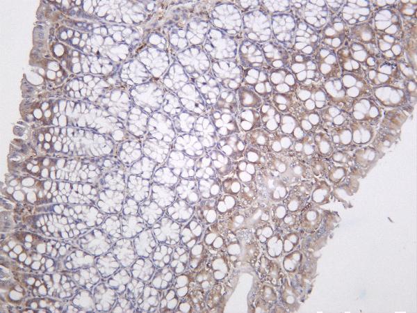
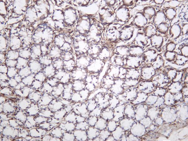
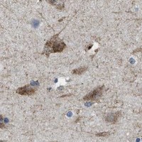
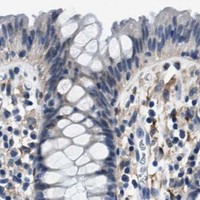
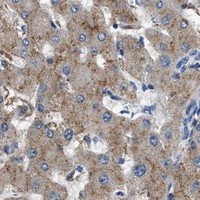
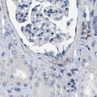


Comprehensive Human Protein Atlas IHC scoring per tissue (reliability: Supported — Medium consistency between antibody staining and RNA expression data.). Rows are taken directly from the HPA tissue chart — click any row's HPA link to view the source.
| Tissue | Cell type | Level | Evidence | Source |
|---|---|---|---|---|
| Lung | Macrophages | High | Protein (IHC) | HPA → |
| Adipose tissue | Adipocytes | Medium | Protein (IHC) | HPA → |
| Caudate | Neuronal cells | Medium | Protein (IHC) | HPA → |
| Cerebral cortex | Neuronal cells | Medium | Protein (IHC) | HPA → |
| Endometrium | Cells in endometrial stroma | Medium | Protein (IHC) | HPA → |
| Tissue | Cell type | Level | Evidence | Source |
|---|---|---|---|---|
| Adrenal gland | Glandular cells | Not detected | Protein (IHC) | HPA → |
| Appendix | Endocrine cells | Not detected | Protein (IHC) | HPA → |
| Bone marrow | Hematopoietic cells | Not detected | Protein (IHC) | HPA → |
| Cervix | Glandular cells | Not detected | Protein (IHC) | HPA → |
| Duodenum | Endocrine cells | Not detected | Protein (IHC) | HPA → |
Troubleshoot LRP1 staining in paraffin sections by checking retrieval, cell compartment, controls and scoring before interpreting changes in signal.
Two anti-LRP1 rabbit monoclonal antibodies cover human, mouse and rat samples (catalog: host, clonality, reactivity). Both have IHC images; M00829 also has an IF image (catalog: image captions).
M00829 lists IHC and IF/ICC applications and shows IHC staining of paraffin-embedded human liver carcinoma (M00829 catalog: applications, IHC caption). M00829-1 lists IHC and IF applications and shows IHC staining of mouse and rat colon and human liver (M00829-1 catalog: applications, IHC captions).
Which to pick: For tissue IHC in paraffin sections, choose M00829 when its human liver carcinoma example fits your sample; the caption does not report the fixative (M00829 IHC caption). For IF/ICC, choose M00829 because it lists both applications and has an IF image (M00829 catalog: applications, IF caption). For IHC across species, M00829-1 has mouse and rat colon and human liver examples, though their processing and fixatives are unreported (M00829-1 IHC captions); both rabbit monoclonals list human, mouse and rat reactivity (catalog: host, clonality, reactivity).