This website uses cookies to ensure you get the best experience on our website.


- Table of Contents
Plan chromogenic IHC for MAGEA4 using testicular spermatogonia as a positive tissue reference (HPA tissue IHC). This guide covers fixation, staining controls and scoring; the supplied tissue evidence does not establish a subcellular pattern (HPA tissue IHC).

Expected localisation, validated protocols, controls and antibodies — the at-a-glance facts below, then the full design guide.
| Expected localisation | Spermatogonia; subcellular pattern unestablished (HPA tissue IHC) | |
| Staining pattern | Staining in testicular spermatogonia; compartment unspecified (HPA tissue IHC) | |
| Antigen retrieval | Citrate pH 6.0 HIER, 95–98 °C, 20 min (rule: cytoplasmic / membrane antigen) | |
| Positive control | Testis | |
| Negative control | Adipose tissue+4 more · see all |
| Fixation | Keep fixation consistent across sections (standard IHC practice; not target-specific); Matched tissue-IHC evidence does not establish the fixation claim. Validate the specimen-specific method before use. (selected-SKU IHC image A07175) | |
| Caveat | Normal spermatogonia can stain positive (HPA tissue IHC) | |
| Regulation | Tumor-associated expression (UniProt) | |
| Isoform / epitope | No annotated isoforms; one 1–317 chain (UniProt) |
The catalog antibody’s IHC-P protocol is accompanied by published MAGEA4 IHC workflows for testis and ovarian cancer samples (PMC6936721; PMC9916968).
| Sample | Tissue sections; selected-image fixative not specified (standard IHC workflow) |
| Fixation | Image fixative and duration unreported (datasheet A07175); verify before use. |
| Sectioning | 4–5 µm sections on charged slides (standard) |
| Deparaffinisation | Xylene, graded ethanol series to water (standard) |
| Antigen retrieval | Heat-induced epitope retrieval in citrate buffer, pH 6.0, 20 min at 95–98 °C (standard rule: cytoplasmic / membrane antigen) |
| Peroxidase block | 3% H2O2, 10 min, room temperature (standard) |
| Blocking | 10% normal serum of the secondary host, 30 min, room temperature (standard) |
| Primary antibody | Rabbit anti-MAGEA4, 5 μg/mL (datasheet A07175) |
| Primary incubation | Overnight at 4 °C (standard) |
| Detection | HRP-polymer secondary, DAB chromogen 5–10 min (standard) |
| Counterstain | Hematoxylin, blue, dehydrate and mount (standard) |
| Expected result | MAGEA4-positive staining in spermatogonia cells of testis (HPA tissue IHC: High). HPA tissue profile: Expression in spermatogonia in seminiferous ducts of testis. No signal in the no-primary control. |
MAGEA4 should stain spermatogonia in testis, where HPA reports high expression with Enhanced tissue IHC reliability (HPA tissue IHC). HPA ICC-IF places MAGEA4 mainly in nuclear speckles, with additional cytosolic localization; that cellular pattern is guidance, not a compartment assignment for paraffin tissue sections (HPA subcellular ICC-IF). UniProt reports no transmembrane segment and does not annotate a subcellular location (UniProt P43358 topology and subcellular record).
| Strong staining in spermatogonia, with nearby cells serving as visual context (HPA tissue IHC). | This matches the HPA testis pattern: spermatogonia are rated high, and tissue IHC reliability is Enhanced (HPA tissue IHC). Assess the named cell population, not just whether the section contains brown signal. |
| Staining appears chiefly at cell borders or in a compartment inconsistent with nuclear speckles or cytosol. | Recheck localization and slide quality before calling it MAGEA4. HPA reports nuclear speckles plus cytosol in ICC-IF, while UniProt annotates no membrane span; neither source establishes an IHC compartment pattern (HPA subcellular ICC-IF; UniProt P43358 topology). |
| Strong staining appears in adipocytes or other cells HPA lists as undetected (HPA tissue IHC). | Treat that pattern as suspect for cross-reactivity or endogenous detection activity, then compare appropriate controls (general IHC practice). An HPA 'not detected' result describes its reported tissue and cell context, not an absolute exclusion across every specimen (HPA tissue IHC). |
| Broad haze or uniform color obscures cell boundaries and the spermatogonial pattern. | Diffuse background prevents a cell-specific call. Review the negative control, blocking, washes and chromogen exposure before scoring (general IHC practice); HPA's Enhanced reliability does not validate an individual noisy run (HPA tissue IHC). |
| No convincing staining is seen in testis spermatogonia. | A negative testis control challenges the run because HPA reports high spermatogonial staining (HPA tissue IHC). Check tissue identity and preservation, antibody preparation, retrieval and detection controls as general IHC practice; the supplied sources do not specify MAGEA4 fixation sensitivity. |
| Tissue and cell context | HPA reports high staining in testis spermatogonia, low staining in placental trophoblastic cells, and no detection in the listed negative cell populations (HPA tissue IHC). These levels support selecting and reading controls by cell type; they do not predict every tumor specimen. |
| IF/ICC Q&A: where should cellular signal appear? | Mainly in nuclear speckles, with additional cytosolic signal (HPA subcellular ICC-IF). This is an ICC-IF observation, so confirm tissue IHC by its spermatogonial pattern rather than demanding visible speckles in a chromogenic section (HPA tissue IHC). |
| Topology and processing | UniProt lists no transmembrane segment, signal peptide or propeptide, and one chain spanning residues 1–317 (UniProt P43358 topology and processing). These annotations give no basis to expect a membrane-restricted or shed-antigen staining pattern. |
| Antibody validation | HPA rates tissue IHC Enhanced for HPA021942 and CAB040535; ICC is Approved for HPA021942, with no ICC status supplied for CAB040535 (HPA antibody records). Apply the validation status to the stated assay, not automatically to another detection method. |
| Situation | Likely cause | Next action |
|---|---|---|
| Testis spermatogonia are negative. | The run may have failed, or the sampled section may lack evaluable spermatogonia; HPA expects high staining in those cells (HPA tissue IHC). | Confirm the cell population on the counterstain and review positive-control tissue, antibody preparation, retrieval and detection steps (general IHC practice). Avoid claiming a MAGEA4-specific fixation effect: none is supplied. |
| Color is present throughout the section, including HPA-undetected cells. | Non-specific antibody binding or endogenous detection activity can produce broad staining (general IHC practice); HPA reports several cell populations as not detected (HPA tissue IHC). | Compare a negative control and inspect blocking, detection chemistry, washes and chromogen exposure (general IHC practice). Score MAGEA4 only where the staining remains cell-specific and controls support it. |
| Only placental trophoblastic cells show faint staining. | HPA rates these cells low, so a faint result there has less separation from background than high-staining testis spermatogonia (HPA tissue IHC). | Compare staining with the negative control and a testis positive control before assigning a positive call (general IHC practice; HPA tissue IHC). Do not use faint placental color alone to establish a failed or successful run. |
| Staining is sharply membrane-restricted. | UniProt lists no transmembrane segment, and HPA ICC-IF reports nuclear speckles plus cytosol (UniProt P43358 topology; HPA subcellular ICC-IF). The supplied tissue IHC record does not settle compartment localization. | Review morphology and negative controls, then seek confirmation with an independently validated IHC antibody if available (general IHC practice; HPA antibody records). Do not score membrane color alone as the expected pattern. |
| An IF/ICC image looks diffuse instead of speckled. | HPA places the main ICC-IF signal in nuclear speckles and an additional signal in cytosol; diffuse fluorescence alone does not reproduce that description (HPA subcellular ICC-IF). | Review nuclear counterstain, optical focus and negative control before interpretation (general IF practice). Use the separate IF/ICC guide for assay setup; this section supplies no IF/ICC protocol. |
| Two slides or antibodies give different tissue patterns. | HPA lists two antibodies with Enhanced IHC validation, but that status does not guarantee identical staining in every specimen or run (HPA antibody records). | Compare the same cell populations and controls, check assay-specific validation, and resolve discordance before scoring (general IHC practice). Preserve the distinction between HPA tissue IHC evidence and ICC-IF localization evidence. |


Comprehensive Human Protein Atlas IHC scoring per tissue (reliability: Enhanced — High consistency between antibody staining and RNA expression data.). Rows are taken directly from the HPA tissue chart — click any row's HPA link to view the source.
| Tissue | Cell type | Level | Evidence | Source |
|---|---|---|---|---|
| Testis | Spermatogonia cells | High | Protein (IHC) | HPA → |
| Tissue | Cell type | Level | Evidence | Source |
|---|---|---|---|---|
| Adipose tissue | Adipocytes | Not detected | Protein (IHC) | HPA → |
| Adrenal gland | Glandular cells | Not detected | Protein (IHC) | HPA → |
| Appendix | Glandular cells | Not detected | Protein (IHC) | HPA → |
| Bone marrow | Hematopoietic cells | Not detected | Protein (IHC) | HPA → |
| Breast | Adipocytes | Not detected | Protein (IHC) | HPA → |
Use matched controls and cell level staining patterns to troubleshoot MAGEA4 chromogenic IHC in paraffin sections.
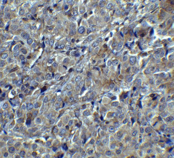
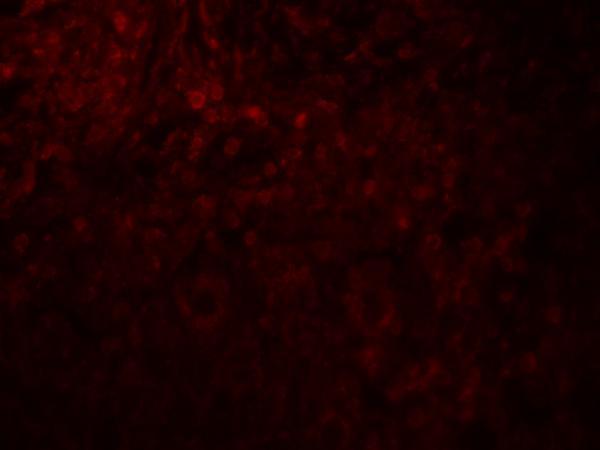
The catalog antibody has IHC and IF data from human breast tumors or tissue and ICC data from HeLa cells (catalog image captions); its listed reactivity is human, mouse and rat (catalog: A07175).
A07175 will render with an IHC image of human breast tumor at 5 μg/mL (A07175 IHC image caption). Its catalog also shows IF in human breast cancer tissue at 20 μg/mL and ICC in HeLa cells at 5 μg/mL (A07175 IF image captions).
Which to pick: Choose A07175 for paraffin-section IHC because IHC-P is listed and its IHC image shows human breast tumor; the image caption does not report the fixative (A07175 application list; A07175 IHC image caption). Choose the same SKU for IF or ICC based on its human breast tissue and HeLa images (A07175 IF image captions). For cross-species work, A07175 lists mouse and rat reactivity, but the supplied IHC and IF images show human samples only (catalog: A07175 reactivity; A07175 image captions).