This website uses cookies to ensure you get the best experience on our website.


- Table of Contents
Plan chromogenic IHC for MDM4 using its mainly nuclear tissue pattern (HPA tissue IHC). Adrenal glandular cells show high staining and duodenal glandular cells show low staining, but staining and RNA have low consistency (HPA tissue IHC).

Expected localisation, validated protocols, controls and antibodies — the at-a-glance facts below, then the full design guide.
| Expected localisation | Tissue: mainly nuclear (HPA tissue IHC); cytoplasm also annotated (UniProt) | |
| Staining pattern | Mainly nuclear staining across tissues (HPA tissue IHC) | |
| Antigen retrieval | EDTA pH 8.0 HIER, heat-mediated (datasheet PB9872) | |
| Positive control | Adrenal gland+4 more · see all | |
| Negative control | None in HPA (detected in all 45 tissues); use no-primary + isotype controls |
| Fixation | Keep fixation consistent across sections. (standard IHC practice; not target-specific) | |
| Caveat | Staining and RNA show low consistency (HPA tissue IHC) | |
| Regulation | Higher expression in thymus (UniProt) | |
| Isoform / epitope | 5 isoforms; epitope coverage unknown; no ectodomain (UniProt) |
The catalog antibody’s IHC-P protocol (datasheet: PB9872) is accompanied by one published MDM4 IHC protocol using tumor xenograft sections (PMC11564821).
| Sample | Paraffin-embedded human lung cancer tissue; fixative not specified (datasheet PB9872) |
| Fixation | Image fixative and duration unreported (datasheet PB9872); verify before use. |
| Sectioning | 4–5 µm sections on charged slides (standard) |
| Deparaffinisation | Xylene, graded ethanol series to water (standard) |
| Antigen retrieval | Heat retrieval: EDTA pH 8.0 (datasheet PB9872); 20 min, 95–100 °C (standard) |
| Peroxidase block | 3% H2O2, 10 min, room temperature (standard) |
| Blocking | 10% goat serum (datasheet PB9872) |
| Primary antibody | Rabbit anti-MDM4, 0.5-1μg/ml (datasheet PB9872) |
| Primary incubation | Overnight at 4 °C (datasheet PB9872) |
| Detection | Streptavidin-biotin complex (SABC), DAB chromogen (datasheet PB9872) |
| Counterstain | Hematoxylin, blue, dehydrate and mount (standard) |
| Expected result | MDM4-positive staining in glandular cells of adrenal gland (HPA tissue IHC: High). HPA tissue profile: Mainly nuclear expression in all tissues. No signal in the no-primary control. |
MDM4 is mainly nuclear across tissues in HPA IHC, with high staining reported in several epithelial, glandular, neuronal and stromal cell groups (HPA tissue IHC). UniProt also lists cytoplasmic localisation and no transmembrane segment (UniProt O15151). HPA rates the tissue IHC pattern Approved but reports low consistency with RNA expression and a splice or transcript discrepancy; interpret intensity with that caution (HPA tissue IHC).
| Predominant nuclear chromogen in adrenal glandular cells or cortical neurons. | This fits HPA's mainly nuclear profile and its High staining calls for those cells (HPA tissue IHC). Score the stained nuclei and their cell identity together; a positive field alone does not establish antibody specificity, given HPA's RNA and staining consistency warning (HPA tissue IHC). |
| Predominantly membranous, extracellular or uniformly cytoplasmic staining with little nuclear signal. | Treat this as a discordant IHC pattern: HPA reports mainly nuclear tissue staining, while UniProt lists nucleus and cytoplasm and no transmembrane segment (HPA tissue IHC; UniProt O15151). Cytoplasmic signal is possible, but a dominant nonnuclear pattern needs scrutiny before scoring it as specific. |
| Strong staining concentrated in an unexpected cell population while expected nuclei remain weak. | Compare cell identities within the same section. HPA reports Low staining in duodenal glandular, urothelial, smooth muscle and marrow hematopoietic cells, but does not call them negative (HPA tissue IHC). A strong reversal raises possible cross-reactivity or endogenous detection activity; morphology alone cannot distinguish those causes. |
| Diffuse chromogen over cells and extracellular spaces, with little nuclear contrast. | This obscures the mainly nuclear pattern expected from HPA tissue IHC (HPA tissue IHC). In general IHC practice, broad background can arise from nonspecific antibody binding, incomplete blocking or detection chemistry. Do not convert diffuse colour into a positive MDM4 score without a cell-resolved nuclear pattern. |
| No nuclear signal in an adrenal gland or bronchus section chosen as a positive comparator. | HPA reports High staining in adrenal glandular and bronchial respiratory epithelial cells (HPA tissue IHC). Check whether those cells are present and whether a parallel control stained. A blank result may reflect the assay or section; HPA's Approved rating and consistency warning do not guarantee every specimen will stain. |
| Tissue and cell selection | HPA reports High staining in adrenal glandular, bronchial respiratory epithelial, cortical neuronal and endometrial stromal cells; several other listed cell groups are Low (HPA tissue IHC). Choose comparators by cell type and nuclear pattern, and avoid treating a Low call as an absent-protein control. |
| Evidence strength | The tissue IHC profile is Approved, with low consistency between antibody staining and RNA expression and a splice or transcript discrepancy (HPA tissue IHC). The listed IHC antibody HPA018919 is Approved; HPA does not list it as Enhanced (HPA antibodies). Interpret unexpected intensity conservatively. |
| Localisation and topology | HPA describes mainly nuclear tissue staining; UniProt lists nuclear and cytoplasmic localisation and no transmembrane segment (HPA tissue IHC; UniProt O15151). A small cytoplasmic component need not invalidate nuclear staining, while a membrane-only pattern lacks support from these records. |
| Isoforms and processing | UniProt lists five MDM4 isoforms, no signal peptide or propeptide, and a 1–490 protein chain (UniProt O15151). These facts support caution about transcript-dependent comparisons; the payload gives no antibody epitope map or evidence that a particular isoform changes IHC staining. |
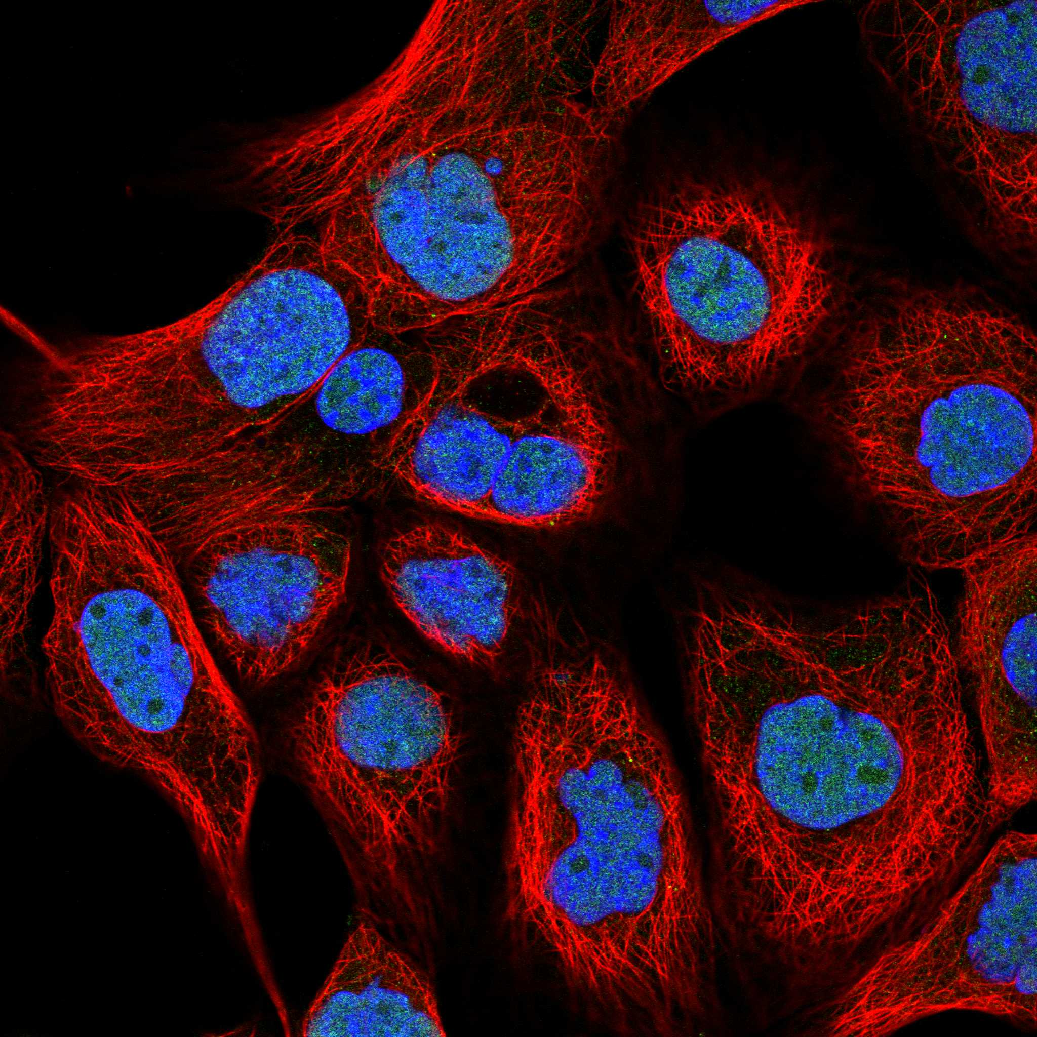
| IF comparison | HPA ICC-IF supports nucleoplasmic localisation, while its tissue IHC profile says mainly nuclear (HPA subcellular; HPA tissue IHC). Use the IF finding as a compartment check only; the supplied records do not establish that IF intensity predicts paraffin-section chromogenic intensity. |
| Situation | Likely cause | Next action |
|---|---|---|
| Expected High cell group is blank. | The expected cells may be absent, or the staining run may have failed; HPA reports High staining for specific cells within listed tissues, not every cell in each section (HPA tissue IHC). | Verify cell identity on the counterstain and inspect a concurrently stained control. Then review the assay's antigen retrieval, primary antibody and detection steps as general IHC checks; no MDM4-specific retrieval or fixation sensitivity is supplied. |
| Only a membrane outline is strong. | That pattern conflicts with mainly nuclear HPA tissue staining and UniProt's lack of a transmembrane segment (HPA tissue IHC; UniProt O15151). | Check localisation at higher magnification against the counterstain, then compare a control section. Treat membrane-only colour as unresolved staining rather than assigning it to MDM4. |
| Low-call cells stain more strongly than the chosen High-call cells. | HPA's Low and High calls are observations with a stated RNA and staining consistency caveat (HPA tissue IHC). Cell misidentification, nonspecific binding or endogenous detection activity are possible general IHC explanations. | Recheck both cell populations and the nuclear pattern; examine appropriate primary-omission and detection controls. Report the discrepancy instead of treating the HPA categories as absolute positive and negative thresholds. |
| Diffuse background hides nuclei. | General IHC causes include nonspecific binding, inadequate blocking or excess detection signal; the supplied HPA and UniProt records do not identify an MDM4-specific background mechanism. | Inspect primary-omission and detection controls, then review blocking, antibody concentration, washes and chromogen development under the assay's established conditions. Score MDM4 only where discrete nuclei remain interpretable. |
| Nuclear staining is present, but intensity differs across specimens. | HPA reports broadly nuclear expression with cell-specific intensity calls and warns of low RNA and staining consistency plus a splice or transcript discrepancy (HPA tissue IHC). | Compare the same cell type and compartment across sections and record intensity separately from localisation. Avoid inferring isoform identity or transcript abundance from chromogen intensity alone. |
| ICC-IF looks nucleoplasmic, but paraffin IHC is weak. | HPA supports nucleoplasmic ICC-IF localisation and reports mainly nuclear tissue IHC, but these records do not establish equivalent signal strength across assays (HPA subcellular; HPA tissue IHC). | Assess each assay with its own controls. For paraffin IHC, review the established retrieval and detection workflow as general practice; do not infer an MDM4-specific fixation effect from the ICC-IF result. |


Comprehensive Human Protein Atlas IHC scoring per tissue (reliability: Approved — Low consistency between antibody staining and RNA expression data. Caution, Splice and/or transcript discrepancy exists.). Rows are taken directly from the HPA tissue chart — click any row's HPA link to view the source.
| Tissue | Cell type | Level | Evidence | Source |
|---|---|---|---|---|
| Adrenal gland | Glandular cells | High | Protein (IHC) | HPA → |
| Bronchus | Respiratory epithelial cells | High | Protein (IHC) | HPA → |
| Cerebellum | Cells in molecular layer | High | Protein (IHC) | HPA → |
| Cerebral cortex | Neuronal cells | High | Protein (IHC) | HPA → |
| Cervix | Glandular cells | High | Protein (IHC) | HPA → |
| Tissue | Cell type | Level | Evidence | Source |
|---|---|---|---|---|
| None in HPA: MDM4 is detected in all 45 scored tissues. Use a no-primary (secondary-only) and an isotype control instead. | ||||
Troubleshoot MDM4 staining in paraffin sections by checking retrieval, nuclear localisation, antibody epitope coverage and controls before comparing signal across samples.
PB9872 has real IHC data from human lung cancer paraffin sections (PB9872 image caption); the catalog lists human and mouse reactivity (catalog: reactivity).
PB9872 is listed for IHC and WB in human and mouse (catalog: applications and reactivity). Its IHC image shows chromogenic staining of a human lung cancer paraffin section (PB9872 image caption).
Which to pick: For tissue IHC, choose PB9872: its own image documents paraffin-section staining at 1 μg/ml with EDTA pH 8.0 retrieval; the fixative is unreported (PB9872 image caption). For IF/ICC, no listed SKU has validation: PB9872 lists IHC and WB and has no IF image (catalog: applications and IF image alts). For cross-species work, PB9872 lists human and mouse reactivity, although its IHC image documents human tissue only (catalog: reactivity; PB9872 image caption).