MMP14 / Matrix metalloproteinase-14 · IHC design guide

Design Immunohistochemistry for MMP14

Plan MMP14 chromogenic IHC in paraffin sections using its observed cytoplasmic tissue staining (HPA tissue IHC) and membrane topology (UniProt). The guide covers fixation, antigen retrieval, antibody selection and interpretation of cell-specific staining.

Evidence assembled Oct 2026 · For research use; verify linked source records and product datasheet before use
Immunohistochemistry protocol sheet for MMP14 (IHC for MMP14): expected localisation Cytoplasmic staining observed (HPA tissue IHC); cell membrane localisation expected (UniProt), antibody A00656, validated IHC image, and IHC protocol steps
Printable MMP14 IHC protocol sheet — expected localisation Cytoplasmic staining observed (HPA tissue IHC); cell membrane localisation expected (UniProt), antibody A00656, controls and protocol steps. Open the full MMP14 IHC guide →

MMP14 Immunohistochemistry Experimental Design Guide

Expected localisation, validated protocols, controls and antibodies — the at-a-glance facts below, then the full design guide.

Must know before staining
Expected localisation Cytoplasmic staining observed (HPA tissue IHC); cell membrane localisation expected (UniProt)
Staining pattern Cytoplasmic staining in several tissues, including adipocytes and glandular cells (HPA tissue IHC)
Antigen retrieval Citrate pH 6 HIER, heat-mediated (datasheet A00656)
Positive control ⓘ Adipose tissue+4 more · see all
Negative control ⓘ Bone marrow+4 more · see all
Important caveats
Reasons your staining may differ from the expected pattern.
Fixation Use consistent fixation across paraffin sections. (standard IHC practice; not target-specific)
Caveat Staining varies by cell type within a tissue (HPA tissue IHC)
Regulation Expression reported in lung tumors (UniProt)
Isoform / epitope No isoforms annotated; extracellular versus cytoplasmic epitopes may differ after propeptide removal (UniProt)
Section 1

Recommended MMP14 IHC & IF Protocols

The catalog antibody’s IHC-P protocol (datasheet A00656) is followed by published MMP14 IHC protocols for prostate tissue, pleural mesothelioma, and rat femur (PMC13303449; PMC2737627; PMC13101557).

Recommended immunohistochemistry (IHC-P) protocol parameters
SampleFormaldehyde-fixed, paraffin-embedded human lung adenocarcinoma tissue (datasheet A00656)
FixationImage formalin-fixed; duration unreported (datasheet A00656); verify before use.
Sectioning4–5 µm sections on charged slides (standard)
DeparaffinisationXylene, graded ethanol series to water (standard)
Antigen retrievalHeat retrieval: Citrate pH 6 (datasheet A00656); 20 min, 95–100 °C (standard)
Peroxidase block3% H2O2, 10 min, room temperature (standard)
Blocking10% normal serum of the secondary host, 30 min, room temperature (standard)
Primary antibodyRabbit anti-MMP14, 1:25 (datasheet A00656)
Primary incubation1 hours at 37°C (datasheet A00656)
DetectionHRP-polymer secondary, DAB chromogen 5–10 min (standard)
CounterstainHematoxylin, blue, dehydrate and mount (standard)
Expected resultMMP14-positive staining in adipocytes of adipose tissue (HPA tissue IHC: Medium). HPA tissue profile: Cytoplasmic expression in several tissues. No signal in the no-primary control.
💡Decision noteStart with heat retrieval in citrate pH 6 (datasheet A00656; PMC2737627); use the published heat cycles when following a paper’s protocol (PMC13303449; PMC2737627).
Section 2

What Is the Expected MMP14 Staining Pattern?

MMP14 is membrane anchored, with an extracellular region and short cytoplasmic tail (UniProt P50281 topology). HPA reports cytoplasmic staining in several tissues, including medium staining in endometrial stromal cells and bronchial basal cells. Its tissue IHC reliability is Approved, with medium consistency between staining and RNA data (HPA).

What am I looking at on my slide?
Medium cytoplasmic stain in endometrial stromal cells (HPA).Matches the observed tissue IHC pattern (HPA: Medium; cytoplasmic expression).
Nuclear-only staining.Suspect artefact; the reported locations are membrane and cytoplasm (UniProt P50281; HPA).
Stain in marrow hematopoietic cells (HPA: Not detected).Investigate cross-reactivity or endogenous detection activity (general IHC practice).
Diffuse stain across cells and empty areas.Suggests background rather than a cell-specific pattern (general IHC practice).
No stain in endometrial stromal cells (HPA: Medium).Check the assay before scoring tissue negative (general IHC practice).
💡Expected MMP14 appearanceExpect medium cytoplasmic stain in endometrial stromal cells (HPA); nuclear-only stain warrants review (UniProt P50281; HPA).
How each factor affects the staining
Membrane topology (UniProt P50281).Extracellular 112–541; transmembrane 542–562; cytoplasmic 563–582.
Protein processing (UniProt P50281).The annotated mature chain spans 112–582 (UniProt P50281).
IHC antibody validation (HPA).HPA051432 and CAB009918 are IHC Approved; tissue IHC has medium RNA consistency (HPA).
ICC/IF Q: Where is MMP14 seen?A: Mainly cytosol; intermediate filaments also reported (HPA: ICC-IF approved).
Why is my staining missing, weak or wrong?
SituationLikely causeNext action
No stain in endometrial stromal cells (HPA: Medium).Detection or retrieval may have failed (general IHC practice).Check controls, then optimize retrieval and detection (general IHC practice).
Weak stain in bronchial basal cells (HPA: Medium).Assay sensitivity may be low (general IHC practice).Titrate the primary antibody using a known-positive section (general IHC practice).
Nuclear-only staining.Possible nonspecific staining (general IHC practice).Review controls and antibody concentration (general IHC practice).
Marrow hematopoietic cells stain (HPA: Not detected).Cross-reactivity or endogenous activity is possible (general IHC practice).Run a no-primary control and review blocking (general IHC practice).
Diffuse chromogen across the section.Inadequate washing or blocking is possible (general IHC practice).Improve washes and review blocking (general IHC practice).
Signal appears without primary antibody.Endogenous detection activity is possible (general IHC practice).Review the detection control and block endogenous activity (general IHC practice).

Sample controls for MMP14 IHC & IF

🧪Run adipose tissue first and look for staining in adipocytes (HPA: Medium in adipocytes). Use bone marrow hematopoietic cells as the negative tissue (HPA: Not detected); any unstained non-adipocyte cells on the adipose slide should show counterstain without chromogen, but their MMP14-negative status is unverified (HPA: adipocyte result only).
Positive control tissue: Adipose tissue (Adipocytes, HPA Medium)
Negative control tissue: Bone marrow (HPA Not detected)
ICC-IF cell lines (HPA subcellular resource): HPA ICC-IF images show MMP14 in BJ [Human fibroblast], HaCaT, U2OS, with annotated localisation: Cytosol (approved) (HPA subcellular).
Technical controls: Include a no-primary, secondary-only control, a host-species- and isotype-matched irrelevant antibody control, and an MMP14 knockout specimen where available (standard IHC controls). Quench endogenous peroxidase and check background from the biotinylated secondary used in the selected protocol (caption: biotinylated secondary).
⚠️Feasibility: A target-specific fixation window and fixation effect are unreported; the selected A00656 caption does not establish a fixative. Heat retrieval in citrate buffer at pH 6 was used, but retrieval dependence was not tested (caption: citrate retrieval). Paraffin IHC has a tissue example, whereas the supplied IF evidence consists of cell-line images, so relative ease of frozen-section IHC or IF is undetermined (caption: paraffin tissue; HPA: ICC-IF cell lines); adipose lipid vacuoles and section tears can complicate interpretation (standard paraffin histology).

HPA tissue IHC evidence for MMP14

Comprehensive Human Protein Atlas IHC scoring per tissue (reliability: Approved — Medium consistency between antibody staining and RNA expression data.). Rows are taken directly from the HPA tissue chart — click any row's HPA link to view the source.

Positive expression · recommended positive controls

TissueCell typeLevelEvidenceSource
Adipose tissue Adipocytes Medium Protein (IHC) HPA →
Breast Adipocytes Medium Protein (IHC) HPA →
Bronchus Basal cells Medium Protein (IHC) HPA →
Cervix Glandular cells Medium Protein (IHC) HPA →
Duodenum Glandular cells Medium Protein (IHC) HPA →

Undetected expression · recommended negative controls

TissueCell typeLevelEvidenceSource
Bone marrow Hematopoietic cells Not detected Protein (IHC) HPA →
Cerebellum Cells in granular layer Not detected Protein (IHC) HPA →
Lymph node Germinal center cells Not detected Protein (IHC) HPA →
Salivary gland Glandular cells Not detected Protein (IHC) HPA →
Spleen Cells in red pulp Not detected Protein (IHC) HPA →
Section 3

Advanced MMP14 IHC Tips

Troubleshoot MMP14 staining in paraffin sections by checking retrieval, cell compartment, controls, and scoring before interpreting chromogenic signal.

What should I change when MMP14 staining is weak after antigen retrieval?
Start with heat-mediated citrate retrieval at pH 6 for paraffin sections (datasheet A00656). The selected tissue image used this retrieval with primary antibody at 1:25 for 1 hour at 37°C; treat those conditions as a starting point for this antibody (datasheet A00656). If signal remains weak, compare modest changes in heating duration on matched sections while keeping detection and exposure to chromogen constant (standard IHC practice). Check a reference section alongside each change because HPA reports medium staining in bronchial basal cells and endometrial stromal cells (HPA tissue IHC). Excessively harsh retrieval can damage section morphology and make cellular localisation harder to judge (standard IHC practice).
Could fixation explain weak or uneven MMP14 staining?
Target-specific sensitivity of MMP14 to fixation is unknown from the supplied evidence; assess fixation as a general IHC variable (standard IHC practice). Compare sections with documented, consistent processing and inspect whether weak signal follows folds, detached areas, or uneven tissue preservation (standard IHC practice). Keep the citrate pH 6 retrieval and antibody incubation constant during that comparison (datasheet A00656). Include a reference tissue section in the same run to distinguish a run-wide loss of staining from a specimen-specific problem (standard IHC practice). Do not infer fixation tolerance from HPA staining patterns or MMP14 membrane topology, which describe expression and structure instead (HPA tissue IHC; UniProt P50281 topology).
Should MMP14 appear at the cell surface or in the cytoplasm?
Evaluate membrane-associated and cytoplasmic staining in their cellular context: MMP14 has an extracellular region at residues 112–541, a transmembrane segment at 542–562, and a short cytoplasmic tail (UniProt P50281 topology). HPA tissue IHC describes cytoplasmic expression in several tissues, so cytoplasmic chromogen alone does not exclude a plausible signal (HPA tissue IHC). Record membrane and cytoplasmic staining separately rather than collapsing them into one positive score (standard IHC practice). Inspect whether signal belongs to stromal or tumor cells, since MMP14 is reported in stromal cells of colon, breast, and head and neck, and in lung tumors (UniProt P50281 tissue specificity). Require convincing cellular boundaries before calling a thin brown rim membranous (standard IHC practice).
How can epitope position affect MMP14 IHC interpretation?
Check the antibody's stated immunogen or epitope before interpreting where chromogen appears; the supplied record does not specify this antibody's epitope (datasheet A00656; UniProt P50281 topology). MMP14 has a signal peptide at residues 1–20 and propeptide at 21–111, while its annotated mature chain spans 112–582 (UniProt P50281 processing). An antibody against an extracellular portion and one against the cytoplasmic tail may report different accessible pools after processing or retrieval (UniProt P50281 topology; standard IHC practice). The supplied record annotates 0 isoforms, so an isoform-specific explanation for discordant staining lacks support here (UniProt P50281 isoforms). Compare epitope information and staining patterns before attributing differences to MMP14 processing (standard IHC practice).
How should I investigate MMP14 localisation by multiplex IF?
Use IF as a separate validation experiment and include a marker for the cell population being scored; HPA reports MMP14 in bronchial basal cells and endometrial stromal cells by tissue IHC (HPA tissue IHC). Choose spectrally separated fluorophores and inspect an unstained section for autofluorescence before assigning a dim channel to MMP14 (standard IF practice). Set permeabilisation according to the known antibody epitope: access to the cytoplasmic tail at residues 563–582 generally requires permeabilisation, whereas surface-accessible extracellular epitopes can be assessed without it (UniProt P50281 topology; standard IF practice). Compare single-stain controls to the multiplex image to detect bleed-through (standard IF practice). HPA reports cytosol as its approved main ICC/IF location, which provides a localisation comparison rather than an IHC scoring rule (HPA subcellular).
What causes diffuse brown background in MMP14 sections?
First compare a no-primary control with the stained section to separate detection background from antibody-associated signal (standard IHC practice). The selected image used 3% BSA for 0.5 hour and an undiluted biotinylated secondary, so reproduce its blocking and detection setup when assessing this protocol (datasheet A00656). For a peroxidase and DAB workflow, verify the routine peroxidase block and inspect any no-primary colour development; these are general detection checks (standard IHC practice). Titrate primary antibody around the image's 1:25 condition and compare matched sections, since excess antibody can increase nonspecific staining (datasheet A00656; standard IHC practice). Judge diffuse colour against cellular localisation, as HPA reports cytoplasmic expression in several tissues (HPA tissue IHC).
How should I quantify MMP14 chromogenic staining across sections? ⚠ ANSWER MARKED FOR VERIFICATION
Define the scored compartment and cell population before measuring signal; MMP14 can be assessed as membrane-associated or cytoplasmic staining, with HPA reporting cytoplasmic tissue expression (UniProt P50281 topology; HPA tissue IHC). For cellular staining, report the percentage of positive cells and an H-score based on the percentages at each intensity grade (standard IHC practice). If the biological question concerns scattered positive cells, report positive-cell density per mm² of viable tissue instead (standard IHC practice). Normalise counts to the number of eligible cells or measured viable area, and apply the same threshold across batches (standard IHC practice). Score stromal and tumor compartments separately where relevant, because both are reported MMP14 expression contexts (UniProt P50281 tissue specificity).
How do I distinguish genuine MMP14 signal from staining artefact?
Call a signal persuasive when it follows intact cells in a plausible compartment and appears reproducibly in matched sections (standard IHC practice). Membrane-associated signal is plausible from MMP14 topology, while cytoplasmic staining is reported by HPA tissue IHC (UniProt P50281 topology; HPA tissue IHC). Check cell identity: HPA reports medium staining in adipocytes and bronchial basal cells, while its bone marrow hematopoietic cells are listed as not detected (HPA tissue IHC). Discount staining restricted to section edges, folds, or necrotic regions unless intact comparable cells show the same pattern (standard IHC practice). Investigate colour in no-primary controls as possible endogenous enzyme or detection-system activity before attributing it to MMP14 (standard IHC practice).
Boster reagents

Best MMP14 / Matrix metalloproteinase-14 IHC Antibodies

Three anti-MMP14 antibodies have IHC images from human or rat tissue (catalog image captions); all list Human, Mouse and Rat reactivity (catalog), and M00656 also lists IF/ICC (catalog applications).

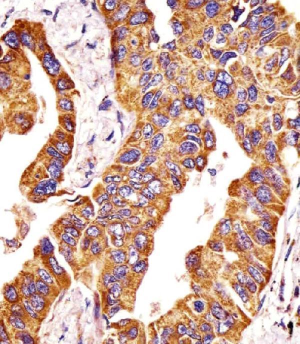
Real IHC data A00656 staining MMP14 in human lung adenocarcinoma tissue sections by Immunohistochemistry (IHC-P -paraformaldehyde-fixed, paraffin-embedded sections). Tissue was fixed with formaldehyde and blocked with 3% BSA for 0. 5 hour at room temperature; antigen retrieval was by heat mediation with a citrate buffer (pH6). Samples were incubated with primary antibody (1/25) for 1 hours at 37°C. A undiluted biotinylated goat polyvalent antibody was used as the secondary antibody.
Anti-MMP14 Antibody (N-term)
Cat # A00656
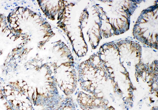
Real IHC data Anti-MMP14 antibody, PA2147, IHC(P) IHC(P): Human Intestinal Cancer Tissue
Anti-Matrix metalloproteinase-14 MMP14 Antibody ®
Cat # PA2147
Real IHC data Immunohistochemical analysis of paraffin-embedded human kdieny, using MMP14 Antibody .
Anti-MMP14/Mt1 Mmp Rabbit Monoclonal Antibody
Cat # M00656

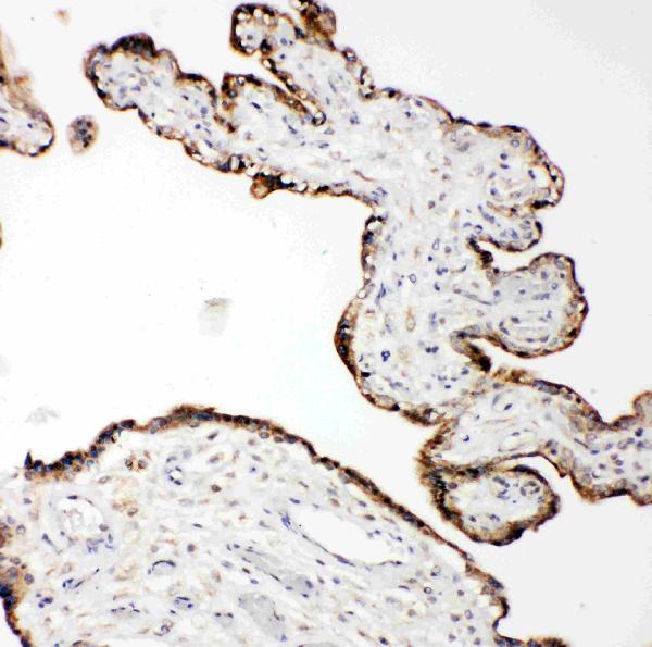
A00656 has an IHC-P image from human lung adenocarcinoma, and PA2147 has IHC-P images from human intestinal cancer, human placenta and rat cardiac muscle (image captions). M00656 lists IHC and IF/ICC applications and has an IHC image from paraffin-embedded human kidney (catalog applications; M00656 image caption).

Which to pick: For tissue IHC, A00656 provides a human lung adenocarcinoma paraffin-section example with citrate pH 6 retrieval (A00656 image caption); PA2147 provides human and rat tissue examples, with fixation unreported (PA2147 image captions). For IF/ICC, choose monoclonal clone BFD-13 M00656 because it lists those applications, although its supplied image shows IHC on paraffin sections and does not report a fixative (M00656 catalog; M00656 image caption). For cross-species work, all three list Human, Mouse and Rat reactivity; A00656 is polyclonal, while M00656 is monoclonal (catalog).

Each figure is that product's own IHC / IF validation image from its datasheet.

References

  1. UniProt Consortium. UniProt entry P50281 (MMP14_HUMAN, Matrix metalloproteinase-14).
  2. Human Protein Atlas. MMP14 tissue IHC expression (reliability: Approved).
  3. Human Protein Atlas. MMP14 subcellular location (ICC-IF): Mainly localized to the cytosol. In addition localized to the intermediate filaments..
  4. Human Protein Atlas. MMP14 antibody validation summary (2 antibodies).
  5. MMP14 expression levels accurately predict the presence of extranodal extensions in oral squamous cell carcinoma: a retrospective cohort study. BMC cancer 2023 — PMC9921360.
  6. High MMP14 and low decorin expression correlate with disease progression and metastasis in Brazilian prostate cancer patients. Journal of molecular histology 2026 — PMC13303449.
  7. Global gene expression profiling of human pleural mesotheliomas: identification of matrix metalloproteinase 14 (MMP-14) as potential tumour target. PloS one 2009 — PMC2737627.
  8. Early intermittent low-dose sclerostin antibody treatment promotes surface bone formation and reduces bone loss, but also decreases osteocyte apoptosis and mechanotransduction in ovariectomized rats: A pilot study. Bone reports 2026 — PMC13101557.
  9. PubMed PMID:8015608 — UniProt-cited evidence.
  10. PubMed PMID:7721107 — UniProt-cited evidence.
  11. PubMed PMID:7708715 — UniProt-cited evidence.