This website uses cookies to ensure you get the best experience on our website.


- Table of Contents
Plan MOB1B staining in paraffin sections using the observed cytoplasmic tissue pattern (HPA tissue IHC). Compare positive glandular cells with adipocytes reported as not detected, while accounting for possible staining of more than one gene product (HPA tissue IHC).

Expected localisation, validated protocols, controls and antibodies — the at-a-glance facts below, then the full design guide.
| Expected localisation | Cytoplasmic tissue staining (HPA tissue IHC); nucleus annotated (UniProt) | |
| Staining pattern | Widespread cytoplasmic staining (HPA tissue IHC) | |
| Antigen retrieval | Tris-EDTA pH 9.0 HIER, heat-mediated (datasheet A09405-2) | |
| Positive control | Adrenal gland+4 more · see all | |
| Negative control | Adipose tissue+1 more · see all |
| Fixation | Keep fixation consistent across sections. (standard IHC practice; not target-specific) | |
| Caveat | Staining may reflect more than one gene product (HPA tissue IHC) | |
| Regulation | No expression regulator specified (UniProt) | |
| Isoform / epitope | 2 isoforms; isoform-specific epitope coverage is unknown (UniProt) |
The catalog antibody protocol uses Tris-EDTA pH 9.0 retrieval (datasheet A09405-2); the published IHC protocols below cover spinal cord and endometrial cancer tissue sections (PMC10877155; PMC10067393).
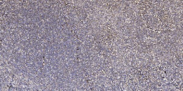
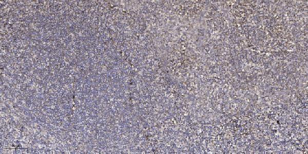
| Sample | Paraffin-embedded human tonsil tissue; fixative not specified (datasheet A09405-2) |
| Fixation | Image fixative and duration unreported (datasheet A09405-2); verify before use. |
| Sectioning | 4–5 µm sections on charged slides (standard) |
| Deparaffinisation | Xylene, graded ethanol series to water (standard) |
| Antigen retrieval | Heat retrieval: Tris-EDTA pH 9.0 (datasheet A09405-2); 20 min, 95–100 °C (standard) |
| Peroxidase block | 3% H2O2, 10 min, room temperature (standard) |
| Blocking | 10% normal serum of the secondary host, 30 min, room temperature (standard) |
| Primary antibody | Rabbit anti-MOB1B, 1:100-1:300 (datasheet A09405-2) |
| Primary incubation | Overnight at 4 °C (standard) |
| Detection | HRP-polymer secondary, DAB chromogen 5–10 min (standard) |
| Counterstain | Hematoxylin, blue, dehydrate and mount (standard) |
| Expected result | MOB1B-positive staining in glandular cells of adrenal gland (HPA tissue IHC: High). HPA tissue profile: Ubiquitous cytoplasmic expression. No signal in the no-primary control. |
In paraffin-section IHC, expect predominantly cytoplasmic staining in many cell types (HPA: ubiquitous cytoplasmic expression; tissue IHC Supported). Glandular cells in colon and adrenal gland and kidney tubular cells are useful high-staining examples (HPA: High). Nuclear localisation is also documented (UniProt Q7L9L4: Cytoplasm; Nucleus), while MOB1B has no transmembrane segment (UniProt Q7L9L4 topology). Interpret specificity cautiously: the HPA tissue profile carries a warning that antibodies may detect proteins from more than one gene (HPA: tissue IHC reliability description).
| Clear cytoplasmic staining in colon glandular cells or kidney tubular cells, with identifiable cell boundaries. | This fits the reported IHC distribution and offers an internal reference for evaluating weaker cells on the same section (HPA: High in colon glandular cells and kidney tubular cells; ubiquitous cytoplasmic expression). It supports a plausible pattern, but staining alone cannot prove that the antibody distinguishes MOB1B from related proteins (HPA: tissue IHC multi-gene caution). |
| Staining is confined to plasma membranes or extracellular material, with little cellular staining. | A membrane-only or extracellular pattern conflicts with the reported cellular locations and warrants an artefact check (HPA: ubiquitous cytoplasmic IHC expression; UniProt Q7L9L4: Cytoplasm; Nucleus; no transmembrane segment). Nuclear staining alone is a less decisive warning because nuclear localisation is documented; compare it with cytoplasmic signal and controls before judging the section. |
| Adipocytes or peripheral nerves stain strongly while nearby expected positive cells do not. | That distribution conflicts with the reported tissue examples (HPA: Not detected in adipocytes and soft-tissue peripheral nerve; High in colon glandular cells). Consider cross-reactivity, section background, or endogenous detection activity as possibilities. These HPA negatives are comparisons for interpretation, not a guarantee that every specimen or assay will be blank. |
| Diffuse colour covers stroma, empty spaces, and cells without a readable cytoplasmic pattern. | Treat the result as uninterpretable until background is controlled; diffuse deposit does not reproduce the reported cellular IHC pattern (HPA: ubiquitous cytoplasmic expression). As a general chromogenic IHC check, compare the no-primary control and inspect blocking, washing, and detection conditions. Those checks are workflow advice, not evidence of a MOB1B-specific background mechanism. |
| A known positive area shows no cellular signal, including colon glands or kidney tubules. | A blank result in those compartments conflicts with reported high staining and may indicate an assay or section problem (HPA: High in colon glandular cells and kidney tubular cells). Check the assay's positive control before calling the specimen negative. The HPA tissue profile does not identify a MOB1B-specific fixation sensitivity or establish why a particular section failed. |
| Compartment and application | For paraffin IHC, use the reported cytoplasmic tissue pattern as the primary comparison (HPA: ubiquitous cytoplasmic expression). For the separate IF/ICC question—can nuclear signal be plausible?—yes: nucleoplasm and nucleoli are supported main locations, with additional cytosol (HPA: subcellular ICC-IF); UniProt also lists Nucleus and Cytoplasm (UniProt Q7L9L4). This IF observation does not supply an IHC protocol option. |
| Cell context | High staining in colon and adrenal glandular cells and kidney tubular cells provides practical IHC reference areas (HPA: High). Adipocytes and soft-tissue peripheral nerves were not detected in the supplied profile (HPA: Not detected). Compare the named cell populations, since a whole-section positive or negative label can hide a mismatch in the cells carrying the signal. |
| Antibody interpretation | The tissue IHC profile is Supported, with medium agreement between staining and RNA expression and a warning about proteins from multiple genes (HPA: reliability description). The listed IHC antibody HPA047088 is Supported; HPA071690 has an ICC approval entry but no supplied IHC status (HPA: antibody validation). These designations support pattern review without establishing absolute MOB1B specificity. |
| Situation | Likely cause | Next action |
|---|---|---|
| Colon glands or kidney tubules are blank. | The observed section disagrees with the supplied high-staining examples; the evidence does not identify a target-specific failure mechanism (HPA: High in colon glandular cells and kidney tubular cells). | Check a positive-control section and the primary-antibody, detection, and counterstain steps as general IHC practice. Record whether the relevant cell type is present and preserved before reporting a negative result; do not assign the failure to MOB1B fixation sensitivity without separate evidence. |
| Only membranes or extracellular spaces carry colour. | That distribution is inconsistent with the reported intracellular locations and absence of a transmembrane segment (HPA: ubiquitous cytoplasmic IHC expression; UniProt Q7L9L4 topology). | Compare the same cell type in a positive reference area, inspect section morphology, and review the no-primary control. Reassess the staining as possible artefact if cellular cytoplasmic signal cannot be demonstrated; membrane colour alone does not match the supplied reference pattern. |
| Adipocytes or peripheral nerves are prominent positives. | These cells were not detected in the supplied HPA tissue examples, so cross-reactivity or endogenous detection activity is worth checking (HPA: Not detected in adipocytes and soft-tissue peripheral nerve). | Check the no-primary control for detection background and compare a named high-staining cell population on a control section (HPA: High in colon glandular cells). Interpret any remaining unexpected signal cautiously because the tissue profile warns of possible detection of proteins from more than one gene (HPA: reliability description). |
| Diffuse chromogen obscures cell boundaries. | The deposit prevents a reliable comparison with the reported cytoplasmic pattern (HPA: ubiquitous cytoplasmic expression). Its cause cannot be assigned from the target evidence alone. | As general IHC practice, inspect no-primary background, blocking, washes, and detection exposure, then repeat with conditions that preserve readable morphology. Score the cellular pattern only after diffuse colour is reduced; the supplied HPA data do not identify a MOB1B-specific background source. |
| A nucleus-heavy IHC result appears inconsistent with the expected cytoplasmic tissue pattern. | The sources describe different application-level observations: cytoplasmic tissue IHC and supported nucleoplasm, nucleoli, and cytosol in ICC-IF (HPA: tissue IHC profile; HPA: subcellular ICC-IF). UniProt also lists both Nucleus and Cytoplasm (UniProt Q7L9L4). | Do not reject nuclear signal solely by compartment. Check whether the named IHC-positive cell types also show a credible cellular pattern and whether controls are clean (HPA: High in colon glandular cells and kidney tubular cells). Use the separate IF/ICC guide for IF methods. |
| Signal is present, but its assignment specifically to MOB1B matters. | The supplied HPA tissue assessment warns that the staining may represent proteins from more than one gene despite its Supported rating (HPA: tissue IHC reliability description). | Report the observed cell type and compartment with that specificity caveat. If a MOB1B-specific conclusion is required, seek independent validation suitable for the research question; the supplied IHC pattern and antibody status alone do not settle gene-level specificity (HPA: tissue IHC reliability description; HPA: antibody validation). |


Comprehensive Human Protein Atlas IHC scoring per tissue (reliability: Supported — Medium consistency between antibody staining and RNA expression data. Caution, targets protein from more than one gene.). Rows are taken directly from the HPA tissue chart — click any row's HPA link to view the source.
| Tissue | Cell type | Level | Evidence | Source |
|---|---|---|---|---|
| Adrenal gland | Glandular cells | High | Protein (IHC) | HPA → |
| Appendix | Glandular cells | High | Protein (IHC) | HPA → |
| Cerebellum | Cells in granular layer | High | Protein (IHC) | HPA → |
| Colon | Glandular cells | High | Protein (IHC) | HPA → |
| Duodenum | Glandular cells | High | Protein (IHC) | HPA → |
Troubleshoot MOB1B staining in paraffin section IHC by checking retrieval, tissue handling, cellular localisation and detection controls before comparing specimens.
The catalog lists MOB1B-labeled antibodies with human tonsil IHC images and human, monkey, and mouse reactivity; IF is listed as an application (catalog: A09405-2, A30515).
A09405-2 and A30515 each show IHC on paraffin-embedded human tonsil; neither caption reports the fixative (catalog: A09405-2 and A30515 IHC captions). A09405-3 shows IHC on formalin-fixed, paraffin-embedded human brain and IF in NIH/3T3 cells, but its catalog target is MOB4A (catalog: A09405-3 title and image captions).
Which to pick: For MOB1B tissue IHC, choose A09405-2 or A30515: both are rabbit polyclonals with IHC listed and their own paraffin-embedded human tonsil images; fixation is unreported in both captions (catalog: A09405-2 and A30515 application lists, dilution data, and IHC captions). For IF or ICC, A30515 lists both applications, while A09405-2 lists IF only; neither has an IF image in this payload (catalog: A30515 and A09405-2 application lists and image records). For human, monkey, or mouse samples, both list reactivity across those species; A09405-3 targets MOB4A and should not be selected to measure MOB1B (catalog: reactivity lists and A09405-3 title).