This website uses cookies to ensure you get the best experience on our website.


- Table of Contents
Plan paraffin-section MOB4 IHC around a general cytoplasmic readout (HPA tissue IHC). Compare high-staining glandular cells with adipocytes reported as not detected, and interpret staining with the cross-gene caution (HPA tissue IHC).

Expected localisation, validated protocols, controls and antibodies — the at-a-glance facts below, then the full design guide.
| Expected localisation | General cytoplasmic tissue staining (HPA tissue IHC) | |
| Staining pattern | Cytoplasmic staining in glandular and epithelial cells (HPA tissue IHC) | |
| Antigen retrieval | Tris-EDTA pH 9.0 HIER, heat-mediated (datasheet A11070) | |
| Positive control | Adrenal gland+4 more · see all | |
| Negative control | Adipose tissue+1 more · see all |
| Fixation | Keep fixation conditions consistent across sections. (standard IHC practice; not target-specific) | |
| Caveat | Staining may include proteins from other genes (HPA tissue IHC) | |
| Regulation | Regulation not annotated (UniProt) | |
| Isoform / epitope | 3 isoforms; no processing annotated; check epitope coverage (UniProt) |
The catalog antibody’s paraffin IHC protocol uses pH 9.0 Tris-EDTA heat retrieval (datasheet A11070). One published larval CNS protocol provides additional staining conditions (PMC6581792).
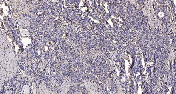
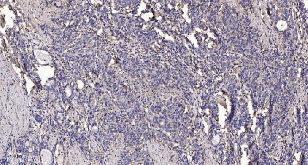
| Sample | Paraffin-embedded human Gastric adenocarcinoma tissue; fixative not specified (datasheet A11070) |
| Fixation | Image fixative and duration unreported (datasheet A11070); verify before use. |
| Sectioning | 4–5 µm sections on charged slides (standard) |
| Deparaffinisation | Xylene, graded ethanol series to water (standard) |
| Antigen retrieval | Heat retrieval: Tris-EDTA pH 9.0 (datasheet A11070); 20 min, 95–100 °C (standard) |
| Peroxidase block | 3% H2O2, 10 min, room temperature (standard) |
| Blocking | 10% normal serum of the secondary host, 30 min, room temperature (standard) |
| Primary antibody | Rabbit anti-MOB4, 1:100-1:300 (datasheet A11070) |
| Primary incubation | Overnight at 4 °C (standard) |
| Detection | HRP-polymer secondary, DAB chromogen 5–10 min (standard) |
| Counterstain | Hematoxylin, blue, dehydrate and mount (standard) |
| Expected result | MOB4-positive staining in glandular cells of adrenal gland (HPA tissue IHC: High). HPA tissue profile: General cytoplasmic expression. No signal in the no-primary control. |
MOB4 staining should be mainly cytoplasmic, with possible perinuclear puncta and Golgi or membrane association (UniProt Q9Y3A3 localisation). High staining is reported in several glandular and epithelial cell types (HPA: tissue IHC). Interpret the pattern cautiously: HPA rates tissue staining Approved with medium RNA agreement and warns that its antibodies may detect proteins from more than one gene (HPA: reliability description). MOB4 has no annotated transmembrane segment (UniProt Q9Y3A3 topology).
| Cytoplasmic staining in colon or duodenal glandular cells, with stronger signal around some nuclei. | This fits the reported high staining in these cells (HPA: High in colon and duodenal glandular cells) and the predicted perinuclear pattern (UniProt Q9Y3A3 localisation). Assess cell type and compartment together; cytoplasmic signal alone does not resolve HPA's multiple-gene detection caveat (HPA: reliability description). |
| Strong nuclear staining dominates while the cytoplasm is faint in an otherwise positive epithelium. | A predominantly nuclear pattern conflicts with the reported general cytoplasmic tissue pattern (HPA: tissue IHC profile) and perinuclear, membrane-associated localisation (UniProt Q9Y3A3 localisation). Investigate detection background and antibody specificity before calling it MOB4; an isolated nuclear signal is insufficient to establish localisation. |
| Prominent staining appears in adipocytes or bone-marrow hematopoietic cells. | These cells are reported as not detected (HPA: adipocytes and bone-marrow hematopoietic cells). Check whether the signal follows cell outlines or nonspecific deposit. Cross-reactivity or endogenous chromogen activity are possibilities to test, rather than proven causes; HPA also cautions that its antibodies may detect proteins from more than one gene (HPA: reliability description). |
| Pale, widespread colour covers cells and surrounding tissue without a clear cytoplasmic boundary. | This is difficult to score against the reported cytoplasmic pattern (HPA: tissue IHC profile). In chromogenic IHC, incomplete blocking, excess detection reagent, or residual endogenous activity can cause diffuse background (general IHC practice). Compare a control processed without primary antibody before assigning the colour to MOB4. |
| No convincing signal appears in bronchial respiratory epithelium or esophageal squamous epithelium. | Both cell types have reported high staining (HPA: bronchus and esophagus). A blank result calls for a run-level check of section integrity, retrieval, primary-antibody application, and detection (general IHC practice). It does not establish absence of MOB4 in that specimen; HPA's reported levels are observations, not a guarantee for every section. |
| Tissue and cell selection | HPA reports high staining across several glandular and epithelial populations, while adipocytes and bone-marrow hematopoietic cells are not detected (HPA: tissue IHC). Use a reported high-staining cell population to judge whether the run worked; the undetected populations are comparison sites, not definitive specificity controls. |
| Compartment and topology | UniProt places MOB4 in cytoplasm, perinuclear puncta, membranes, and Golgi stacks, with no transmembrane segment (UniProt Q9Y3A3 localisation and topology). Perinuclear enrichment can fit the record; membrane association does not imply a surface-only stain. Score the dominant compartment in the relevant cells. |
| Antibody interpretation | Two listed antibodies have Approved IHC status, but neither is marked IHC Enhanced in the supplied record (HPA: HPA044125 and CAB034082 antibody status). HPA reports medium agreement with RNA and cautions about detection of proteins from more than one gene (HPA: reliability description). A matching stain supports interpretation without proving MOB4-specific binding. |
| Isoforms and processing | Three isoforms are listed; the supplied record gives no isoform-specific staining or epitope map (UniProt Q9Y3A3 isoforms). It also lists no signal peptide, propeptide, or glycosylation sites (UniProt Q9Y3A3 processing). Do not infer an isoform-specific stain or a shed-product pattern from these annotations. |
| Retrieval and detection | The supplied sources give no MOB4-specific retrieval setting or fixation-sensitivity result (HPA: supplied tissue IHC; UniProt Q9Y3A3). Retrieval, blocking, and chromogen controls remain general paraffin-IHC workflow decisions (general IHC practice); their effects on MOB4 staining must be checked in the actual run. |
| Situation | Likely cause | Next action |
|---|---|---|
| A reported high-staining epithelium is blank. | A failed retrieval, omitted primary antibody, or detection problem can produce a blank IHC run (general IHC practice); the expected high cell-level staining comes from HPA (HPA: bronchus or esophagus). | Check a reported positive cell population on the same run, then verify reagent application, retrieval record, and chromogen development (general IHC practice). Do not score a blank section as a biological negative until the run control works. |
| Colour concentrates in nuclei instead of cytoplasm. | Dominant nuclear staining conflicts with the reported cytoplasmic tissue pattern (HPA: tissue IHC profile) and UniProt's perinuclear and Golgi localisation (UniProt Q9Y3A3 localisation). | Inspect a no-primary control and compare cell boundaries with the counterstain (general IHC practice). Reassess detection background and antibody specificity before accepting a nuclear localisation. |
| Adipocytes or bone-marrow hematopoietic cells stain strongly. | Both are listed as not detected (HPA: tissue IHC). Endogenous chromogen activity or antibody cross-reactivity are testable explanations (general IHC practice; HPA: multiple-gene detection caution). | Compare no-primary and detection controls, and examine whether a reported high-staining epithelial population shows the expected cytoplasm (general IHC practice; HPA: tissue IHC profile). Treat persistent staining in these cells cautiously. |
| The entire section has weak brown haze. | Diffuse colour lacks the reported cell-associated cytoplasmic pattern (HPA: tissue IHC profile). Inadequate blocking, excessive detection signal, or residual endogenous activity can contribute (general IHC practice). | Compare the no-primary control, inspect wash and blocking steps, and adjust detection conditions within the assay workflow (general IHC practice). Score only cell-associated signal above local background. |
| A second antibody gives a different cell pattern. | The two listed antibodies are IHC Approved, while HPA reports medium RNA agreement and possible detection from more than one gene (HPA: antibody status and reliability description). Agreement is therefore useful evidence, and disagreement needs investigation. | Compare the same tissue compartments and cell types under matched scoring criteria; review each antibody's validation and controls before choosing a pattern (general IHC practice). Do not describe either reagent as IHC Enhanced from this record (HPA: supplied antibody status). |
| IF/ICC Q&A: Do perinuclear fluorescent puncta validate MOB4 localisation? | Perinuclear puncta are consistent with UniProt's localisation annotation, but the supplied HPA subcellular record has no main location or ICC-IF images (UniProt Q9Y3A3 localisation; HPA: subcellular record). | Treat puncta as a localisation hypothesis, not an independently validated IF/ICC result. Use the separate IF/ICC guide for assay design; this IHC section supplies no IF/ICC protocol (HPA: subcellular record). |


Comprehensive Human Protein Atlas IHC scoring per tissue (reliability: Approved — Medium consistency between antibody staining and RNA expression data. Caution, targets protein from more than one gene.). Rows are taken directly from the HPA tissue chart — click any row's HPA link to view the source.
| Tissue | Cell type | Level | Evidence | Source |
|---|---|---|---|---|
| Adrenal gland | Glandular cells | High | Protein (IHC) | HPA → |
| Appendix | Glandular cells | High | Protein (IHC) | HPA → |
| Bronchus | Respiratory epithelial cells | High | Protein (IHC) | HPA → |
| Colon | Glandular cells | High | Protein (IHC) | HPA → |
| Duodenum | Glandular cells | High | Protein (IHC) | HPA → |
Troubleshoot MOB4 staining in paraffin sections by checking retrieval, compartmental pattern, controls, and scoring before drawing biological conclusions.
Two anti-MOB4 antibodies have real IHC images from human gastric adenocarcinoma paraffin sections (catalog IHC captions). Both list IF and human, mouse, and rat reactivity (catalog applications and reactivity).
A11070 will render with an IHC image from human gastric adenocarcinoma paraffin sections; IF is listed, and human, mouse, and rat reactivity is reported (A11070 catalog IHC caption, applications, and reactivity). A19781 will render with its own IHC image from human gastric adenocarcinoma paraffin sections; IF and ICC are listed, and human, mouse, and rat reactivity is reported (A19781 catalog IHC caption, applications, and reactivity).
Which to pick: For tissue IHC, either SKU has a paraffin-section image using primary antibody at 1:200 after Tris-EDTA retrieval at pH 9.0 (A11070 and A19781 IHC captions); neither caption reports the fixative. For IF/ICC, choose A19781 because both applications are listed and it is described as polyclonal (A19781 catalog applications and dilution_raw); A11070 lists IF, but neither SKU has an IF image in the payload (catalog applications and image alts). For mouse or rat samples, both list reactivity with those species, while each supplied IHC image shows only human tissue (catalog reactivity and IHC captions).