This website uses cookies to ensure you get the best experience on our website.


- Table of Contents
Plan MTTP staining in paraffin sections using the documented cytoplasmic pattern in intestinal glandular cells and hepatocytes (HPA tissue IHC). The guide covers antibody dilution, detection and interpretation while accounting for uncertain tissue-staining reliability (HPA tissue IHC).

Expected localisation, validated protocols, controls and antibodies — the at-a-glance facts below, then the full design guide.
| Expected localisation | Cytoplasmic tissue staining (HPA tissue IHC); ER and Golgi location (UniProt) | |
| Staining pattern | Glandular cells and hepatocytes show cytoplasmic staining (HPA tissue IHC) | |
| Antigen retrieval | EDTA pH 8.0 HIER, heat-mediated (datasheet A01715-1) | |
| Positive control | Duodenum+2 more · see all | |
| Negative control | Adipose tissue+4 more · see all |
| Fixation | Keep paraffin-section fixation consistent across samples. (standard IHC practice; not target-specific) | |
| Caveat | Presumed off-target binding; staining reliability is uncertain (HPA tissue IHC) | |
| Regulation | Expression regulation is not specified (UniProt) | |
| Isoform / epitope | 2 isoforms; mature chain starts at residue 19; epitope coverage unknown (UniProt) |
The catalog antibody protocol is followed by three published MTTP IHC protocols (datasheet A01715-1; PMC11951013; PMC9534973; PMC11055869).
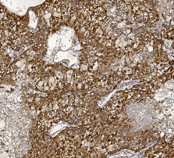
| Sample | Paraffin-embedded human liver cancer tissue; fixative not specified (datasheet A01715-1) |
| Fixation | Image fixative and duration unreported (datasheet A01715-1); verify before use. |
| Sectioning | 4–5 µm sections on charged slides (standard) |
| Deparaffinisation | Xylene, graded ethanol series to water (standard) |
| Antigen retrieval | Heat retrieval: EDTA pH 8.0 (datasheet A01715-1); 20 min, 95–100 °C (standard) |
| Peroxidase block | 3% H2O2, 10 min, room temperature (standard) |
| Blocking | 10% goat serum (datasheet A01715-1) |
| Primary antibody | Rabbit anti-MTTP, 2-5μg/ml (datasheet A01715-1) |
| Primary incubation | Overnight at 4 °C (datasheet A01715-1) |
| Detection | Streptavidin-biotin complex (SABC), DAB chromogen (datasheet A01715-1) |
| Counterstain | Hematoxylin, blue, dehydrate and mount (standard) |
| Expected result | MTTP-positive staining in glandular cells of duodenum (HPA tissue IHC: High). HPA tissue profile: Cytoplasmic expression in liver and small intestine. No signal in the no-primary control. |
MTTP is an endoplasmic reticulum protein also annotated in the Golgi, with no transmembrane segment (UniProt P55157). In paraffin-section IHC, expect cytoplasmic staining in intestinal glandular cells and hepatocytes (HPA tissue IHC). Interpret the pattern cautiously: HPA rates its tissue IHC profile Uncertain because staining and RNA data have low consistency, and presumed off-target staining was disregarded (HPA tissue IHC).
| Strong cytoplasmic staining in duodenal or small-intestinal glandular cells; moderate staining in hepatocytes. | This matches HPA's observed High glandular-cell and Medium hepatocyte staining (HPA tissue IHC). A fine intracellular pattern is compatible with ER localization (UniProt P55157); IHC alone does not prove that every stained structure is ER. |
| Dominant nuclear or cell-surface staining, with little cytoplasmic signal in expected positive cells. | This does not fit MTTP's ER/Golgi annotation or the reported cytoplasmic tissue profile (UniProt P55157; HPA tissue IHC). Check the antibody and detection controls before assigning that compartment to MTTP. |
| Strong staining in adipocytes or another cell population outside the reported positive pattern. | HPA reports adipocytes in adipose tissue as Not detected and disregarded presumed off-target staining (HPA tissue IHC). Consider cross-reactivity; signal that persists without primary antibody may reflect endogenous detection activity (standard IHC practice). |
| Broad, nearly uniform color across tissue, including extracellular spaces or expected negative cells. | This obscures the cell-specific cytoplasmic pattern reported by HPA (HPA tissue IHC). Evaluate background with a no-primary control and review blocking, washes, and chromogen development (standard IHC practice). |
| No detectable staining in duodenal or small-intestinal glandular cells on a processed positive-control section. | Those cells are reported High by HPA, so an absent signal warrants a technical check (HPA tissue IHC). Review the catalog antibody's IHC-P instructions, retrieval, dilution, and detection controls (standard IHC practice). |
| Tissue and cell selection | Duodenal and small-intestinal glandular cells are reported High; hepatocytes are Medium (HPA tissue IHC). Include a reported positive tissue when interpreting a weak experimental section. |
| Intracellular location and processing | MTTP has a signal peptide at residues 1–18, a mature chain at 19–894, and no transmembrane segment (UniProt P55157). These annotations support an intracellular interpretation; they do not identify the antibody epitope. |
| Tissue IHC confidence | HPA labels the tissue profile Uncertain and one listed antibody's IHC validation Uncertain (HPA tissue IHC; HPA antibodies: HPA054862). Treat an isolated unexpected stain as provisional. |
| Isoforms and antibody coverage | UniProt lists 2 MTTP isoforms (UniProt P55157). The supplied record gives no epitope or isoform coverage for the catalog antibody, so it cannot explain a discrepant stain by isoform choice. |
| IF/ICC Q&A: What localization should be expected? | HPA supports ER localization in ICC-IF and calls cytosol uncertain; UniProt also annotates Golgi (HPA subcellular; UniProt P55157). Use the separate IF/ICC guide for that application. |
| Situation | Likely cause | Next action |
|---|---|---|
| Positive-control intestinal glandular cells are blank. | The result conflicts with reported High staining, but HPA's tissue profile is Uncertain (HPA tissue IHC). A failed IHC step remains possible (standard IHC practice). | Confirm the catalog antibody's IHC-P procedure, then check retrieval, primary dilution, and detection with appropriate run controls (standard IHC practice). |
| Hepatocytes stain weakly beside strong intestinal glandular cells. | HPA reports Medium hepatocyte staining versus High intestinal glandular-cell staining (HPA tissue IHC). | Score the two cell types against their respective reported levels; keep exposure and chromogen development comparable across sections (standard IHC practice). |
| Adipocytes show strong staining. | HPA reports adipose adipocytes as Not detected, and its overall tissue profile is Uncertain (HPA tissue IHC). Cross-reactivity or detection background is plausible (standard IHC practice). | Compare a no-primary control and a known-positive section in the same run before interpreting the adipocyte signal (standard IHC practice). |
| Color appears predominantly nuclear or along plasma membranes. | That distribution conflicts with ER/Golgi annotation and HPA's cytoplasmic tissue profile (UniProt P55157; HPA tissue IHC). | Recheck morphology and counterstain, then compare primary-free and positive-control sections; do not score the unexpected compartment as MTTP without further validation (standard IHC practice). |
| Most cells and empty spaces acquire diffuse chromogen. | Non-cell-specific color prevents assessment of HPA's reported cell-specific pattern (HPA tissue IHC). Excess background can arise during detection (standard IHC practice). | Inspect the no-primary control; review blocking, wash stringency, and chromogen development using standard IHC controls (standard IHC practice). |
| The stain differs from an ICC-IF image. | HPA's ICC-IF record supports ER localization but marks cytosol uncertain; its tissue IHC reliability is also Uncertain (HPA subcellular; HPA tissue IHC). | Compare each result within its own application and controls. For paraffin IHC, prioritize the observed tissue and cell pattern while retaining HPA's reliability caveat (HPA tissue IHC). |


Comprehensive Human Protein Atlas IHC scoring per tissue (reliability: Uncertain — Low consistency between antibody staining and RNA expression data. Presumed off target binding observed and disregarded.). Rows are taken directly from the HPA tissue chart — click any row's HPA link to view the source.
| Tissue | Cell type | Level | Evidence | Source |
|---|---|---|---|---|
| Duodenum | Glandular cells | High | Protein (IHC) | HPA → |
| Small intestine | Glandular cells | High | Protein (IHC) | HPA → |
| Liver | Hepatocytes | Medium | Protein (IHC) | HPA → |
| Tissue | Cell type | Level | Evidence | Source |
|---|---|---|---|---|
| Adipose tissue | Adipocytes | Not detected | Protein (IHC) | HPA → |
| Adrenal gland | Glandular cells | Not detected | Protein (IHC) | HPA → |
| Appendix | Glandular cells | Not detected | Protein (IHC) | HPA → |
| Bone marrow | Hematopoietic cells | Not detected | Protein (IHC) | HPA → |
| Breast | Adipocytes | Not detected | Protein (IHC) | HPA → |
Troubleshoot MTTP staining in paraffin sections by checking retrieval, compartment, cell type, controls, and scoring before interpreting differences in signal.
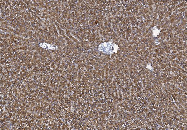
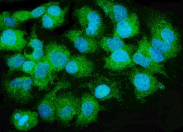
A01715-1 has IHC images from human liver cancer and rat liver paraffin sections, plus IF data from HEPG2 cells (catalog image captions).
A01715-1 will render with its human liver cancer paraffin-section IHC figure; a second catalog caption documents rat liver paraffin-section IHC (catalog IHC image captions). The same SKU lists IF and ICC applications and has an IF image from HEPG2 cells (catalog applications and IF image caption).
Which to pick: Choose A01715-1 for human or rat paraffin-section IHC: its captions document EDTA heat retrieval at pH 8.0 and 2 μg/ml antibody; the fixative is unreported (catalog IHC image captions). For IF/ICC, A01715-1 has a HEPG2 cell IF image using 5 μg/ml antibody (catalog IF image caption). For cross-species planning, the catalog lists Human, Monkey and Rat reactivity, while the supplied IHC images document Human and Rat only; no clone type is supplied (catalog reactivity, IHC image captions and clone field).