This website uses cookies to ensure you get the best experience on our website.


- Table of Contents
Plan NLGN2 paraffin IHC around cortical neuropil and cerebellar cytoplasm/membrane staining (HPA tissue IHC). This guide covers fixation, antigen retrieval, controls and chromogenic scoring, with 5 μg/mL as the catalog antibody’s IHC starting concentration (datasheet).

Expected localisation, validated protocols, controls and antibodies — the at-a-glance facts below, then the full design guide.
| Expected localisation | Cortical neuropil; cerebellar cytoplasm/membrane (HPA tissue IHC) | |
| Staining pattern | Cortical neuropil; enterocyte microvilli; Sertoli cells (HPA tissue IHC) | |
| Antigen retrieval | Citrate pH 6.0 HIER, 95–98 °C, 20 min (rule: cytoplasmic / membrane antigen) | |
| Positive control | Cerebellum+4 more · see all | |
| Negative control | Adipose tissue+4 more · see all |
| Fixation | Matched tissue-IHC evidence does not establish the fixation claim. Validate the specimen-specific method before use. (selected-SKU IHC image A06595) | |
| Caveat | Staining–RNA consistency is medium (HPA tissue IHC) | |
| Regulation | Brain exceeds islet beta cells (UniProt) | |
| Isoform / epitope | No isoforms annotated; extracellular vs cytoplasmic epitope matters (UniProt) |
The catalog antibody’s IHC-P protocol is supplemented by published NLGN2 staining methods for breast tumor sections and rat hippocampus (PMC8595097; PMC3579928).
| Sample | Tissue sections; selected-image fixative not specified (standard IHC workflow) |
| Fixation | Image fixative and duration unreported (datasheet A06595); verify before use. |
| Sectioning | 4–5 µm sections on charged slides (standard) |
| Deparaffinisation | Xylene, graded ethanol series to water (standard) |
| Antigen retrieval | Heat-induced epitope retrieval in citrate buffer, pH 6.0, 20 min at 95–98 °C (standard rule: cytoplasmic / membrane antigen) |
| Peroxidase block | 3% H2O2, 10 min, room temperature (standard) |
| Blocking | 10% normal serum of the secondary host, 30 min, room temperature (standard) |
| Primary antibody | Rabbit anti-NLGN2, 5 μg/mL (datasheet A06595) |
| Primary incubation | Overnight at 4 °C (standard) |
| Detection | HRP-polymer secondary, DAB chromogen 5–10 min (standard) |
| Counterstain | Hematoxylin, blue, dehydrate and mount (standard) |
| Expected result | NLGN2-positive staining in molecular layer cells - cytoplasm/membrane of cerebellum (HPA tissue IHC: Medium). HPA tissue profile: Selective expression in CNS. No signal in the no-primary control. |
NLGN2 is a transmembrane protein with an extracellular region and cytoplasmic tail; brain localization includes postsynaptic membranes, while retinal localization includes presynaptic membranes (UniProt Q8NFZ4 topology and subcellular location). In paraffin IHC, expect staining in cortical neuropil and cerebellar molecular layer cells, with additional reported staining in enterocyte microvilli and Sertoli cells (HPA tissue IHC). HPA rates its tissue IHC reliability Enhanced, with medium consistency between staining and RNA data (HPA tissue IHC).
| Cortical neuropil stains at medium intensity; cerebellar molecular layer cells show cytoplasmic or membrane staining (HPA tissue IHC). | This matches the reported brain IHC pattern (HPA tissue IHC). Evaluate the stained structures in their tissue context: NLGN2 is associated with neuronal membranes and synapses, but the HPA cerebellar description also includes cytoplasm (UniProt Q8NFZ4 subcellular location; HPA tissue IHC). |
| Signal is predominantly nuclear, or an organelle-like pattern replaces the expected neuropil and membrane-associated staining (HPA tissue IHC; UniProt Q8NFZ4 subcellular location). | Treat this as discordant with the supplied IHC expectation and investigate specificity or staining artefact (HPA tissue IHC; general IHC practice). HPA separately reports mitochondrial ICC-IF staining; that observation alone does not establish the expected chromogenic IHC pattern (HPA subcellular ICC-IF; HPA tissue IHC). |
| Adipocytes or adrenal glandular cells stain strongly (HPA tissue IHC: both cell types not detected). | These are discordant findings in the named cell types, so assess antibody cross-reactivity and endogenous chromogenic detection activity (HPA tissue IHC; general IHC practice). Avoid treating an entire tissue as negative solely because HPA lists one cell type as not detected (HPA tissue IHC). |
| Colour spreads across tissue compartments and the surrounding slide without a clear cellular pattern (general IHC practice). | Diffuse background cannot establish NLGN2 localization. Review blocking, antibody concentration, washing and detection controls before scoring specific staining (general IHC practice); compare any retained pattern with the cell-level HPA observations (HPA tissue IHC). |
| A well-preserved cerebral cortex or cerebellum section has no detectable target signal (HPA tissue IHC: medium staining in the specified structures). | A missing positive-control pattern leaves this IHC run uninterpretable for negative samples (general IHC practice). Check tissue sampling and assay performance first; absence in one section does not overturn the reported tissue pattern (HPA tissue IHC; general IHC practice). |
| Membrane topology | NLGN2 spans residues 678–698, with extracellular residues 15–677 and cytoplasmic residues 699–835 (UniProt Q8NFZ4 topology). Interpret a membrane-associated pattern in light of this architecture; the supplied record does not identify the catalog antibody's epitope. |
| Choice of comparison tissue and cell | HPA reports medium staining in cortical neuropil, cerebellar molecular layer cells, enterocyte microvilli and Sertoli cells, but low staining in hippocampal and caudate neuronal cells (HPA tissue IHC). Compare the specified cells and structures, since a faint hippocampal result alone is a weak failure criterion. |
| Antibody-specific evidence | HPA lists HPA069278 as IHC Enhanced and HPA055321 as ICC Approved, without assigning both statuses to one antibody (HPA antibodies). HPA describes tissue IHC reliability as Enhanced with medium staining–RNA consistency (HPA tissue IHC); check which evidence applies to the antibody actually used. |
| IF/ICC Q: Does HPA mitochondrial staining define the expected IHC result? | A: No. HPA reports approved mitochondrial localization in ICC-IF, while its tissue IHC observations describe neuropil, cytoplasmic or membrane, and microvillar patterns (HPA subcellular ICC-IF; HPA tissue IHC). The listed ICC and IHC validation statuses belong to different antibodies (HPA antibodies). |
| Epitope and fixation limits | The supplied topology identifies extracellular and cytoplasmic regions, but no catalog-antibody epitope or target-specific fixation sensitivity is supplied (UniProt Q8NFZ4 topology; supplied evidence scope). Select antigen retrieval from the IHC-validated antibody's protocol and assess it with controls (general IHC practice). |
| Situation | Likely cause | Next action |
|---|---|---|
| No stain appears in cortical neuropil or cerebellar molecular layer cells (HPA tissue IHC). | The expected positive-control pattern is absent; tissue sampling or assay performance may explain the result (HPA tissue IHC; general IHC practice). | Confirm the relevant structures are present, then review the catalog antibody's IHC-P protocol, retrieval, dilution and detection controls before interpreting test sections (general IHC practice). |
| The hippocampus is faint while cortex shows a clear neuropil pattern (HPA tissue IHC). | HPA reports low staining in hippocampal neuronal cells and medium staining in cortical neuropil (HPA tissue IHC). | Score each region against its own reported cell-level pattern; retain cortex as the stronger tissue comparison and avoid calling faint hippocampal staining an assay failure by itself (HPA tissue IHC). |
| Adipocytes or adrenal glandular cells show conspicuous chromogenic colour (HPA tissue IHC: not detected). | Cross-reactivity, nonspecific binding or endogenous detection activity are possible explanations (general IHC practice); the observed cell types conflict with the HPA entries (HPA tissue IHC). | Inspect reagent-omission and detection controls, review blocking, and compare staining in the named cells with a reported positive structure (general IHC practice; HPA tissue IHC). |
| Diffuse colour obscures neuropil and cell borders (general IHC practice). | Excess antibody or detection background can reduce contrast between specific staining and surrounding tissue (general IHC practice). | Review the antibody's IHC-P dilution guidance, blocking, washing and detection exposure; accept a result only when the reported cell-level distribution can be judged (general IHC practice; HPA tissue IHC). |
| A mitochondrial-looking pattern dominates the chromogenic section (HPA subcellular ICC-IF; HPA tissue IHC). | HPA's mitochondrial assignment comes from ICC-IF, whereas its tissue IHC evidence describes other structures and uses separately listed antibody validation (HPA subcellular ICC-IF; HPA tissue IHC; HPA antibodies). | Check antibody identity and IHC validation, then compare with cortical neuropil or cerebellar molecular layer staining before assigning NLGN2 localization (HPA antibodies; HPA tissue IHC; general IHC practice). |
| Colon or duodenum shows staining concentrated at enterocyte microvilli (HPA tissue IHC). | This is a reported medium IHC pattern in those cells (HPA tissue IHC); UniProt also reports NLGN2 protein detection in colon (UniProt Q8NFZ4 tissue specificity). | Record the microvillar compartment and intensity, and use a reported brain structure as an additional comparison when assessing assay performance (HPA tissue IHC; general IHC practice). |


Comprehensive Human Protein Atlas IHC scoring per tissue (reliability: Enhanced — Medium consistency between antibody staining and RNA expression data.). Rows are taken directly from the HPA tissue chart — click any row's HPA link to view the source.
| Tissue | Cell type | Level | Evidence | Source |
|---|---|---|---|---|
| Cerebellum | Molecular layer cells - cytoplasm/membrane | Medium | Protein (IHC) | HPA → |
| Cerebral cortex | Neuropil | Medium | Protein (IHC) | HPA → |
| Colon | Enterocytes - Microvilli | Medium | Protein (IHC) | HPA → |
| Duodenum | Enterocytes - Microvilli | Medium | Protein (IHC) | HPA → |
| Testis | Sertoli cells | Medium | Protein (IHC) | HPA → |
| Tissue | Cell type | Level | Evidence | Source |
|---|---|---|---|---|
| Adipose tissue | Adipocytes | Not detected | Protein (IHC) | HPA → |
| Adrenal gland | Glandular cells | Not detected | Protein (IHC) | HPA → |
| Appendix | Endocrine cells | Not detected | Protein (IHC) | HPA → |
| Bone marrow | Hematopoietic cells | Not detected | Protein (IHC) | HPA → |
| Breast | Adipocytes | Not detected | Protein (IHC) | HPA → |
Troubleshoot chromogenic NLGN2 staining in paraffin sections by checking retrieval, compartment, controls and scoring before interpreting the signal.
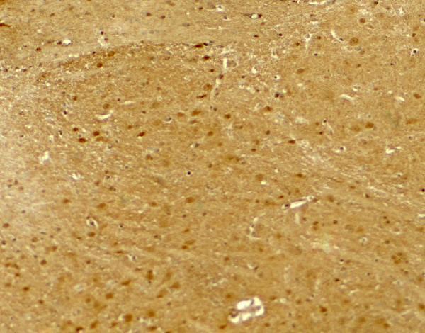
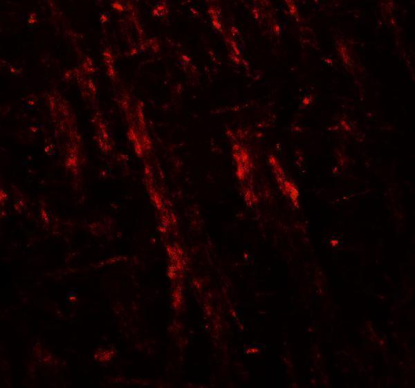
A06595 has mouse brain IHC and IF images (catalog image captions); IHC-P and IF are listed applications, with Human, Mouse and Rat reactivity (catalog applications/reactivity).
A06595 has an IHC image from mouse brain tissue at 5 μg/mL (catalog IHC image caption). Its IF image is also from mouse brain tissue, at 20 μg/mL (catalog IF image caption); Human, Mouse and Rat are listed as reactive species (catalog reactivity).
Which to pick: Choose A06595 for tissue IHC: IHC-P is listed, and its own IHC image shows mouse brain tissue at 5 μg/mL (catalog applications; A06595 IHC image caption). For fluorescence work, A06595 has an IF image from mouse brain at 20 μg/mL; ICC validation is unreported (A06595 IF image caption; catalog applications). A06595 lists Human, Mouse and Rat reactivity, though the supplied IHC image documents Mouse only; its fixative is unreported (catalog reactivity; A06595 IHC image caption).