This website uses cookies to ensure you get the best experience on our website.


- Table of Contents
Plan NME2 staining in paraffin sections using the catalog antibody and the reported cytoplasmic tissue pattern (HPA tissue IHC). Kidney tubule cells show high staining, while adipocytes are reported as not detected; interpret results with the atlas warning that staining may reflect more than one gene (HPA tissue IHC).

Expected localisation, validated protocols, controls and antibodies — the at-a-glance facts below, then the full design guide.
| Expected localisation | General cytoplasmic staining in tissue (HPA tissue IHC) | |
| Staining pattern | General cytoplasmic staining (HPA tissue IHC) | |
| Antigen retrieval | Citrate pH 6 HIER, heat-mediated (datasheet PA1830) | |
| Positive control | Endometrium+4 more · see all | |
| Negative control | Adipose tissue+3 more · see all |
| Fixation | Keep fixation conditions consistent across sections. (standard IHC practice; not target-specific) | |
| Caveat | Staining may reflect proteins from more than one gene (HPA tissue IHC) | |
| Regulation | Expression regulation unreported (UniProt) | |
| Isoform / epitope | 2 isoforms (1, 3); epitope impact unknown (UniProt) |
The catalog antibody’s IHC-P protocol (datasheet: PA1830) is accompanied by published NME2 IHC parameters from three articles (PMC4336288; PMC12150935; PMC8572706).
| Sample | Tissue sections; selected-image fixative not specified (standard IHC workflow) |
| Fixation | Image fixative and duration unreported (datasheet PA1830); verify before use. |
| Sectioning | 4–5 µm sections on charged slides (standard) |
| Deparaffinisation | Xylene, graded ethanol series to water (standard) |
| Antigen retrieval | Heat retrieval: Citrate pH 6, 20 min (datasheet PA1830) |
| Peroxidase block | 3% H2O2, 10 min, room temperature (standard) |
| Blocking | 10% goat serum (datasheet PA1830) |
| Primary antibody | Rabbit anti-NME2, 0.5-1μg/ml (datasheet PA1830) |
| Primary incubation | Overnight at 4 °C (datasheet PA1830) |
| Detection | Streptavidin-biotin complex (SABC), DAB chromogen (datasheet PA1830) |
| Counterstain | Hematoxylin, blue, dehydrate and mount (standard) |
| Expected result | NME2-positive staining in glandular cells of endometrium (HPA tissue IHC: Medium). HPA tissue profile: General cytoplasmic expression. No signal in the no-primary control. |
NME2 staining should be mainly cytoplasmic, with nuclear and peripheral cell-projection localisation also reported (HPA: general cytoplasmic IHC profile; UniProt P22392 localisation). Strong staining is reported in kidney tubules, urothelium and several glandular epithelia (HPA: High in those cells). NME2 has no transmembrane segment (UniProt P22392 topology). Interpret the pattern cautiously: HPA rates tissue IHC Approved, with medium RNA–staining consistency, pending external verification and a warning that the antibody targets protein from more than one gene.
| Cytoplasmic staining in kidney tubular cells, urothelium or prostate glandular cells. | This fits the reported compartment and high-staining cell types (HPA: general cytoplasmic profile; High in kidney tubules, urothelium and prostate glands). Score the relevant cells separately from neighboring tissue; staining alone does not establish that every detected molecule is NME2 (HPA: antibody targets protein from more than one gene). |
| Predominantly crisp cell-surface or extracellular staining, with little cytoplasmic signal. | An exclusively surface or extracellular pattern does not fit the expected general cytoplasmic IHC profile (HPA: tissue IHC profile). NME2 has no transmembrane segment, although UniProt also reports localisation at peripheral ruffles and lamellipodia (UniProt P22392 topology and localisation). Review the signal as a possible artefact before calling it positive. |
| Strong staining in adipocytes or muscle cells while the expected positive cells stain weakly. | HPA reports NME2 as Not detected in adipocytes, cardiomyocytes, skeletal myocytes and smooth muscle cells (HPA: tissue IHC). Investigate cross-reactivity or endogenous chromogenic activity (HPA: antibody targets protein from more than one gene; general IHC practice). These cells are comparison points, not proof of universal biological absence. |
| Diffuse brown haze across cells, stroma and the section background. | A haze without cell-specific localisation cannot be scored as the reported cytoplasmic pattern (HPA: tissue IHC profile). Check nonspecific detection and background with appropriate controls, then reassess blocking and washing (general IHC practice). Do not infer NME2 abundance from background intensity. |
| No staining in kidney tubular cells or another reported high-staining cell population. | The result conflicts with an HPA high-staining example, but a single blank section cannot distinguish technical failure from sample variation (HPA: High in kidney tubules; Approved IHC with medium RNA–staining consistency). Check section quality, the validated IHC workflow and a control section before interpreting it as absent expression (general IHC practice). |
| Which compartment is expected? | HPA describes mainly cytoplasmic tissue staining, while UniProt also lists nucleus, perinuclear cytoplasm, ruffles and lamellipodia (HPA: tissue IHC profile; UniProt P22392 localisation). A nuclear component can therefore be plausible; an isolated nuclear pattern needs review against the cytoplasmic IHC profile. |
| Which tissue comparisons are informative? | Kidney tubules, lymph-node non-germinal-center cells, respiratory epithelium, several glandular epithelia and urothelium are reported High; adipocytes and muscle cells are Not detected (HPA: tissue IHC). Compare defined cell populations, since UniProt describes NME2 as ubiquitously expressed (UniProt P22392 tissue specificity). |
| How certain is antibody-specific attribution? | HPA rates tissue IHC Approved but reports medium consistency with RNA data, pending external verification, and detection of protein from more than one gene (HPA: tissue IHC reliability). Its antibody list assigns IHC Approved to HPA041113; ICC support for other listed antibodies does not establish their IHC performance (HPA: antibody validation). |
| Do sequence annotations predict a retrieval setting? | Two isoforms are listed, with no annotated signal peptide, propeptide, glycosylation sites or modified residues (UniProt P22392 isoforms and processing). The supplied evidence gives no epitope position or target-specific fixation sensitivity, so it cannot justify an NME2-specific retrieval condition; follow the antibody's validated IHC instructions (general IHC practice). |
| IF/ICC Q&A: what localisation should an IF image show? | Mainly cytosolic fluorescence is the HPA-supported ICC-IF result (HPA: Cytosol, supported). UniProt also reports nuclear and peripheral localisation (UniProt P22392 localisation). Assess an IF image using its own antibody and controls; HPA's supported ICC antibodies are listed separately from its IHC Approved antibody (HPA: antibody validation). |
| Situation | Likely cause | Next action |
|---|---|---|
| A reported high-staining tissue is blank. | The section or detection workflow may have failed; HPA's High designation is an observation, not a guarantee for every specimen (HPA: tissue IHC; general IHC practice). | Run a known-positive section alongside it and check the antibody-specific IHC instructions, reagent activity and detection steps (general IHC practice). |
| Most compartments have uniform brown background. | Nonspecific binding, residual detection reagent or endogenous peroxidase activity can obscure cellular staining in chromogenic IHC (general IHC practice). | Inspect a no-primary control; review blocking, washing and endogenous-peroxidase control steps before scoring (general IHC practice). |
| A sharp membrane rim is the only signal. | An exclusively membrane-bound pattern conflicts with the general cytoplasmic profile and the lack of a transmembrane segment (HPA: tissue IHC profile; UniProt P22392 topology). | Check the positive control for cytoplasmic signal and compare the rim with background and no-primary controls (general IHC practice). |
| Adipocytes or muscle cells stain more strongly than nearby expected-positive cells. | This reverses HPA's reported Not detected pattern in those cell types; off-pattern signal may include cross-reactivity or endogenous chromogenic activity (HPA: tissue IHC and antibody reliability; general IHC practice). | Identify the stained cell type, inspect no-primary controls and seek agreement with an independently validated antibody before attributing the signal to NME2 (general IHC practice). |
| Only nuclei stain strongly. | Nuclear localisation is reported by UniProt, but HPA describes general tissue staining as cytoplasmic (UniProt P22392 localisation; HPA: tissue IHC profile). | Keep the nuclear observation distinct from the expected cytoplasmic score; compare with a reported high-staining tissue and review counterstain and controls (HPA: tissue IHC; general IHC practice). |
| The staining pattern varies across antibody preparations. | Antibody-specific recognition can differ, and HPA cautions that its tissue IHC antibody targets protein from more than one gene (HPA: antibody reliability). | Record the antibody identity and validation status, then compare the same cell populations under controlled IHC conditions (HPA: antibody validation; general IHC practice). |


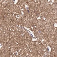
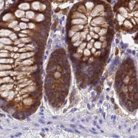

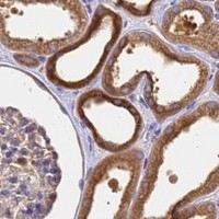


Comprehensive Human Protein Atlas IHC scoring per tissue (reliability: Approved — Medium consistency between antibody staining and RNA expression data. Pending external verification. Caution, targets protein from more than one gene.). Rows are taken directly from the HPA tissue chart — click any row's HPA link to view the source.
| Tissue | Cell type | Level | Evidence | Source |
|---|---|---|---|---|
| Endometrium | Glandular cells | Medium | Protein (IHC) | HPA → |
| Kidney | Cells in tubules | High | Protein (IHC) | HPA → |
| Lymph node | Non-germinal center cells | High | Protein (IHC) | HPA → |
| Nasopharynx | Respiratory epithelial cells | High | Protein (IHC) | HPA → |
| Prostate | Glandular cells | High | Protein (IHC) | HPA → |
| Tissue | Cell type | Level | Evidence | Source |
|---|---|---|---|---|
| Adipose tissue | Adipocytes | Not detected | Protein (IHC) | HPA → |
| Heart muscle | Cardiomyocytes | Not detected | Protein (IHC) | HPA → |
| Skeletal muscle | Myocytes | Not detected | Protein (IHC) | HPA → |
| Smooth muscle | Smooth muscle cells | Not detected | Protein (IHC) | HPA → |
Use matched tissue controls and compartment-specific scoring to troubleshoot NME2 staining in paraffin sections (HPA tissue IHC; UniProt P22392 localisation).
PA1830 has IHC images from human intestinal cancer tissue, rat cerebellum, and mouse intestine, plus IF/ICC images from U20S and HELA cells (PA1830 image captions).
The rendered SKU is PA1830; its IHC(P) captions show human intestinal cancer tissue, rat cerebellum, and mouse intestine (PA1830 IHC image captions). Its IF/ICC captions show U20S and HELA cells, and the catalog lists Human, Mouse, and Rat reactivity (PA1830 IF/ICC image captions; catalog: PA1830 reactivity).
Which to pick: For tissue IHC, choose PA1830: its own captions document paraffin sections, but do not report the fixative (PA1830 IHC image captions). For IF/ICC, PA1830 has cell image captions and lists both applications (PA1830 IF/ICC image captions; catalog: PA1830 applications). For cross-species IHC, PA1830 has Human, Mouse, and Rat reactivity and IHC captions for each; M01762-1 is listed as a rabbit monoclonal with the same reactivity and IHC/IF applications, but has no supplied IHC/IF image captions (catalog: PA1830 and M01762-1).