This website uses cookies to ensure you get the best experience on our website.


- Table of Contents
Plan chromogenic OAT IHC using high enterocyte staining in duodenum and small intestine as a reference (HPA tissue IHC). This guide covers paraffin-section antibody conditions (datasheet A01126-2) and scoring of granular cytoplasmic staining (HPA tissue IHC).

Expected localisation, validated protocols, controls and antibodies — the at-a-glance facts below, then the full design guide.
| Expected localisation | Granular cytoplasm in tissue (HPA tissue IHC); mitochondrial matrix expected (UniProt) | |
| Staining pattern | Most cells show granular cytoplasmic staining (HPA tissue IHC) | |
| Antigen retrieval | EDTA pH 8.0 HIER, heat-mediated (datasheet A01126-2) | |
| Positive control | Duodenum+4 more · see all | |
| Negative control | Adipose tissue+4 more · see all |
| Fixation | Keep formalin fixation consistent across sections. (standard IHC practice; not target-specific) | |
| Caveat | Hepatocytes stain low while intestinal enterocytes stain high (HPA tissue IHC) | |
| Regulation | Intestine-enhanced RNA expression (HPA tissue RNA) | |
| Isoform / epitope | 2 isoforms; N-termini differ; epitope unknown (UniProt) |
The catalog antibody has an IHC-P protocol (datasheet: A01126-2). The published examples cover liver sections (PMC9056925) and lung tissue with BALF cells (PMC10907606).
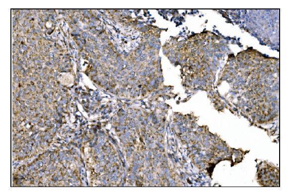
| Sample | Paraffin-embedded human lung cancer tissue; fixative not specified (datasheet A01126-2) |
| Fixation | Image fixative and duration unreported (datasheet A01126-2); verify before use. |
| Sectioning | 4–5 µm sections on charged slides (standard) |
| Deparaffinisation | Xylene, graded ethanol series to water (standard) |
| Antigen retrieval | Heat retrieval: EDTA pH 8.0 (datasheet A01126-2); 20 min, 95–100 °C (standard) |
| Peroxidase block | 3% H2O2, 10 min, room temperature (standard) |
| Blocking | 10% goat serum (datasheet A01126-2) |
| Primary antibody | Rabbit anti-OAT, 2-5μg/ml (datasheet A01126-2) |
| Primary incubation | Overnight at 4 °C (datasheet A01126-2) |
| Detection | Streptavidin-biotin complex (SABC), DAB chromogen (datasheet A01126-2) |
| Counterstain | Hematoxylin, blue, dehydrate and mount (standard) |
| Expected result | OAT-positive staining in enterocytes of duodenum (HPA tissue IHC: High). HPA tissue profile: Cytoplasmic expression with a granular pattern in most cells. No signal in the no-primary control. |
OAT is a mitochondrial matrix enzyme with no transmembrane segment (UniProt P04181). In paraffin section IHC, expect granular cytoplasmic staining, strongest in duodenal and small intestinal enterocytes (HPA: High in both). HPA rates its tissue staining Enhanced, reflecting high consistency between antibody staining and RNA expression (HPA: reliability description). Assess the named cell population rather than assigning one intensity to an entire tissue.
| Granular cytoplasmic staining is strong in intestinal enterocytes. | This matches the reported tissue pattern and High enterocyte staining (HPA: tissue IHC). The granular appearance is consistent with a mitochondrial matrix protein (UniProt P04181: subcellular location). Judge intensity against nearby cells and the same detection run; HPA levels describe observed staining, not a required numerical score. |
| Signal is mainly nuclear, membranous, or uniformly fills the cytoplasm. | This departs from the granular cytoplasmic IHC profile (HPA: tissue IHC). Check morphology, counterstain, and controls before calling it OAT. HPA also reports an additional nucleoplasmic location in ICC-IF (HPA: subcellular); that observation alone does not make predominantly nuclear chromogenic IHC an expected tissue pattern. |
| Strong signal appears in an HPA-listed undetected cell population. | For example, adipocytes are Not detected in adipose tissue (HPA: tissue IHC). Consider cross-reactivity or endogenous chromogenic activity and compare with a no-primary control (general IHC practice). The HPA designation applies to the specified cells; it does not establish that every cell in that section must be blank. |
| A broad, diffuse deposit covers cells and surrounding tissue. | This obscures the reported granular cytoplasmic pattern (HPA: tissue IHC). Review blocking, washes, detection chemistry, and the no-primary control (general IHC practice). Do not score diffuse color as OAT merely because the section includes an HPA-positive tissue. |
| Enterocytes show no discernible signal in a designated positive section. | Duodenal and small intestinal enterocytes are High in the HPA tissue record (HPA: tissue IHC). Recheck section identity, primary antibody performance, retrieval, and detection with a working positive control (general IHC practice). A failed run cannot establish that OAT is absent from the tested specimen. |
| Cell population and tissue | HPA reports High enterocyte staining in duodenum and small intestine, Medium staining in several other specified cell populations, and Low hepatocyte staining (HPA: tissue IHC). Choose and score controls by cell type; a weak liver section is a poor benchmark for the expected enterocyte intensity. |
| Intracellular location | OAT resides in the mitochondrial matrix and has no transmembrane segment (UniProt P04181). Expect a granular cytoplasmic IHC pattern (HPA: tissue IHC). HPA's additional nucleoplasmic ICC-IF finding is method-specific context, not evidence for a predominantly nuclear tissue IHC call (HPA: subcellular). |
| Antibody validation | HPA lists Enhanced IHC status for HPA040098 and CAB033576 (HPA: antibodies). Its overall tissue reliability is Enhanced (HPA: tissue IHC). These support comparison with the published pattern but do not prove that an unlisted catalog antibody, dilution, or detection system behaves identically. |
| Protein processing and isoforms | UniProt lists hepatic-form residues 26–439, renal-form residues 36–439, and two isoforms (UniProt P04181). If staining differs across specimens, check the chosen antibody's documented epitope before proposing a form-specific explanation; no epitope or form-specific IHC performance is supplied. |
| Retrieval and detection setup | Retrieval, primary concentration, blocking, and chromogen development require run controls and local optimization (general IHC practice). Neither source supplies an OAT-specific retrieval condition or fixation-sensitivity result; do not infer one from mitochondrial location, modifications, or HPA tissue intensity (UniProt P04181; HPA: tissue IHC). |
| Situation | Likely cause | Next action |
|---|---|---|
| High enterocyte control is blank. | The run may have failed at primary binding, retrieval, or detection (general IHC practice); HPA reports High staining in these cells (HPA: tissue IHC). | Verify the control's identity and preserved morphology, then check reagent activity and each detection step using the antibody's documented IHC-P instructions (general IHC practice). Do not infer an OAT-specific fixation failure from this result. |
| Granular color is too faint to distinguish from counterstain. | Insufficient developed signal or heavy counterstain can hide a cellular pattern (general IHC practice). | Compare with the High enterocyte control (HPA: tissue IHC); adjust development or counterstain under a controlled run (general IHC practice). Record the change before comparing specimen intensities. |
| No-primary control develops the same color as test sections. | Primary-independent background or endogenous detection activity is plausible (general IHC practice). | Check detection-only reagents and apply the appropriate endogenous-activity block for the enzyme system in use (general IHC practice). Reassess the reported granular cytoplasmic pattern only after background is controlled (HPA: tissue IHC). |
| Strong color appears in adipocytes or other undetected HPA cell types. | Cross-reactivity or detection background is possible; adipocytes are Not detected in HPA's tissue record (HPA: tissue IHC). | Compare the exact cell population with its HPA designation and a no-primary control (HPA: tissue IHC; general IHC practice). If the discrepancy persists, verify with an independently validated antibody (general IHC practice). |
| The signal is predominantly nuclear. | This conflicts with the granular cytoplasmic tissue pattern (HPA: tissue IHC), although an additional nucleoplasmic ICC-IF location is reported (HPA: subcellular). | Inspect focus, counterstain, and control sections; compare localization with a second validated reagent if available (general IHC practice). Report the compartment and method explicitly rather than converting the ICC-IF observation into an IHC expectation. |
| All cells have diffuse brown staining that conceals cell boundaries. | Excess detection development, inadequate washing, or nonspecific reagent binding may produce diffuse deposit (general IHC practice). | Compare no-primary and positive controls, then optimize washing, blocking, and chromogen development (general IHC practice). Score OAT only where the cellular pattern remains interpretable against HPA's granular cytoplasmic profile (HPA: tissue IHC). |
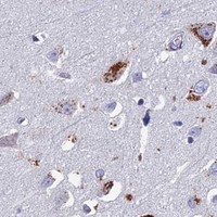


Comprehensive Human Protein Atlas IHC scoring per tissue (reliability: Enhanced — High consistency between antibody staining and RNA expression data.). Rows are taken directly from the HPA tissue chart — click any row's HPA link to view the source.
| Tissue | Cell type | Level | Evidence | Source |
|---|---|---|---|---|
| Duodenum | Enterocytes | High | Protein (IHC) | HPA → |
| Small intestine | Enterocytes | High | Protein (IHC) | HPA → |
| Adrenal gland | Glandular cells | Medium | Protein (IHC) | HPA → |
| Breast | Glandular cells | Medium | Protein (IHC) | HPA → |
| Bronchus | Respiratory epithelial cells | Medium | Protein (IHC) | HPA → |
| Tissue | Cell type | Level | Evidence | Source |
|---|---|---|---|---|
| Adipose tissue | Adipocytes | Not detected | Protein (IHC) | HPA → |
| Appendix | Endocrine cells | Not detected | Protein (IHC) | HPA → |
| Cerebellum | Cells in granular layer | Not detected | Protein (IHC) | HPA → |
| Colon | Endocrine cells | Not detected | Protein (IHC) | HPA → |
| Epididymis | Glandular cells | Not detected | Protein (IHC) | HPA → |
For OAT IHC in paraffin sections, troubleshoot retrieval, staining pattern, and controls together; interpret chromogenic signal against its expected mitochondrial distribution.
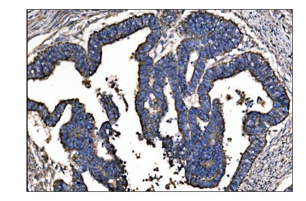
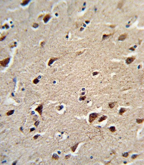
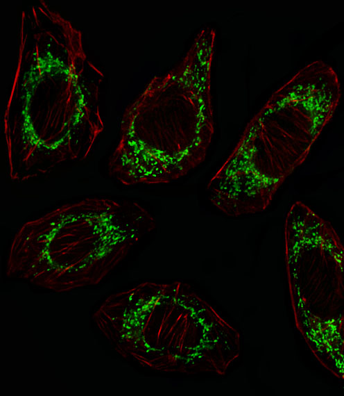
Both antibodies have human tissue IHC images (catalog IHC captions); A01126-1 also has an A549 cell IF image (A01126-1 IF caption). Both list human, mouse, and rat reactivity (catalog).
A01126-2 shows IHC in paraffin-embedded human lung cancer, rectal cancer, testicular cancer, and small intestine (A01126-2 IHC captions). A01126-1 shows DAB IHC in formalin-fixed, paraffin-embedded human brain and IF in A549 cells (A01126-1 IHC and IF captions).
Which to pick: For tissue IHC, choose A01126-2 when its broader set of human paraffin-section examples is useful; its IHC captions do not report a fixative (A01126-2 IHC captions). For IF/ICC, choose A01126-1: IF is listed at 1:10–1:50, with an A549 cell image at 1:25 (A01126-1 catalog and IF caption). For mouse or rat work, A01126-1 is a polyclonal candidate listing IHC-P, IF, and both species, but its supplied IHC and IF images show human samples only (A01126-1 catalog, IHC caption, and IF caption).