PAK2 / Serine/threonine-protein kinase PAK 2 · IHC design guide

Design Immunohistochemistry for PAK2

Use lymph node non-germinal center cells as a high staining control for cytoplasmic PAK2 in paraffin sections (HPA tissue IHC). This guide covers chromogenic IHC planning with the catalog antibody’s 2–5 μg/mL range (datasheet A01419-4) and cleavage dependent interpretation (UniProt).

Evidence assembled Oct 2026 · For research use; verify linked source records and product datasheet before use
Immunohistochemistry protocol sheet for PAK2 (IHC for PAK2): expected localisation General cytoplasmic staining (HPA tissue IHC), antibody A01419-4, validated IHC image, and IHC protocol steps
Printable PAK2 IHC protocol sheet — expected localisation General cytoplasmic staining (HPA tissue IHC), antibody A01419-4, controls and protocol steps. Open the full PAK2 IHC guide →

PAK2 Immunohistochemistry Experimental Design Guide

Expected localisation, validated protocols, controls and antibodies — the at-a-glance facts below, then the full design guide.

Must know before staining
Expected localisation General cytoplasmic staining (HPA tissue IHC)
Staining pattern Cytoplasmic; high in lymph node non-germinal center cells (HPA tissue IHC)
Antigen retrieval EDTA pH 8.0 HIER, heat-mediated (datasheet A01419-4)
Positive control ⓘ Lymph node+4 more · see all
Negative control ⓘ Adipose tissue+4 more · see all
Important caveats
Reasons your staining may differ from the expected pattern.
Fixation Keep fixation conditions consistent across sections. (standard IHC practice; not target-specific)
Caveat Apoptotic p34 can enter nuclei; signal depends on epitope (UniProt)
Regulation Expression regulation unspecified (UniProt)
Isoform / epitope No annotated isoforms; cleavage changes p27/p34 epitope coverage (UniProt)
Section 1

Recommended PAK2 IHC & IF Protocols

The catalog antibody uses EDTA pH 8.0 retrieval (datasheet A01419-4). The published IHC protocols below cover pancreatic tissue, gastric tissue, and unspecified sections (PMC6425405; PMC3975179; PMC6802609).

Recommended immunohistochemistry (IHC-P) protocol parameters
SampleParaffin-embedded human appendiceal adenocarcinoma tissue; fixative not specified (datasheet A01419-4)
FixationImage fixative and duration unreported (datasheet A01419-4); verify before use.
Sectioning4–5 µm sections on charged slides (standard)
DeparaffinisationXylene, graded ethanol series to water (standard)
Antigen retrievalHeat retrieval: EDTA pH 8.0 (datasheet A01419-4); 20 min, 95–100 °C (standard)
Peroxidase block3% H2O2, 10 min, room temperature (standard)
Blocking10% goat serum (datasheet A01419-4)
Primary antibodyRabbit anti-PAK2, 2-5 μg/ml (datasheet A01419-4)
Primary incubationOvernight at 4 °C (datasheet A01419-4)
DetectionStreptavidin-biotin complex (SABC), DAB chromogen (datasheet A01419-4)
CounterstainHematoxylin, blue, dehydrate and mount (standard)
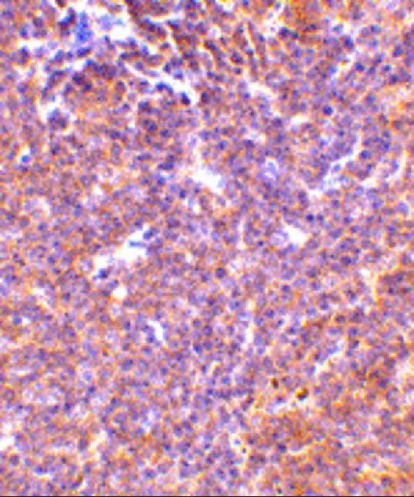
Expected resultPAK2-positive staining in non-germinal center cells of lymph node (HPA tissue IHC: High). HPA tissue profile: General cytoplasmic expression, most abundant in immune tissues. No signal in the no-primary control.
💡Decision noteStart with EDTA pH 8.0 for the catalog antibody (datasheet A01419-4). Use citrate pH 6.0 when reproducing the pancreatic or gastric protocols (PMC6425405; PMC3975179).
Section 2

What Is the Expected PAK2 Staining Pattern?

PAK2 is broadly expressed and should give predominantly cytoplasmic staining in tissue IHC, with strong signal in lymph-node non-germinal-center cells and moderate signal in several glandular or respiratory epithelial populations (HPA tissue IHC). HPA rates the tissue staining Approved, with medium consistency against RNA data (HPA tissue IHC). Nuclear and perinuclear localization are also possible (UniProt Q13177 subcellular); PAK2 has no transmembrane segment (UniProt Q13177 topology).

What am I looking at on my slide?
Strong cytoplasmic signal in lymph-node non-germinal-center cells, with weaker staining in surrounding populations.This fits the reported high staining in those cells and the broader cytoplasmic tissue pattern (HPA tissue IHC). Compare cell populations within the section; uniform intensity across every cell would be less consistent with this example.
Moderate cytoplasmic staining in appendix, colon, duodenum, cervix, or gallbladder glandular cells, or bronchial respiratory epithelium.Each listed population has medium staining in the supplied tissue observations (HPA tissue IHC). Intensity is relative to that tissue and assay; a moderate result does not imply equal abundance across organs.
Signal confined to extracellular material or an unrelated structure, without convincing cellular staining.That distribution does not match the reported cytoplasmic tissue pattern or PAK2's intracellular locations (HPA tissue IHC; UniProt Q13177 subcellular). Consider deposit, detection artifact, or nonspecific binding before scoring it as PAK2.
Strong staining in a population reported as not detected, such as adipocytes or bone-marrow hematopoietic cells.Treat this as unexpected for the cited examples (HPA tissue IHC). Check cell identification, cross-reactivity, and endogenous detection activity; an HPA not-detected call is an assay observation, not proof that expression is impossible.
No staining in lymph-node non-germinal-center cells while the counterstain and tissue morphology remain visible.The missing signal conflicts with the high-staining example (HPA tissue IHC). First investigate the IHC assay and controls; an individual negative section cannot by itself establish loss of PAK2 expression.
💡Expected PAK2 appearanceCall a section positive when lymph-node non-germinal-center cells show strong, predominantly cytoplasmic staining, with the expected cell boundaries and morphology; isolated extracellular color or indiscriminate staining of HPA not-detected populations is suspect (HPA tissue IHC; UniProt Q13177 subcellular).
How each factor affects the staining
Tissue and cell selectionLymph-node non-germinal-center cells provide a high-staining reference; several glandular epithelia are medium, whereas adipocytes and selected glial populations are reported not detected (HPA tissue IHC). Choose and score the named cell population, rather than treating an entire organ as uniformly positive or negative.
CompartmentGeneral tissue IHC expression is cytoplasmic (HPA tissue IHC). UniProt also lists nuclear, perinuclear, and membrane localization, with MYO18A-linked positioning of a PAK2 complex at the inner cell membrane (UniProt Q13177 subcellular). Nuclear staining therefore needs context, not automatic rejection.
Processing and epitope positionUniProt lists full-length PAK2 and cleavage products PAK-2p27 and PAK-2p34; the p34 fragment can translocate to the nucleus during apoptosis (UniProt Q13177 processing/function). The antibody epitope is not supplied, so this section cannot predict which forms it detects or attribute nuclear staining to cleavage.
IHC antibody evidenceOne listed antibody has Approved IHC status; another has Supported ICC status but no listed IHC rating (HPA antibodies). Approved tissue staining has medium consistency with RNA data (HPA tissue IHC). These labels support cautious pattern comparison, not an independent claim of IHC Enhanced validation.
Antigen retrievalRetrieval is a general paraffin-IHC optimization variable; the supplied sources give no PAK2-specific retrieval condition or fixation sensitivity. If signal is absent, assess retrieval alongside controls, without claiming that fixation masks a particular PAK2 epitope.
IF/ICC Q&A: should the IF image look cytoplasmic?HPA ICC-IF reports mainly nucleoplasm and nuclear speckles, with additional cytosol and focal-adhesion staining (HPA subcellular). That differs in emphasis from the tissue IHC profile (HPA tissue IHC). Interpret each application against its own evidence; use the separate IF/ICC guide for its workflow.
Why is my staining missing, weak or wrong?
SituationLikely causeNext action
No chromogenic signal in the lymph-node reference population.The result misses a high-staining HPA example; assay failure or unsuitable detection remains possible (HPA tissue IHC).Check the positive control, antibody application, retrieval, and detection steps as general IHC practice. Do not infer a PAK2-specific fixation effect from this result.
Color appears throughout the section, including blank spaces or extracellular material.Diffuse deposition does not fit the cellular cytoplasmic tissue profile (HPA tissue IHC). Background from detection chemistry or inadequate washing is possible (general IHC practice).Inspect reagent-only and no-primary controls; review blocking, wash steps, and development time as general IHC practice before assigning a PAK2 score.
Strong signal appears in adipocytes or bone-marrow hematopoietic cells.Those cells are reported not detected in the supplied HPA tissue observations; cross-reactivity or endogenous detection activity may explain unexpected color (HPA tissue IHC).Confirm cell identity and compare a no-primary control. Interpret any persistent cell-specific signal cautiously because the HPA result is an observed pattern, not an absolute biological negative.
Only nuclear staining is visible in a tissue IHC section.The dominant HPA tissue pattern is cytoplasmic, but UniProt allows nuclear PAK2 and HPA ICC-IF emphasizes nuclear structures (HPA tissue IHC; UniProt Q13177 subcellular; HPA subcellular).Check whether staining is cellular and reproducible in the expected positive population. Report the compartment explicitly; do not assign cleavage or artifact without supporting controls.
A glandular tissue stains faintly compared with lymph node.Several named glandular populations are medium or low, while lymph-node non-germinal-center cells are high (HPA tissue IHC).Score the specified cell population against an on-run positive reference. Avoid increasing development solely to force all tissues to match the lymph-node intensity.
A second antibody produces a different compartment pattern.The listed antibodies have different application evidence: one Approved for IHC and one Supported for ICC, with no listed IHC rating for the latter (HPA antibodies). Epitope locations are unavailable.Compare only within each antibody's validated application, review controls, and record the disagreement. Do not assume both antibodies recognize the same PAK2 forms.

Sample controls for PAK2 IHC & IF

🧪Run lymph node first: non-germinal center cells should stain (HPA: High). Use adipose tissue adipocytes as the negative reference (HPA: Not detected); within the lymph node slide, treat morphologically identifiable unstained cells as internal negative references without assigning a specific cell type (HPA: no lymph node cell type designated negative).
Positive control tissue: Lymph node (Non-germinal center cells, HPA High)
Negative control tissue: Adipose tissue (HPA Not detected)
ICC-IF cell lines (HPA subcellular resource): HPA ICC-IF images show PAK2 in A-431, MCF-7, U2OS, RPTEC/TERT1, hTERT-RPE1 (serum starved), with annotated localisation: Nucleoplasm (supported), Nuclear speckles (supported) (HPA subcellular).
Technical controls: Include a no-primary, secondary-only control; an isotype control matched to the rabbit primary’s clonality; and a PAK2 knockout sample or peptide competition if the immunizing peptide is available (A01419-4 caption: rabbit primary; standard IHC practice). For lymph node, quench endogenous peroxidase and check for endogenous biotin background before scoring the biotin/SABC-DAB signal (A01419-4 caption: biotinylated secondary and SABC-DAB; standard IHC practice).
⚠️Feasibility: A target-specific fixation window or fixation effect is unreported, and the selected A01419-4 paraffin-section caption does not report a fixative (A01419-4 caption). That caption uses heat retrieval in EDTA at pH 8.0, but does not establish retrieval dependence in lymph node (A01419-4 caption). The supplied evidence does not establish frozen sections or IF as easier; endogenous peroxidase in lymph node myeloid cells can confound chromogenic scoring (standard IHC practice).

HPA tissue IHC evidence for PAK2

Comprehensive Human Protein Atlas IHC scoring per tissue (reliability: Approved — Medium consistency between antibody staining and RNA expression data.). Rows are taken directly from the HPA tissue chart — click any row's HPA link to view the source.

Positive expression · recommended positive controls

TissueCell typeLevelEvidenceSource
Lymph node Non-germinal center cells High Protein (IHC) HPA →
Appendix Glandular cells Medium Protein (IHC) HPA →
Bronchus Respiratory epithelial cells Medium Protein (IHC) HPA →
Cervix Glandular cells Medium Protein (IHC) HPA →
Colon Glandular cells Medium Protein (IHC) HPA →

Undetected expression · recommended negative controls

TissueCell typeLevelEvidenceSource
Adipose tissue Adipocytes Not detected Protein (IHC) HPA →
Bone marrow Hematopoietic cells Not detected Protein (IHC) HPA →
Caudate Glial cells Not detected Protein (IHC) HPA →
Cerebellum Cells in granular layer Not detected Protein (IHC) HPA →
Cerebral cortex Glial cells Not detected Protein (IHC) HPA →
Section 3

Advanced PAK2 IHC Tips

Use the catalog antibody’s paraffin-section conditions as the IHC starting point, then judge PAK2 staining by cell type, compartment, and controls.

What retrieval should I try first if PAK2 staining is weak in paraffin sections?
Start with heat-mediated antigen retrieval in EDTA buffer at pH 8.0 (datasheet A01419-4). The documented paraffin-section image used this retrieval before incubation with 2 μg/ml primary antibody overnight at 4°C, so repeat those conditions when investigating weak staining (datasheet A01419-4). If signal remains weak, compare a small retrieval time series on matched sections while holding antibody concentration and detection constant; excessive heating can damage tissue morphology (standard IHC practice). Score glandular cells in appendix alongside a negative-primary control, because their reported staining is medium and background can otherwise be mistaken for improvement (HPA: Appendix glandular cells, Medium).
Could fixation explain variable PAK2 staining between sections?
The selected paraffin-section caption does not state a fixative, and target-specific PAK2 fixation sensitivity is unknown (datasheet A01419-4). Record each specimen’s fixative, fixation duration, processing history, and section age before comparing staining, since these variables can alter antigen accessibility in IHC (standard IHC practice). Run differently processed sections through the same pH 8.0 EDTA retrieval and 2 μg/ml primary-antibody conditions, then compare signal within the same cell population (datasheet A01419-4; standard IHC practice). Do not infer a PAK2-specific fixation effect from its cytoplasmic or nuclear distribution or its modified residues; those annotations describe the protein, not fixation performance (UniProt Q13177: subcellular location and modified residues).
How should I assess nuclear staining when PAK2 is usually cytoplasmic in tissue IHC?
Assess cytoplasmic staining first, since the tissue IHC profile describes general cytoplasmic expression, especially in immune tissues (HPA: tissue IHC profile). Nuclear staining is biologically plausible: PAK2 is annotated in cytoplasm and nucleus, and its cleavage product PAK-2p34 can translocate to the nucleus during apoptosis (UniProt Q13177: subcellular location and processing). However, nuclear speckles and nucleoplasm are reported from subcellular IF observations, so their appearance alone does not validate a paraffin-section IHC pattern (HPA: subcellular location). Compare nuclear and cytoplasmic signal in intact cells on matched sections, with a negative-primary control and preserved morphology, before assigning a compartment-specific score (standard IHC practice).
Can this stain distinguish full-length PAK2 from its apoptotic cleavage products?
No epitope location is supplied for the catalog antibody, so its staining cannot be assigned to full-length PAK2 or a specific cleavage product from this caption alone (datasheet A01419-4). The record lists no isoforms but does list full-length residues 2–524, PAK-2p27 residues 2–212, and PAK-2p34 residues 213–524 (UniProt Q13177: isoforms and processing). Because cleavage separates these regions, an antibody’s documented epitope would determine which forms it can recognize (UniProt Q13177: processing; standard immunostaining principle). Treat altered nuclear staining as a hypothesis about processing and check epitope documentation or an orthogonal assay before interpreting it as PAK-2p34 (UniProt Q13177: apoptotic cleavage).
How can I investigate PAK2 localisation by IF alongside the tissue IHC result?
On the separate IF/ICC guide, multiplex PAK2 with a cell-type marker chosen for the population being examined, such as an immune-cell marker in lymph node, where non-germinal center cells stain highly (HPA: Lymph node non-germinal center cells, High). Choose a fluorophore whose emission is distinct from the marker and from tissue autofluorescence, and include single-label and no-primary controls to inspect bleed-through and background (standard IF practice). Permeabilise cells to access cytosolic and nuclear epitopes; PAK2 has no transmembrane segment and is reported in both compartments (UniProt Q13177: topology and subcellular location). Evaluate nuclear speckles, nucleoplasm, cytosol, and focal adhesions as possible IF patterns without assuming that the paraffin-section IHC conditions transfer to IF (HPA: subcellular location; datasheet A01419-4).
What should I check if DAB appears outside plausible PAK2-positive cells?
First inspect a section processed without primary antibody and compare its DAB deposit with the test section; the documented method uses a biotinylated secondary, avidin-biotin complex, and DAB (datasheet A01419-4). Include a peroxidase-blocking step and check for endogenous biotin-related signal when using that detection system, as these are general chromogenic IHC controls (standard IHC practice). If diffuse staining persists, titrate the primary around the documented 2 μg/ml concentration and reassess blocking and washing while keeping exposure and development comparable (datasheet A01419-4; standard IHC practice). Judge residual signal against cell-resolved staining, since reported PAK2 tissue expression varies by cell population rather than uniformly across a section (HPA: tissue IHC profile).
How should I quantify PAK2 IHC across sections with different cell mixtures? ⚠ ANSWER MARKED FOR VERIFICATION
Define the scored compartment and cell population before comparing sections; tissue IHC describes chiefly cytoplasmic expression and reports different levels among cell types (HPA: tissue IHC profile). For glandular cells, record the percentage at each intensity and calculate an H-score as 1 × % weak + 2 × % moderate + 3 × % strong, using the same thresholds across slides (standard IHC scoring practice). For dispersed immune cells, report positive cells per mm² of viable tissue or the percentage positive among annotated immune cells, and state which denominator you used (standard IHC quantification practice). Exclude folds, necrosis, and damaged edges, and normalize comparisons to the same cell class and sampled area (standard IHC practice).
When is a strong PAK2 stain convincing rather than an IHC artefact?
A convincing result follows intact cell boundaries and a plausible compartment, with cytoplasmic tissue staining supported by the reported profile and nuclear localisation also possible for PAK2 (HPA: tissue IHC profile; UniProt Q13177: subcellular location). Compare cell identity: high staining in lymph-node non-germinal center cells and medium staining in appendix glandular cells are reported reference patterns, whereas adipocytes are listed as undetected (HPA: tissue IHC cell-level results). Treat staining concentrated at section edges, in necrosis, or in a no-primary control as suspect, and inspect for endogenous peroxidase signal before scoring DAB (standard IHC practice). Interpret discrepancies cautiously because the tissue IHC profile is rated Approved with medium consistency against RNA expression (HPA: tissue IHC reliability).
Boster reagents

Best PAK2 / Serine/threonine-protein kinase PAK 2 IHC Antibodies

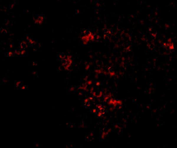
Anti-PAK2 IHC images cover human paraffin sections and mouse and rat tissue (catalog: A01419-4 IHC captions); IF images cover MCF-7 cells and mouse spleen cells (catalog: IF captions).

Real IHC data IHC analysis of PAK2 using anti-PAK2 antibody (A01419-4). PAK2 was detected in a paraffin-embedded section of human appendiceal adenocarcinoma tissue. Heat mediated antigen retrieval was performed in EDTA buffer (pH 8.0, epitope retrieval solution). The tissue section was blocked with 10% goat serum. The tissue section was then incubated with 2 μg/ml rabbit anti-PAK2 Antibody (A01419-4) overnight at 4°C. Biotinylated goat anti-rabbit IgG was used as secondary antibody and incubated for 30 minutes at 37°C. The tissue section was developed using Strepavidin-Biotin-Complex (SABC) (Catalog # SA1022) with DAB as the chromogen.
Anti-PAK2 Antibody ®
Cat # A01419-4
Real IHC data Immunohistochemistry Validation of PAK2 in Mouse Spleen Tissue Immunohistochemical analysis of paraffin-embedded mouse kidney tissue using anti-PAK2 antibody (A01419-2) at 10 μg/ml. Tissue was fixed with formaldehyde and blocked with 10% serum for 1 h at RT; antigen retrieval was by heat mediation with a citrate buffer (pH6). Samples were incubated with primary antibody overnight at 4˚C. A goat anti-rabbit IgG H&L (HRP) at 1/250 was used as secondary. Counter stained with Hematoxylin.
Anti-PAK2 Antibody
Cat # A01419-2

A01419-4 has IHC images from human appendiceal adenocarcinoma and lung cancer, plus mouse and rat skeletal muscle, and an IF image from MCF-7 cells (catalog: A01419-4 image captions). A01419-2 has an IHC image described as mouse kidney in the caption body and an IF image from mouse spleen cells (catalog: A01419-2 image captions).

Which to pick: For tissue IHC across human, mouse and rat samples, choose A01419-4: its captions show paraffin sections retrieved with EDTA at pH 8.0; the fixative is unreported (catalog: A01419-4 IHC captions). For IF/ICC, A01419-4 has an MCF-7 cell IF image, while A01419-2 has a mouse spleen cell IF image (catalog: IF captions). A01419-2 offers a mouse kidney paraffin IHC example with fixation and citrate retrieval at pH 6; its listed reactivity includes human, mouse and rat, but its stated IHC validation is in mouse samples (catalog: A01419-2 IHC caption and dilution notes). The selected A01419-4 tissue-IHC caption documents paraffin sections, but does not specify the fixative (selected-SKU IHC image A01419-4).

Each figure is that product's own IHC / IF validation image from its datasheet.

References

  1. UniProt Consortium. UniProt entry Q13177 (PAK2_HUMAN, Serine/threonine-protein kinase PAK 2).
  2. Human Protein Atlas. PAK2 tissue IHC expression (reliability: Approved).
  3. Human Protein Atlas. PAK2 subcellular location (ICC-IF): Mainly localized to the nucleoplasm and nuclear speckles. In addition localized to the cytosol and focal adhesion sites..
  4. Human Protein Atlas. PAK2 antibody validation summary (2 antibodies).
  5. P21 activated kinase 2 promotes pancreatic cancer growth and metastasis. Oncology letters 2019 — PMC6425405.
  6. Activation of P21-activated protein kinase 2 is an independent prognostic predictor for patients with gastric cancer. Diagnostic pathology 2014 — PMC3975179.
  7. Human Umbilical Cord Mesenchymal Stem Cells Ameliorate Hepatic Stellate Cell Activation and Liver Fibrosis by Upregulating MicroRNA-455-3p through Suppression of p21-Activated Kinase-2. BioMed research international 2021 — PMC7932777.
  8. MicroRNA-455-3p promotes TGF-β signaling and inhibits osteoarthritis development by directly targeting PAK2. Experimental & molecular medicine 2019 — PMC6802609.
  9. PubMed PMID:15489334 — UniProt-cited evidence.
  10. PubMed PMID:7744004 — UniProt-cited evidence.
  11. PubMed PMID:7556080 — UniProt-cited evidence.