This website uses cookies to ensure you get the best experience on our website.


- Table of Contents
Plan chromogenic paraffin IHC for PDIA3 with the catalog antibody at 0.5–1 μg/mL (datasheet PB9772). Assess cytoplasmic staining in glandular epithelia and compare it with cardiomyocytes, where staining was not detected (HPA tissue IHC).

Expected localisation, validated protocols, controls and antibodies — the at-a-glance facts below, then the full design guide.
| Expected localisation | Cytoplasmic tissue staining (HPA tissue IHC); ER lumen (UniProt) | |
| Staining pattern | Cytoplasmic staining, highest in glandular epithelia (HPA tissue IHC) | |
| Antigen retrieval | Citrate pH 6 HIER, heat-mediated (datasheet PB9772) | |
| Positive control | Cerebellum+4 more · see all | |
| Negative control | Heart muscle+1 more · see all |
| Fixation | Keep fixation consistent across sections (standard IHC practice; not target-specific) | |
| Caveat | Cardiomyocytes and skeletal myocytes may lack detectable staining (HPA tissue IHC) | |
| Regulation | No specific expression regulator reported (UniProt) | |
| Isoform / epitope | No isoforms listed; mature chain starts at residue 25 (UniProt) |
The catalog antibody’s IHC-P protocol (datasheet: PB9772) is followed by published PDIA3 staining protocols for cervical carcinoma, gallbladder carcinoma, and lung cancer tissue (PMC11117164; PMC3691597; PMC5722536).
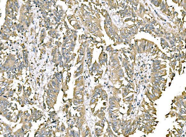
| Sample | Paraffin-embedded Human Lung Cancer tissue; fixative not specified (datasheet PB9772) |
| Fixation | Image fixative and duration unreported (datasheet PB9772); verify before use. |
| Sectioning | 4–5 µm sections on charged slides (standard) |
| Deparaffinisation | Xylene, graded ethanol series to water (standard) |
| Antigen retrieval | Heat retrieval: Citrate pH 6, 20 min (datasheet PB9772) |
| Peroxidase block | 3% H2O2, 10 min, room temperature (standard) |
| Blocking | 10% goat serum (datasheet PB9772) |
| Primary antibody | Rabbit anti-PDIA3, 0.5-1μg/ml (datasheet PB9772) |
| Primary incubation | Overnight at 4 °C (datasheet PB9772) |
| Detection | Streptavidin-biotin complex (SABC), DAB chromogen (datasheet PB9772) |
| Counterstain | Hematoxylin, blue, dehydrate and mount (standard) |
| Expected result | PDIA3-positive staining in processes in granular layer of cerebellum (HPA tissue IHC: High). HPA tissue profile: Cytoplasmic expression in all tissues with highest levels in glandular epithelia. No signal in the no-primary control. |
PDIA3 is an endoplasmic reticulum (ER) luminal protein with no transmembrane segment (UniProt P30101 topology). In paraffin section IHC, expect predominantly cytoplasmic staining, especially in glandular epithelia (HPA: tissue IHC, Enhanced reliability). HPA reports strong staining in several other cell types, including kidney tubular cells and cerebral cortical neurons (HPA: High). Assess the stained cell population as well as its compartment.
| Cytoplasmic staining in glandular cells, with a stronger signal than nearby unstained areas. | This fits the broad tissue profile: HPA describes cytoplasmic expression with the highest levels in glandular epithelia (HPA: tissue IHC). An ER-associated distribution is consistent with the protein's location, although chromogenic IHC may not resolve an ER network (UniProt P30101; HPA: subcellular ICC-IF). |
| Prominent staining confined to nuclei or cell surfaces, without a convincing cytoplasmic component. | Treat this as a localization mismatch requiring review of controls and staining conditions. The established main location is the ER (HPA: subcellular ICC-IF; UniProt P30101). UniProt also reports PDIA3 in melanosome fractions, so an unusual pattern needs context rather than an automatic rejection (UniProt P30101). |
| Strong staining in cardiomyocytes or skeletal myocytes while an expected positive cell population is weak. | Question specificity or endogenous detection activity: HPA reports PDIA3 as not detected in cardiomyocytes and skeletal myocytes (HPA: tissue IHC). Compare a no-primary control and a known-positive tissue on the same run; the HPA categories describe observed staining, not an absolute biological absence. |
| Uniform chromogen across cells, stroma, and blank spaces, obscuring cell boundaries. | This is more consistent with nonspecific background or detection-system activity than a cell-associated PDIA3 pattern. Interpret the section only after controls distinguish background from cytoplasmic signal; HPA's tissue profile concerns cell staining and does not validate a uniform haze (HPA: tissue IHC). |
| No signal in glandular cells of endometrium or in kidney tubular cells. | A negative run is suspect when these HPA high-staining populations are present (HPA: High in endometrial glandular cells; High in kidney tubular cells). Check tissue preservation and assay controls, then optimize the IHC workflow. Their reported high staining does not establish PDIA3-specific fixation sensitivity. |
| Cell population and tissue selection | The positive control must contain the scored cell type: HPA rates endometrial and epididymal glandular cells, kidney tubular cells, and lung type I alveolar cells High, but cardiomyocytes and skeletal myocytes Not detected (HPA: tissue IHC). A whole-section average can conceal these differences. |
| ER localization and optical resolution | PDIA3 resides in the ER lumen without a transmembrane segment (UniProt P30101 topology), and ICC-IF assigns the ER as its enhanced main location (HPA: subcellular ICC-IF). Interpret a chromogenic cytoplasmic pattern at cellular scale; do not require an individually resolved reticular network in paraffin sections. |
| Protein processing and isoforms | UniProt annotates a signal peptide at residues 1–24, a mature chain at 25–505, and no listed isoforms (UniProt P30101). These annotations can guide review of an antibody's stated epitope, but the supplied record gives no catalog antibody epitope or PDIA3-specific retrieval requirement. |
| Antibody validation | HPA assigns Enhanced IHC validation to HPA002645, HPA003230, CAB011199, and CAB075744 (HPA: antibodies). This supports comparison with HPA tissue patterns, but validation of those antibodies does not by itself establish the performance of an unspecified catalog antibody. |
| Antigen retrieval and endogenous activity | Retrieval choice and checks for endogenous detection activity are general paraffin IHC workflow decisions. The supplied UniProt and HPA records report no PDIA3-specific retrieval condition or fixation effect; assess changes with on-run positive and no-primary controls rather than attributing a result to PDIA3 chemistry. |
| Situation | Likely cause | Next action |
|---|---|---|
| Expected-positive glandular or tubular cells are blank. | The run may have insufficient detectable signal, or the relevant cells may be absent from the section. HPA rates endometrial glandular and kidney tubular cells High (HPA: tissue IHC); the record does not identify a PDIA3-specific cause of signal loss. | Confirm the cell population on the counterstain, verify the positive control, and review antibody dilution, retrieval, and detection as general IHC parameters. Reassess only after the positive control stains. |
| Cardiomyocytes or skeletal myocytes stain strongly. | This conflicts with HPA's Not detected categories for those cell types (HPA: tissue IHC). Nonspecific antibody binding or endogenous detection activity are possible general IHC explanations, not demonstrated causes in this section. | Compare a no-primary control and an expected-positive tissue processed in the same run. If the control remains clean, review antibody specificity and score the observed cell types explicitly. |
| Only nuclei show a convincing signal. | A nuclear-only pattern does not fit PDIA3's ER location (UniProt P30101; HPA: subcellular ICC-IF). The evidence does not identify which staining step caused the mismatch. | Check the no-primary control and cellular morphology, then repeat with a validated positive control. Do not score isolated nuclear chromogen as the expected PDIA3 pattern. |
| Diffuse staining makes the cytoplasm hard to score. | Excess background can mask cell-associated signal in chromogenic IHC. HPA reports a cytoplasmic tissue pattern, not uniform section-wide staining (HPA: tissue IHC). | Inspect a no-primary control, review blocking and detection steps, and adjust antibody concentration only as a general IHC optimization. Compare cell-associated staining with background on the same slide. |
| A tissue looks negative despite reports of widespread expression. | HPA's broad cytoplasmic profile includes cell-specific exceptions: cardiomyocytes and skeletal myocytes are Not detected, while smooth muscle cells are Low (HPA: tissue IHC). A section can also lack the reported positive cell population. | Identify the tissue and scored cells before calling assay failure. Include a documented High population as the run control, and report the local result without converting HPA's categories into absolute absence. |
| Can ICC-IF images define an IHC protocol or settle a disputed chromogenic pattern? | ICC-IF supports ER localization in cultured cells (HPA: subcellular ICC-IF); it does not specify a paraffin IHC retrieval or detection procedure. The two applications have different specimen and readout conditions. | Use the ICC-IF result as a compartment check only. Judge paraffin IHC against HPA tissue staining and an on-run positive control; consult the separate IF/ICC guide for that application. |




Comprehensive Human Protein Atlas IHC scoring per tissue (reliability: Enhanced — High consistency between antibody staining and RNA expression data.). Rows are taken directly from the HPA tissue chart — click any row's HPA link to view the source.
| Tissue | Cell type | Level | Evidence | Source |
|---|---|---|---|---|
| Cerebellum | Processes in granular layer | High | Protein (IHC) | HPA → |
| Cerebral cortex | Neuronal cells | High | Protein (IHC) | HPA → |
| Duodenum | Endocrine cells | High | Protein (IHC) | HPA → |
| Endometrium | Glandular cells | High | Protein (IHC) | HPA → |
| Epididymis | Glandular cells | High | Protein (IHC) | HPA → |
Troubleshoot PDIA3 staining in paraffin sections by checking retrieval, compartment pattern, controls and scoring before interpreting chromogenic signal.
PB9772 and M01464-4 have IHC images from human paraffin sections (catalog IHC captions); PB9772 also has an IF/ICC image from U20S cells (PB9772 IF caption).
PB9772 will render with its human lung cancer paraffin-section IHC image (PB9772 IHC caption); its catalog lists IHC, IF and ICC applications and Human, Mouse and Rat reactivity (PB9772 catalog). M01464-4 will render with its human rectal cancer paraffin-section IHC image (M01464-4 IHC caption); its catalog lists IHC and Human reactivity (M01464-4 catalog).
Which to pick: For human paraffin-section IHC, M01464-4 is a mouse monoclonal with a rectal cancer tissue image, while PB9772 is a rabbit antibody with a lung cancer tissue image (M01464-4 and PB9772 catalogs and IHC captions). For IF/ICC or cross-species testing, choose PB9772: its catalog lists IF/ICC and Human, Mouse and Rat reactivity, and its IF caption documents U20S cells (PB9772 catalog and IF caption). The fixative is unreported for both illustrated paraffin sections (PB9772 and M01464-4 IHC captions).