PHB2 / Prohibitin-2 · IHC design guide

Design Immunohistochemistry for PHB2

Plan PHB2 paraffin-section IHC around its granular cytoplasmic tissue pattern (HPA tissue IHC). Compare high-staining glandular cells with low-staining adipocytes when assessing signal (HPA tissue IHC).

Evidence assembled Oct 2026 · For research use; verify linked source records and product datasheet before use
Immunohistochemistry protocol sheet for PHB2 (IHC for PHB2): expected localisation Granular cytoplasmic staining in tissue (HPA tissue IHC), antibody M03315, validated IHC image, and IHC protocol steps
Printable PHB2 IHC protocol sheet — expected localisation Granular cytoplasmic staining in tissue (HPA tissue IHC), antibody M03315, controls and protocol steps. Open the full PHB2 IHC guide →

PHB2 Immunohistochemistry Experimental Design Guide

Expected localisation, validated protocols, controls and antibodies — the at-a-glance facts below, then the full design guide.

Must know before staining
Expected localisation Granular cytoplasmic staining in tissue (HPA tissue IHC)
Staining pattern Widespread granular cytoplasmic staining (HPA tissue IHC)
Antigen retrieval EDTA pH 8.0 HIER, heat-mediated (datasheet M03315)
Positive control ⓘ Adrenal gland+4 more · see all
Negative control ⓘ None in HPA (detected in all 45 tissues); use no-primary + isotype controls
Important caveats
Reasons your staining may differ from the expected pattern.
Fixation Keep fixation consistent across sections (standard IHC practice; not target-specific); optimize empirically.
Caveat Adipocytes may stain weakly despite broad tissue expression (HPA tissue IHC)
Regulation Staining-intensity regulation is unreported (UniProt)
Isoform / epitope 2 isoforms; epitope differences are unreported (UniProt)
Section 1

Recommended PHB2 IHC & IF Protocols

The catalog antibody’s IHC-P protocol is followed by three published PHB2 chromogenic IHC protocols (datasheet M03315; PMC5343968; PMC3958913; PMC5567385).

Recommended immunohistochemistry (IHC-P) protocol parameters
SampleParaffin-embedded human gallbladder adenocarcinoma tissue; fixative not specified (datasheet M03315)
FixationImage fixative and duration unreported (datasheet M03315); verify before use.
Sectioning4–5 µm sections on charged slides (standard)
DeparaffinisationXylene, graded ethanol series to water (standard)
Antigen retrievalHeat retrieval: EDTA pH 8.0 (datasheet M03315); 20 min, 95–100 °C (standard)
Peroxidase block3% H2O2, 10 min, room temperature (standard)
Blocking10% goat serum (datasheet M03315)
Primary antibodyMouse monoclonal (clone 2B5) anti-PHB2, 2μg/ml (datasheet M03315)
Primary incubationOvernight at 4 °C (datasheet M03315)
DetectionStreptavidin-biotin complex (SABC), DAB chromogen (datasheet M03315)
CounterstainHematoxylin, blue, dehydrate and mount (standard)
Expected resultPHB2-positive staining in glandular cells of adrenal gland (HPA tissue IHC: High). HPA tissue profile: Ubiquitous cytoplasmic expression with a granular pattern. No signal in the no-primary control.
💡Decision noteStart with heat-mediated EDTA retrieval at pH 8.0 for the catalog antibody (datasheet M03315); the cited excerpts do not specify a retrieval recipe.
Section 2

What Is the Expected PHB2 Staining Pattern?

PHB2 is chiefly associated with the mitochondrial inner membrane and can also occur in the cytoplasm, nucleus and cell membrane; UniProt annotates no transmembrane segment (UniProt Q99623 localization and topology). In paraffin tissue sections, expect granular cytoplasmic staining across many cell types, with strong staining in selected cells (HPA: ubiquitous granular cytoplasmic profile; tissue IHC reliability Supported, with medium consistency against RNA expression).

What am I looking at on my slide?
Granular cytoplasmic staining in glandular cells or cardiomyocytes.This fits the reported tissue pattern and mitochondrial localization (HPA: granular cytoplasm; UniProt Q99623 localization). Adrenal, breast and endometrial glandular cells, and heart cardiomyocytes, are recorded as High (HPA: tissue IHC). Judge the pattern within identified cells, since a strongly stained structure alone does not establish which cell type produced the signal.
Predominantly nuclear staining, or a smooth membrane outline, with little granular cytoplasm.Reconsider antibody specificity and slide conditions: neither is the dominant tissue IHC appearance (HPA: granular cytoplasm). Nuclear and cell membrane localization are annotated, so those compartments alone cannot prove artefact (UniProt Q99623 localization). A nuclear pattern may also depend on cell state: UniProt describes mostly nuclear PHB2 in differentiated primary myoblasts (UniProt Q99623).
Strong staining confined to an unexpected cell population, while expected cells lack signal.Check cell identification and controls before calling cross-reactivity or endogenous detection activity (general IHC practice). PHB2 has low tissue specificity, and HPA lists no negative tissue group; unexpected cells are therefore not automatically true negatives (HPA: low tissue specificity; negative list empty). Compare the cellular pattern with the reported granular cytoplasm (HPA: tissue IHC).
Diffuse color across cells and surrounding section, obscuring granules.Treat this as background until a clean cellular pattern is resolved (general IHC practice). Uneven blocking, excess antibody or endogenous detection activity are possible workflow causes, not established PHB2 properties (general IHC practice). Reassess with appropriate control sections and detection controls; diffuse color cannot by itself confirm mitochondrial PHB2 (HPA: granular cytoplasmic tissue pattern; mitochondrial ICC-IF localization).
No discernible signal in a section selected for strong PHB2 staining.First confirm that the relevant cells are present: HPA reports High staining in bronchial respiratory epithelium, appendix, colon and duodenal endocrine cells, and several other listed populations (HPA: tissue IHC). Then review the assay and controls (general IHC practice). HPA's Supported rating and medium RNA agreement make these useful comparators, not guarantees for every specimen (HPA: reliability).
💡Expected PHB2 appearanceCall a positive result when identified cells show distinct granular cytoplasmic staining, often strong in HPA High populations; isolated diffuse color or a dominant smooth outline needs control review (HPA: tissue IHC; general IHC practice).
How each factor affects the staining
Cell population and comparator tissueChoose a documented High population as a positive comparator, such as breast glandular cells or heart cardiomyocytes (HPA: tissue IHC). Adipocytes, chondrocytes and epididymal glandular cells are listed as Low, not absent; they are weak comparators rather than proven negative controls (HPA: tissue IHC).
Compartment and cell stateMitochondrial localization supports a granular cytoplasmic reading (UniProt Q99623 localization; HPA: mitochondrial ICC-IF). UniProt also records nuclear and other locations, including a shift toward nuclear localization in differentiated primary myoblasts; assess unusual compartments in their cellular context (UniProt Q99623).
Antibody evidence and interpretation limitHPA lists IHC as Supported for HPA039874, CAB014889 and CAB026335; it does not list these IHC results as Enhanced (HPA: antibody validation). The tissue profile itself has medium consistency with RNA data, so staining pattern and controls remain relevant to each specimen (HPA: tissue reliability).
Detection chemistryWith chromogenic IHC, endogenous activity or nonspecific detection can contribute color independent of target binding; use an appropriate detection control when the pattern is diffuse or unexpected (general IHC practice). This is a general workflow consideration, not an HPA or UniProt claim about PHB2.
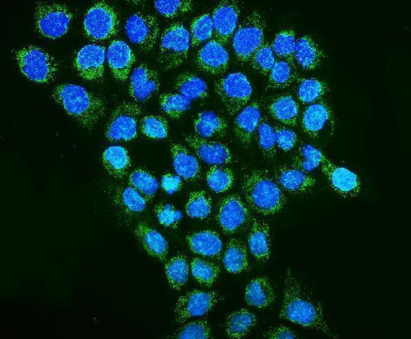
IF/ICC Q&A: should mitochondrial fluorescence appear?Yes: HPA summarizes ICC-IF localization as mitochondrial, and HPA039874 has Enhanced ICC evidence (HPA: subcellular summary; antibody validation). Interpret that as support for an IF localization check, not as an IHC-P protocol or evidence that every tissue section will show a resolved mitochondrial network.
Why is my staining missing, weak or wrong?
SituationLikely causeNext action
High-background chromogen masks the expected granules.Nonspecific detection, excess primary antibody or endogenous activity may obscure cellular staining (general IHC practice).Review the detection control, blocking and antibody titration, then look again for cell-bound granules (general IHC practice; HPA: granular cytoplasmic profile).
A documented High cell population appears unstained.The relevant cells may be absent from the section, or a staining step may have failed (general IHC practice).Verify cell identity and a positive comparator; review retrieval, primary antibody and detection steps as general IHC checks. HPA High is an observed level, not a guarantee for each specimen (HPA: tissue IHC).
Staining is mainly nuclear in a tissue section.It differs from the dominant tissue IHC profile, although nuclear PHB2 is annotated (HPA: granular cytoplasm; UniProt Q99623 localization).Confirm the cellular context and inspect controls and granular cytoplasmic signal before assigning the nuclear color to PHB2 (general IHC practice; HPA: tissue IHC).
Only Low-listed cells stain faintly.Weak staining may agree with the reported Low level; those cells are not documented as negative (HPA: adipocytes, chondrocytes and epididymal glandular cells Low).Compare a documented High population on a suitable control section before interpreting a faint result as assay failure (HPA: tissue IHC; general IHC practice).
Unexpected cells stain strongly while expected cells do not.Cell misidentification, cross-reactivity or endogenous detection activity are possibilities; widespread PHB2 expression also limits inference from cell type alone (general IHC practice; HPA: low tissue specificity).Recheck morphology, compare the reported granular pattern and inspect detection controls; avoid calling an unlisted cell population PHB2-negative by default (HPA: tissue IHC; general IHC practice).
IF/ICC shows diffuse fluorescence instead of a mitochondrial pattern.The result differs from HPA's mitochondrial ICC-IF summary, but does not establish a specific cause (HPA: subcellular summary).Check IF controls and compare with a mitochondrial marker as a general localization check; use the separate IF/ICC guide for assay design (general IF practice; HPA: mitochondrial ICC-IF localization).

Sample controls for PHB2 IHC & IF

🧪Run adrenal gland first and score its glandular cells, which HPA rates High for PHB2 (HPA: High in adrenal glandular cells). HPA lists no negative tissue and detects PHB2 in all 45 scored tissues, so no-primary and isotype sections carry the negative comparison; no cell population within the positive slide is established as PHB2-negative, and weaker staining in other cells should be treated only as a relative comparison (HPA: no negative rows; detected in all 45 scored tissues).
Positive control tissue: Adrenal gland (Glandular cells, HPA High)
Negative control tissue: None in HPA: PHB2 is detected in all 45 scored tissues. Use a no-primary (secondary-only) and an isotype control instead.
ICC-IF cell lines (HPA subcellular resource): HPA ICC-IF images show PHB2 in U-251MG, U2OS, A-431, U2OS, siRNA 1 (10x), U2OS, siRNA 2 (10x), U2OS, scrambled (10x), NIH 3T3, with annotated localisation: Mitochondria (enhanced) (HPA subcellular).
Technical controls: Include a no-primary, secondary-only section and a mouse IgG isotype control matched to the catalog antibody’s clonality if known; use PHB2 knockout material as a biological specificity control when available (caption: mouse primary antibody; standard IHC control practice). For the adrenal section, block endogenous peroxidase and check for endogenous biotin background because the reported detection uses a biotin-based complex and DAB (caption: biotinylated secondary, SABC and DAB; standard chromogenic IHC practice).
⚠️Feasibility: The selected M03315 paraffin-section caption does not report a fixative, and no target-specific fixation window or fixation effect is reported in the supplied evidence (caption: fixative unreported). Its staining used heat retrieval in EDTA at pH 8.0, but the evidence does not establish whether retrieval is required for every PHB2 antibody or specimen (caption: EDTA pH 8.0 heat retrieval). The supplied evidence does not establish that frozen sections or tissue IF are easier; HPA ICC-IF images show mitochondrial localization, while endogenous biotin or peroxidase background should be checked in the selected adrenal section rather than assumed to be a demonstrated tissue-specific artefact (HPA: mitochondria enhanced; caption: SABC and DAB; standard IHC control practice).

HPA tissue IHC evidence for PHB2

Comprehensive Human Protein Atlas IHC scoring per tissue (reliability: Supported — Medium consistency between antibody staining and RNA expression data.). Rows are taken directly from the HPA tissue chart — click any row's HPA link to view the source.

Positive expression · recommended positive controls

TissueCell typeLevelEvidenceSource
Adrenal gland Glandular cells High Protein (IHC) HPA →
Appendix Endocrine cells High Protein (IHC) HPA →
Breast Glandular cells High Protein (IHC) HPA →
Bronchus Respiratory epithelial cells High Protein (IHC) HPA →
Colon Endocrine cells High Protein (IHC) HPA →

Undetected expression · recommended negative controls

TissueCell typeLevelEvidenceSource
None in HPA: PHB2 is detected in all 45 scored tissues. Use a no-primary (secondary-only) and an isotype control instead.
Section 3

Advanced PHB2 IHC Tips

Troubleshoot PHB2 staining in paraffin sections by checking retrieval, compartment pattern, controls and scoring before interpreting biological differences.

How should I retrieve PHB2 antigen in paraffin sections?
Start with heat-mediated retrieval in EDTA at pH 8.0 for this antibody (datasheet M03315). The reported paraffin-section example used that retrieval before overnight incubation at 4°C with 2 µg/mL antibody (datasheet M03315). Keep section thickness, heating and cooling consistent across cases so differences in retrieval do not masquerade as differences in PHB2 staining (standard IHC practice). If signal is weak, first check reagent activity and the positive control, then adjust heating duration on matched sections while monitoring tissue damage (standard IHC practice). Record any departure from the reported retrieval conditions alongside the staining result (datasheet M03315; standard IHC practice).
Could fixation explain weak or variable PHB2 staining?
PHB2-specific sensitivity to fixation is unknown from the supplied evidence; the catalog image identifies paraffin-embedded tissue but does not state its fixative (datasheet M03315). Record the fixative, fixation duration and processing history for each specimen before comparing staining intensity (standard IHC practice). Excessive or uneven fixation can alter antigen accessibility in IHC generally, so compare similarly processed sections and include a known positive section in each run (standard IHC practice). If a specimen stains weakly, repeat staining with the stated EDTA pH 8.0 retrieval before attributing the result to PHB2 biology (datasheet M03315; standard IHC practice). Do not infer fixation tolerance from tissue staining patterns or PHB2 sequence annotations (HPA tissue IHC; UniProt Q99623).
Which staining compartments should I expect for PHB2?
Expect predominantly granular cytoplasmic staining in tissue sections, consistent with the reported ubiquitous tissue pattern and mitochondrial location (HPA tissue IHC; HPA subcellular). Assess whether granules occupy plausible cytoplasmic regions around intact nuclei before calling a section positive (HPA tissue IHC; standard IHC practice). PHB2 also has reported nuclear, cytoplasmic and cell-membrane locations, so a nonmitochondrial pattern needs biological context rather than automatic dismissal (UniProt Q99623 subcellular location). In primary myoblasts, the reported distribution shifts toward the nucleus with differentiation; that observation does not establish the same shift in every tissue (UniProt Q99623 subcellular location). Compare cellular compartments on the same slide with a no-primary control when the pattern is unexpected (standard IHC practice).
How do isoforms and epitope access affect PHB2 IHC?
PHB2 has 2 annotated isoforms, but the supplied antibody evidence does not map its epitope or establish isoform coverage (UniProt Q99623 isoforms; datasheet M03315). Interpret staining as antibody-detected PHB2 signal, not as an isoform-specific measurement, unless independent validation establishes that distinction (UniProt Q99623 isoforms; standard IHC practice). PHB2 is associated with the mitochondrial inner membrane despite having no annotated transmembrane segment, so sequence topology alone cannot identify which side of the membrane contains this antibody’s epitope (UniProt Q99623 topology and subcellular location). Acetylation and phosphorylation sites are annotated, but their effects on recognition by this antibody are unreported (UniProt Q99623 modified residues; datasheet M03315). If compartment patterns differ, compare matched retrieval conditions and an independent antibody with a mapped epitope (standard IHC practice).
How should I investigate PHB2 localisation by IF alongside IHC?
Use IF as a separate localisation check: the available subcellular images identify mitochondria as PHB2’s main location (HPA subcellular). Multiplex PHB2 with a mitochondrial marker and, where cell identity matters, a marker for the expected cell type; check each channel separately before interpreting overlap (HPA subcellular; standard IF practice). Choose a fluorophore in a channel with low tissue autofluorescence, often farther toward the red, and include unstained tissue to measure residual fluorescence (standard IF practice). Permeabilisation may be needed to reach an intracellular epitope, but its required strength and the epitope’s side of the inner membrane are unknown for this antibody (UniProt Q99623 topology; datasheet M03315). Optimize IF fixation and permeabilisation in that assay rather than transferring the paraffin-section conditions (datasheet M03315; standard IF practice).
What causes diffuse or misleading brown PHB2 staining?
Compare diffuse brown signal with the expected granular cytoplasmic pattern before treating it as PHB2 staining (HPA tissue IHC). The reported chromogenic example used a biotinylated secondary, avidin-biotin detection and DAB, so secondary-only and no-primary controls can help identify detection background (datasheet M03315; standard IHC practice). Apply a peroxidase block and examine controls for residual endogenous enzyme activity; this is a general DAB workflow check, not PHB2-specific evidence (standard IHC practice). The reported example also used 10% goat serum blocking and 2 µg/mL primary antibody, which provide documented starting conditions for this SKU (datasheet M03315). If background persists, titrate antibody on matched sections while preserving the same retrieval and detection conditions (standard IHC practice).
How should I score PHB2 staining across specimens? ⚠ ANSWER MARKED FOR VERIFICATION
Define the cell population and compartment before scoring, because PHB2 has reported mitochondrial and other cellular locations (UniProt Q99623 subcellular location; HPA subcellular). For chromogenic sections, report the percentage of positive cells and intensity by compartment, or calculate an H-score from prespecified intensity categories (standard IHC practice). If counting positive cells, express counts per mm² of viable tissue or normalize positive cells to the total eligible cells in the same region (standard IHC practice). Apply the same thresholds, imaging settings and region-selection rules across sections, excluding folds, necrosis and edge artefacts (standard IHC practice). Interpret modest intensity differences cautiously because tissue staining has supported reliability with medium RNA–protein consistency (HPA tissue IHC).
When is PHB2 staining convincing rather than artefactual?
A convincing result shows cellular staining with a granular cytoplasmic component that fits the reported tissue pattern, alongside clean negative controls (HPA tissue IHC; standard IHC practice). High staining is reported in adrenal glandular cells, bronchial respiratory epithelial cells and heart cardiomyocytes, providing documented tissue contexts for comparison (HPA tissue IHC). Predominantly nuclear staining requires context because PHB2 can localize to nuclei, with a differentiation-associated nuclear shift reported specifically in primary myoblasts (UniProt Q99623 subcellular location). Treat staining confined to section edges, necrotic areas or damaged cells as suspect, and use a no-primary control to assess DAB signal from endogenous enzyme activity (standard IHC practice). Reconcile an unexpected compartment or cell distribution with morphology and an independent assay before assigning PHB2-specific biology (standard IHC practice).
Boster reagents

Best PHB2 / Prohibitin-2 IHC Antibodies

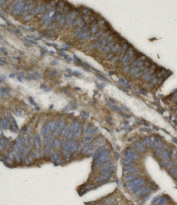
Anti-PHB2 antibodies have IHC images from human tumors and mouse and rat intestine, plus an IF image from A431 cells (catalog image captions).

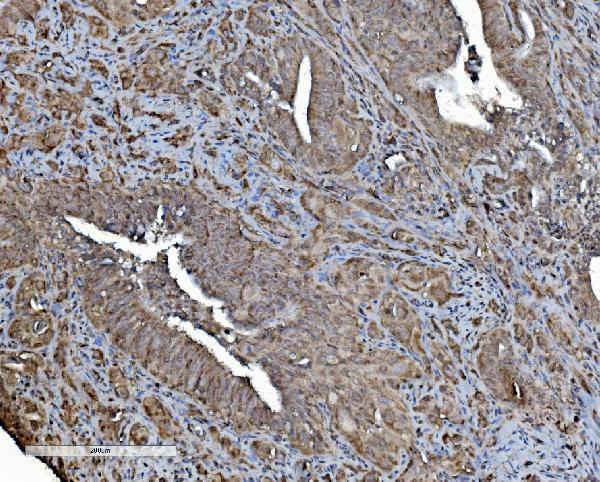
Real IHC data IHC analysis of REA/PHB2 using anti-REA/PHB2 antibody (M03315). REA/PHB2 was detected in paraffin-embedded section of human gallbladder adenocarcinoma tissue. Heat mediated antigen retrieval was performed in EDTA buffer (pH8.0, epitope retrieval solution). The tissue section was blocked with 10% goat serum. The tissue section was then incubated with 2μg/ml mouse anti-REA/PHB2 Antibody (M03315) overnight at 4°C. Biotinylated goat anti-mouse IgG was used as secondary antibody and incubated for 30 minutes at 37°C. The tissue section was developed using Strepavidin-Biotin-Complex (SABC) (Catalog # SA1021) with DAB as the chromogen.
Anti-REA/PHB2 Antibody ® (monoclonal, 2B5)
Cat # M03315
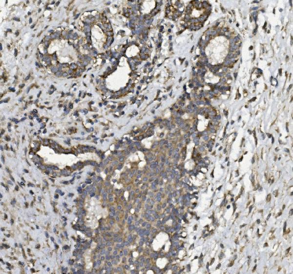
Real IHC data IHC analysis of REA/PHB2 using anti REA/PHB2 antibody (A03315-3). REA/PHB2 was detected in paraffin-embedded section of human mammary cancer tissue. Heat mediated antigen retrieval was performed in EDTA buffer (pH8.0, epitope retrieval solution). The tissue section was blocked with 10% goat serum. The tissue section was then incubated with 2μg/ml rabbit anti-REA/PHB2 Antibody (A03315-3) overnight at 4°C. Biotinylated goat anti-rabbit IgG was used as secondary antibody and incubated for 30 minutes at 37°C. The tissue section was developed using Strepavidin-Biotin-Complex (SABC) (Catalog # SA1022) with DAB as the chromogen.
Anti-REA/PHB2 Antibody ®
Cat # A03315-3

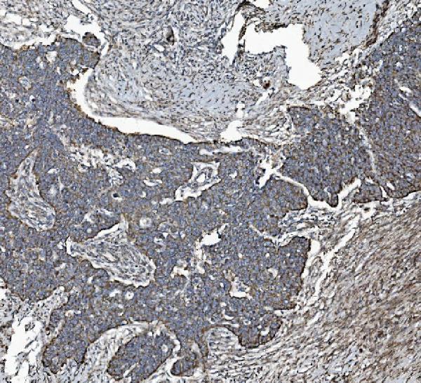
M03315 has IHC images from paraffin sections of human gallbladder, ovarian, renal, and lung cancers (M03315 IHC captions). A03315-3 has IHC images from human mammary and rectal cancers and mouse and rat intestine, plus an IF image from A431 cells (A03315-3 image captions).

Which to pick: For human tissue IHC, M03315 is a mouse monoclonal, clone 2B5, with human paraffin-section examples (catalog; M03315 IHC captions). For cross-species tissue IHC, A03315-3 has paraffin-section images from human, mouse, and rat samples; its own IHC captions report EDTA pH 8 retrieval and 2 μg/ml primary antibody (A03315-3 IHC captions). Choose A03315-3 for IF/ICC because those applications are listed and its IF image shows A431 cells; neither antibody’s IHC caption reports the tissue fixative (catalog; A03315-3 IF caption; IHC captions).

Each figure is that product's own IHC / IF validation image from its datasheet.

References

  1. UniProt Consortium. UniProt entry Q99623 (PHB2_HUMAN, Prohibitin-2).
  2. Human Protein Atlas. PHB2 tissue IHC expression (reliability: Supported).
  3. Human Protein Atlas. PHB2 subcellular location (ICC-IF): Localized to the mitochondria..
  4. Human Protein Atlas. PHB2 antibody validation summary (3 antibodies).
  5. Role of PHB2 as a potential biomarker in pan-cancer: a multi-database analysis. Scientific reports 2025 — PMC12227697.
  6. Proteome Characteristics of Non-Alcoholic Steatohepatitis Liver Tissue and Associated Hepatocellular Carcinomas. International journal of molecular sciences 2017 — PMC5343968.
  7. Expression of PHB2 in rat brain cortex following traumatic brain injury. International journal of molecular sciences 2014 — PMC3958913.
  8. Olfactory bulb neuroproteomics reveals a chronological perturbation of survival routes and a disruption of prohibitin complex during Alzheimer's disease progression. Scientific reports 2017 — PMC5567385.
  9. PubMed PMID:9074930 — UniProt-cited evidence.
  10. PubMed PMID:10359819 — UniProt-cited evidence.
  11. PubMed PMID:10931946 — UniProt-cited evidence.