PJA1 / E3 ubiquitin-protein ligase Praja-1 · IHC design guide

Design Immunohistochemistry for PJA1

Plan chromogenic PJA1 IHC in paraffin sections with the catalog antibody at a starting concentration of 5 μg/mL (datasheet: IHC-P). Assess nuclear staining, including high staining in cerebral cortex neurons, while accounting for low consistency between antibody staining and RNA expression (HPA tissue IHC).

Evidence assembled Oct 2026 · For research use; verify linked source records and product datasheet before use
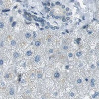
Immunohistochemistry protocol sheet for PJA1 (IHC for PJA1): expected localisation Nuclear staining across tissues (HPA tissue IHC), antibody A11179, validated IHC image, and IHC protocol steps
Printable PJA1 IHC protocol sheet — expected localisation Nuclear staining across tissues (HPA tissue IHC), antibody A11179, controls and protocol steps. Open the full PJA1 IHC guide →

PJA1 Immunohistochemistry Experimental Design Guide

Expected localisation, validated protocols, controls and antibodies — the at-a-glance facts below, then the full design guide.

Must know before staining
Expected localisation Nuclear staining across tissues (HPA tissue IHC)
Staining pattern Nuclear staining in many cell types (HPA tissue IHC)
Antigen retrieval Citrate pH 6.0 HIER, 95–98 °C, 20 min (rule: cytoplasmic / membrane antigen)
Positive control ⓘ Adrenal gland+4 more · see all
Negative control ⓘ Liver
Important caveats
Reasons your staining may differ from the expected pattern.
Fixation Keep fixation consistent across sections (standard IHC practice; not target-specific)
Caveat Staining and RNA expression show low consistency (HPA tissue IHC)
Regulation Brain expression peaks in cortex (UniProt)
Isoform / epitope 3 isoforms; epitope coverage is unspecified (UniProt)
Section 1

Recommended PJA1 IHC & IF Protocols

The catalog antibody’s IHC-P protocol is followed by three published PJA1 chromogenic IHC protocols using human or mouse tissue (PMC10084124; PMC5620009; PMC7818255).

Recommended immunohistochemistry (IHC-P) protocol parameters
SampleTissue sections; selected-image fixative not specified (standard IHC workflow)
FixationImage fixative and duration unreported (datasheet A11179); verify before use.
Sectioning4–5 µm sections on charged slides (standard)
DeparaffinisationXylene, graded ethanol series to water (standard)
Antigen retrievalHeat-induced epitope retrieval in citrate buffer, pH 6.0, 20 min at 95–98 °C (standard rule: cytoplasmic / membrane antigen)
Peroxidase block3% H2O2, 10 min, room temperature (standard)
Blocking10% normal serum of the secondary host, 30 min, room temperature (standard)
Primary antibodyRabbit anti-PJA1, 5 μg/mL (datasheet A11179)
Primary incubationOvernight at 4 °C (standard)
DetectionHRP-polymer secondary, DAB chromogen 5–10 min (standard)
CounterstainHematoxylin, blue, dehydrate and mount (standard)
Expected resultPJA1-positive staining in glandular cells of adrenal gland (HPA tissue IHC: High). HPA tissue profile: Ubiquitous nuclear expression. No signal in the no-primary control.
💡Decision noteStart with citrate pH 6.0 HIER at 95–98 °C for 20 min (page retrieval rule); the published human tissue protocol also uses citrate pH 6.0 (PMC10084124).
Section 2

What Is the Expected PJA1 Staining Pattern?

PJA1 is reported as ubiquitously nuclear in HPA tissue IHC, with approved nucleoplasm and nucleoli localisation in ICC-IF (HPA). Expect nuclear staining in the listed positive cell populations, including cerebral cortex neurons and cerebellar molecular-layer cells (HPA: High). PJA1 has no transmembrane segment or signal peptide (UniProt Q8NG27). Interpret intensity cautiously: HPA rates its tissue IHC profile Approved but reports low consistency with RNA expression and pending external verification (HPA).

What am I looking at on my slide?
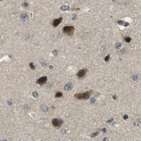
Distinct nuclear staining in cerebral cortex neuronal cells or cerebellar molecular-layer cells.This fits HPA's High IHC staining in these populations and its ubiquitous nuclear tissue profile (HPA). Nucleoplasm and nucleoli are approved ICC-IF locations, offering a compartment-level comparison, although ICC-IF does not independently verify the intensity of a paraffin IHC result (HPA).
Predominantly membranous or extracellular staining, with little nuclear signal.This conflicts with the HPA nuclear profile and approved nucleoplasm/nucleoli ICC-IF locations (HPA). PJA1 lacks a transmembrane segment and signal peptide (UniProt Q8NG27). Treat the pattern as possible artefact or nonspecific detection; check controls before assigning it to PJA1 (general IHC practice).
Strong staining in liver cholangiocytes, especially if expected nuclear staining is absent elsewhere.HPA reports PJA1 as Not detected in cholangiocytes, so this cell-specific result warrants scrutiny; it does not make the entire liver a negative control (HPA). Cross-reactivity or endogenous detection activity are possible explanations, to be distinguished with appropriate controls (general IHC practice).
Diffuse colour across nuclei, cytoplasm and tissue spaces without clear cell boundaries.A diffuse field cannot be scored confidently against HPA's nuclear profile or its cell-specific High and Low categories (HPA). Check background using a no-primary control and review blocking, washes and detection conditions before interpreting intensity (general IHC practice).
No detectable staining in cerebral cortex neuronal cells on a processed section.HPA reports High staining in those cells, making them a useful positive-pattern check, but its IHC profile has low consistency with RNA data (HPA). Confirm tissue preservation and controls, then review the catalog antibody's IHC-P conditions and detection workflow (general IHC practice).
💡Expected PJA1 appearanceA convincing positive shows clear nuclear signal in HPA-listed High populations such as cerebral cortex neurons; isolated strong membrane staining or colour in HPA-undetected cholangiocytes merits investigation as a possible false positive (HPA; UniProt Q8NG27 topology).
How each factor affects the staining
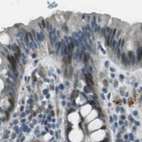
Cell and tissue selectionHPA lists High staining in adrenal, bronchial, cervical, colonic, esophageal and gallbladder epithelial or glandular populations, as well as selected brain cells (HPA). Low staining in breast or seminal-vesicle glandular cells makes those populations weaker checks (HPA).
Confidence in tissue interpretationThe tissue IHC profile is Approved, yet HPA reports low consistency with RNA expression and says external verification is pending (HPA). Its antibody HPA000595 is Approved for IHC, not reported as Enhanced in the supplied validation record (HPA).
Expression evidence across sourcesUniProt reports expression in several brain regions, highest in cerebral cortex (UniProt Q8NG27); HPA shows a broader nuclear IHC profile and tissue-enhanced RNA in epididymis (HPA). Use observed IHC cell patterns for slide comparison without treating the sources as an exact intensity scale.
Isoforms and processingUniProt lists three PJA1 isoforms, no signal peptide or propeptide, and a single annotated chain spanning residues 1–643 (UniProt Q8NG27). No epitope location is supplied, so isoform-specific staining cannot be predicted from this record.
Can ICC-IF help interpret localisation?Yes. HPA approves nucleoplasm and nucleoli localisation and lists ICC-IF images from A-431, U-251MG and U2OS (HPA). These images can inform a compartment comparison; IF/ICC experimental conditions belong in its separate guide.
Why is my staining missing, weak or wrong?
SituationLikely causeNext action
Cortex neurons show no signal, while the slide otherwise develops normally.The result disagrees with HPA's High neuronal staining, but the cause cannot be assigned from that observation alone (HPA).Check a positive-pattern section and controls; review section quality, retrieval conditions and the catalog antibody's IHC-P instructions before repeating (general IHC practice).
Signal is mainly cytoplasmic or membrane associated.The compartment differs from HPA's nuclear tissue profile and approved nucleoplasm/nucleoli ICC-IF locations (HPA).Compare with no-primary and positive-pattern controls; reassess detection background and score only supported nuclear staining (general IHC practice; HPA).
Broad brown background obscures individual cells.Background detection can prevent a meaningful comparison with HPA's cell-specific staining categories (general IHC practice; HPA).Run a no-primary control, check endogenous peroxidase blocking for chromogenic detection, and review blocking, washes and development time (general IHC practice).
Liver cholangiocytes appear strongly positive.HPA records PJA1 as Not detected in cholangiocytes; cross-reactivity or endogenous activity remain possibilities, not established diagnoses (HPA; general IHC practice).Verify cell identity and compare a no-primary control with a known High population before treating the cholangiocyte signal as specific (HPA; general IHC practice).
Breast or seminal-vesicle glandular cells stain weakly.HPA classifies these populations as Low, so weak signal alone does not establish assay failure (HPA).Compare the same run with an HPA High population, such as cerebral cortex neurons or colon glandular cells, before adjusting detection conditions (HPA; general IHC practice).
Paraffin IHC and ICC-IF appear to disagree.HPA supports nuclear tissue IHC and nucleoplasm/nucleoli ICC-IF, but the supplied records do not establish identical staining intensity across methods (HPA).Compare compartment and cell identity first, then inspect each method's own controls; keep IF/ICC protocol choices with the separate IF/ICC guide (HPA; general IHC practice).

Sample controls for PJA1 IHC & IF

🧪Run cerebral cortex first and require staining in neuronal cells (HPA: High in cerebral-cortex neuronal cells). Use liver cholangiocytes as the negative comparator (HPA: Not detected in liver cholangiocytes); neighboring cortical cells without detectable staining should remain at background, although the supplied HPA rows do not identify a specific negative cortical cell type.
Positive control tissue: Adrenal gland (Glandular cells, HPA High)
Negative control tissue: Liver (HPA Not detected)
ICC-IF cell lines (HPA subcellular resource): HPA ICC-IF images show PJA1 in A-431, U-251MG, U2OS, with annotated localisation: Nucleoplasm (approved), Nucleoli (approved) (HPA subcellular).
Technical controls: Include no-primary (secondary-only) and primary-antibody controls matched to the primary antibody’s host species and, where applicable, isotype; use PJA1 knockout tissue or immunizing-peptide competition as a biological specificity control if available (standard IHC practice). Quench endogenous peroxidase for chromogenic detection in cortex (standard IHC practice).
⚠️Feasibility: A PJA1-specific fixation window or fixation effect is unreported, and the selected A11179 mouse-brain IHC caption does not state a fixative (caption: fixative not stated). Antigen-retrieval dependence is also unreported; the caption reports 5 µg/ml for its mouse-brain image, so retrieval and concentration need empirical optimization for the chosen paraffin sections (caption: 5 µg/ml). HPA has ICC-IF images but does not establish that IF or frozen sections are easier than paraffin IHC (HPA: ICC-IF images in A-431, U-251MG and U2OS); assess cortical lipofuscin autofluorescence if using IF (standard neural-tissue IF practice).

HPA tissue IHC evidence for PJA1

Comprehensive Human Protein Atlas IHC scoring per tissue (reliability: Approved — Low consistency between antibody staining and RNA expression data. Pending external verification.). Rows are taken directly from the HPA tissue chart — click any row's HPA link to view the source.

Positive expression · recommended positive controls

TissueCell typeLevelEvidenceSource
Adrenal gland Glandular cells High Protein (IHC) HPA →
Bronchus Respiratory epithelial cells High Protein (IHC) HPA →
Cerebellum Cells in molecular layer High Protein (IHC) HPA →
Cerebral cortex Neuronal cells High Protein (IHC) HPA →
Cervix Glandular cells High Protein (IHC) HPA →

Undetected expression · recommended negative controls

TissueCell typeLevelEvidenceSource
Liver Cholangiocytes Not detected Protein (IHC) HPA →
Section 3

Advanced PJA1 IHC Tips

Troubleshoot PJA1 staining in paraffin sections by checking retrieval, compartment, controls and scoring before interpreting a chromogenic signal.

What retrieval should I start with for PJA1 in paraffin sections?
Start with citrate buffer at pH 6.0 for heat induced epitope retrieval at 95–98 °C for 20 min (page retrieval setting). Let sections cool in buffer, then compare a retrieved section with a section processed without retrieval to assess whether the signal depends on heating (standard IHC practice). If staining is weak, test a second retrieval condition on matched sections while holding antibody concentration and detection constant; excessive heating can damage tissue or raise background (standard IHC practice). Score cellular staining separately from damaged edges, and use a positive control section because PJA1 staining may vary by tissue and cell type (HPA tissue IHC).
How should I troubleshoot weak PJA1 staining when fixation details are uncertain?
Target specific fixation sensitivity for PJA1 is unknown from the supplied evidence, so record the fixative and fixation duration for each specimen rather than assigning weak staining to a particular fixation effect (supplied evidence). The mouse brain image documents staining with catalog antibody A11179 at 5 µg/ml, but its caption does not state a fixative (A11179 tissue IHC caption). Compare sections prepared with known processing histories using the same pH 6.0 retrieval and detection workflow (page retrieval setting; standard IHC practice). Include a positive control and an omitted primary control to distinguish loss of detectable antigen from staining contributed by the detection system (standard IHC practice).
Where should PJA1 staining appear, and how should unexpected compartments be checked?
Assess nucleoplasmic and nucleolar staining separately: those locations are approved in HPA cell imaging, while UniProt supplies no subcellular annotation for PJA1 (HPA subcellular; UniProt Q8NG27). The protein has no annotated transmembrane segment or signal peptide, so a crisp membrane only pattern warrants checking antibody specificity and tissue morphology (UniProt Q8NG27 topology and processing; standard IHC practice). HPA describes ubiquitous nuclear tissue staining, but warns of low consistency with RNA expression and pending external verification (HPA tissue IHC). Compare the questioned pattern with a positive control, an omitted primary control and cell specific morphology before treating it as biological localisation (standard IHC practice).
Could an uncharacterised epitope or PJA1 isoform explain inconsistent staining?
PJA1 has 3 annotated isoforms, but the supplied evidence does not identify the catalog antibody’s epitope or establish which isoforms it detects (UniProt Q8NG27; supplied antibody evidence). Check the antibody’s immunogen or epitope documentation before claiming that a stained section measures every isoform (standard IHC practice). UniProt lists phosphorylation at residues 265, 277, 365 and 367, without evidence here that any modification changes this antibody’s IHC staining (UniProt Q8NG27 modified residues; supplied antibody evidence). Compare matched positive and negative controls under identical retrieval and detection conditions; interpret discordant staining as unresolved until epitope coverage and specificity are established (standard IHC practice).
How can IF help investigate a disputed PJA1 IHC pattern?
Use IF as a separate localisation check and multiplex PJA1 with a marker for the cell population under study, such as a validated neuronal marker when examining cerebral cortex (HPA: High in cerebral cortex neuronal cells; standard IF practice). Choose a fluorophore channel away from the specimen’s strongest autofluorescence, and inspect unstained tissue in each channel before interpreting overlap (standard IF practice). Permeabilise cells to allow antibody access to the nucleoplasm and nucleoli, while optimising detergent exposure to preserve morphology (HPA subcellular; standard IF practice). HPA provides ICC/IF images from A-431, U-251MG and U2OS, but those observations do not establish the catalog antibody’s IF performance (HPA subcellular; supplied antibody evidence).
What should I change when PJA1 chromogenic staining has high background?
Inspect an omitted primary control to identify staining from the detection system, and check whether background follows section edges, folds or damaged tissue (standard IHC practice). Apply a peroxidase block before chromogenic detection and use a blocking step appropriate to the secondary reagent; these are general IHC steps, not PJA1 specific findings (standard IHC practice). Titrate the primary antibody on matched sections while keeping retrieval at citrate pH 6.0, 95–98 °C for 20 min (page retrieval setting; standard IHC practice). Preserve a credible positive control while lowering diffuse signal, since HPA’s reported nuclear pattern has limited agreement with RNA expression (HPA tissue IHC).
How should I quantify PJA1 staining across paraffin sections? ⚠ ANSWER MARKED FOR VERIFICATION
Define the scored cell population and compartment before analysis; nucleoplasmic and nucleolar signal are the reported PJA1 locations in cell imaging (HPA subcellular; standard IHC practice). For chromogenic sections, report an H-score from staining intensity and the percentage of positive cells, or report positive cell density per mm² of viable tissue (standard IHC practice). Normalise counts to the number of eligible cells or measured viable tissue area, and keep thresholds, retrieval and illumination consistent across compared sections (standard IHC practice). Record background from an omitted primary control and analyse regions without folds or necrosis separately from intact tissue (standard IHC practice).
When is a PJA1 positive section convincing rather than artefactual?
A convincing signal follows intact cell morphology, exceeds the omitted primary control and includes a plausible nuclear component, consistent with reported nucleoplasmic and nucleolar localisation (HPA subcellular; standard IHC practice). HPA reports high staining in cerebral cortex neuronal cells and cerebellar molecular layer cells, while its tissue antibody has low consistency with RNA expression and awaits external verification (HPA tissue IHC). Treat membrane only staining, signal confined to section edges or necrotic areas, and staining reproduced by the omitted primary control as unresolved or artefactual (UniProt Q8NG27 topology; standard IHC practice). Check endogenous enzyme staining with the detection control before assigning brown chromogen to PJA1 (standard IHC practice).
Boster reagents

Best PJA1 / E3 ubiquitin-protein ligase Praja-1 IHC Antibodies

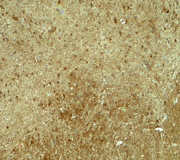
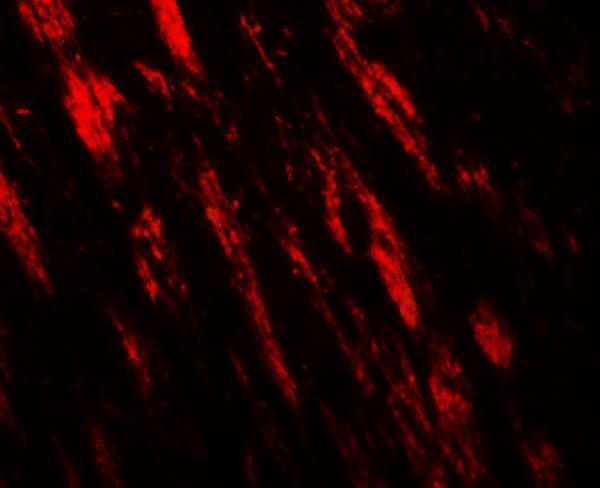
A11179 has real IHC and IF images from mouse brain tissue (IHC and IF image captions). Listed reactivity covers human, mouse, and rat (catalog reactivity).

Real IHC data Immunohistochemistry of PJA1 in mouse brain tissue with PJA1 antibody at 5 μg/ml.
Anti-PJA1 Antibody
Cat # A11179

A11179 will render with an IHC image of mouse brain tissue stained at 5 μg/mL (IHC image caption). Its IF image shows mouse brain tissue stained at 20 μg/mL (IF image caption).

Which to pick: For paraffin-section IHC, choose A11179: IHC-P is listed, and its own IHC caption documents mouse brain staining; the caption does not report a fixative (catalog applications; IHC image caption). For IF, A11179 has a mouse brain image, while ICC is not listed as an application (IF image caption; catalog applications). For work across species, A11179 lists human, mouse, and rat reactivity, although both images show mouse brain; clonality is unreported (catalog reactivity; IHC and IF image captions; catalog clone field).

Each figure is that product's own IHC / IF validation image from its datasheet.

References

  1. UniProt Consortium. UniProt entry Q8NG27 (PJA1_HUMAN, E3 ubiquitin-protein ligase Praja-1).
  2. Human Protein Atlas. PJA1 tissue IHC expression (reliability: Approved).
  3. Human Protein Atlas. PJA1 subcellular location (ICC-IF): Localized to the nucleoplasm and nucleoli..
  4. Human Protein Atlas. PJA1 antibody validation summary (1 antibodies).
  5. Praja1 RING-finger E3 ubiquitin ligase is a common suppressor of neurodegenerative disease-associated protein aggregation. Neuropathology : official journal of the Japanese Society of Neuropathology 2022 — PMC10084124.
  6. Targeting the E3 Ubiquitin Ligase PJA1 Enhances Tumor-Suppressing TGFβ Signaling. Cancer research 2020 — PMC7704101.
  7. PRAJA is overexpressed in glioblastoma and contributes to neural precursor development. Genes & cancer 2017 — PMC5620009.
  8. Praja1 RING-finger E3 ubiquitin ligase suppresses neuronal cytoplasmic TDP-43 aggregate formation. Neuropathology : official journal of the Japanese Society of Neuropathology 2020 — PMC7818255.
  9. PubMed PMID:12036302 — UniProt-cited evidence.
  10. PubMed PMID:15772651 — UniProt-cited evidence.
  11. PubMed PMID:15489334 — UniProt-cited evidence.