PRPF31 / U4/U6 small nuclear ribonucleoprotein Prp31 · IHC design guide

Design Immunohistochemistry for PRPF31

Plan PRPF31 staining in paraffin sections using its widespread nuclear tissue pattern (HPA tissue IHC). This guide covers fixation, antibody dilution and interpretation of staining in positive tissues.

Evidence assembled Oct 2026 · For research use; verify linked source records and product datasheet before use
Immunohistochemistry protocol sheet for PRPF31 (IHC for PRPF31): expected localisation Nuclear in tissue (HPA tissue IHC); speckles and Cajal bodies (UniProt), antibody A30534, validated IHC image, and IHC protocol steps
Printable PRPF31 IHC protocol sheet — expected localisation Nuclear in tissue (HPA tissue IHC); speckles and Cajal bodies (UniProt), antibody A30534, controls and protocol steps. Open the full PRPF31 IHC guide →

PRPF31 Immunohistochemistry Experimental Design Guide

Expected localisation, validated protocols, controls and antibodies — the at-a-glance facts below, then the full design guide.

Must know before staining
Expected localisation Nuclear in tissue (HPA tissue IHC); speckles and Cajal bodies (UniProt)
Staining pattern Widespread nuclear staining across cell types (HPA tissue IHC)
Antigen retrieval Tris-EDTA pH 9.0 HIER, heat-mediated (datasheet A30534)
Positive control ⓘ Adipose tissue+4 more · see all
Negative control ⓘ None in HPA (detected in all 44 tissues); use no-primary + isotype controls
Important caveats
Reasons your staining may differ from the expected pattern.
Fixation Keep fixation consistent across paraffin sections (standard IHC practice; not target-specific)
Caveat Hematopoietic cells can show high nuclear staining (HPA tissue IHC)
Regulation Broad expression; low tissue specificity (HPA tissue IHC)
Isoform / epitope 4 isoforms; epitope differences are unspecified (UniProt)
Section 1

Recommended PRPF31 IHC & IF Protocols

The catalog antibody's paraffin IHC protocol (datasheet A30534) is paired with a published mouse retinal cryosection protocol (PMC6938640).

Recommended immunohistochemistry (IHC-P) protocol parameters
SampleParaffin-embedded human cervical carcinoma tissue; fixative not specified (datasheet A30534)
FixationImage fixative and duration unreported (datasheet A30534); verify before use.
Sectioning4–5 µm sections on charged slides (standard)
DeparaffinisationXylene, graded ethanol series to water (standard)
Antigen retrievalHeat retrieval: Tris-EDTA pH 9.0 (datasheet A30534); 20 min, 95–100 °C (standard)
Peroxidase block3% H2O2, 10 min, room temperature (standard)
Blocking10% normal serum of the secondary host, 30 min, room temperature (standard)
Primary antibodyRabbit anti-PRPF31, 1:50 recommended; image 1:200 (datasheet A30534)
Primary incubationOvernight at 4 °C (standard)
DetectionHRP-polymer secondary, DAB chromogen 5–10 min (standard)
CounterstainHematoxylin, blue, dehydrate and mount (standard)
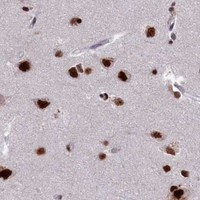
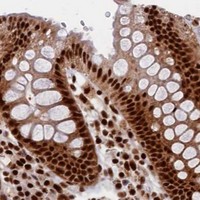
Expected resultPRPF31-positive staining in adipocytes of adipose tissue (HPA tissue IHC: High). HPA tissue profile: Ubiquitous nuclear expression. No signal in the no-primary control.
💡Decision noteFor paraffin sections, start with heat-mediated Tris-EDTA retrieval at pH 9.0 (datasheet A30534). The published cryosection protocol does not state a retrieval step (PMC6938640).
Section 2

What Is the Expected PRPF31 Staining Pattern?

PRPF31 should appear mainly in nuclei across many cell types in paraffin tissue sections: HPA reports ubiquitous nuclear expression, with Supported tissue IHC reliability and high staining in the listed positive cell populations (HPA tissue IHC). UniProt places PRPF31 in nuclear speckles and Cajal bodies and reports no transmembrane segment (UniProt Q8WWY3).

What am I looking at on my slide?
Brown nuclear staining in adipocytes, glandular cells, hematopoietic cells, respiratory epithelial cells, glial cells, or cerebellar granular-layer cells.This fits the reported compartment and listed high-staining populations (HPA tissue IHC: High; HPA tissue IHC: ubiquitous nuclear expression). Judge intensity against the same run's background and controls; the HPA High category is not a numeric score for an individual slide (general IHC practice).
Predominantly cytoplasmic, membranous, or extracellular deposit with little nuclear staining.This conflicts with the reported nuclear pattern and lack of a transmembrane segment (HPA tissue IHC; UniProt Q8WWY3). Treat the deposit as suspect; check whether detection chemistry, nonspecific binding, or tissue pigment explains it before interpreting it as PRPF31 (general IHC practice).
A conspicuous signal appears in one unexpected cell population while nuclei in nearby listed positive cells are weak or blank.Check cell identification and possible cross-reactivity or endogenous detection activity (general IHC practice). PRPF31 is reported as ubiquitous, and this payload lists no negative cell population; an unlisted cell type alone cannot establish a false positive (UniProt Q8WWY3; HPA tissue IHC).
Broad brown haze covers tissue, including spaces outside nuclei, and obscures cell boundaries.Diffuse haze prevents a reliable nuclear call (general IHC practice; HPA tissue IHC: nuclear expression). Review blocking, washing, detection reagent exposure, and the negative reagent control; do not score haze as PRPF31 expression (general IHC practice).
No convincing nuclear signal appears in a section containing a listed high-staining cell population.A technical failure or unsuitable antibody conditions are possible (general IHC practice). Confirm tissue identity and control performance, then review the antibody's IHC-P instructions. HPA High describes observed staining in specified cells; it does not guarantee every preparation will stain (HPA tissue IHC: High).
💡Expected PRPF31 appearanceCall the IHC result positive when discernible nuclear chromogen is present in the relevant cells, including listed HPA High populations; cytoplasmic or extracellular color without corresponding nuclear signal is suspect (HPA tissue IHC: ubiquitous nuclear expression and High; general IHC practice).
How each factor affects the staining
Nuclear distributionHPA describes ubiquitous nuclear tissue staining, while UniProt places PRPF31 predominantly in speckles and Cajal bodies (HPA tissue IHC; UniProt Q8WWY3). Routine chromogenic IHC may show a nuclear signal without resolving those structures (general IHC practice).
Choice of reference tissueThe supplied HPA entries report High staining in eight listed tissue and cell combinations and provide no negative or low entries (HPA tissue IHC). Use a listed population as a positive reference; do not designate an unlisted tissue as negative from this record.
Antibody validationHPA labels rabbit polyclonal HPA041939 as IHC Supported; HPA061873 has ICC Supported status, with no IHC status supplied (HPA antibodies). These labels apply to the respective antibody and application, not automatically to another antibody.
Isoforms and epitope coverageUniProt lists four PRPF31 isoforms, but the supplied record gives no antibody epitope or isoform-specific IHC data (UniProt Q8WWY3). If patterns differ between antibodies, check their documented epitope coverage before assigning an isoform explanation (general IHC practice).
IF/ICC Q&A: Should IF show the same compartment?Yes: HPA reports supported nucleoplasmic ICC-IF localization, with images from HEK293, PC-3, and U2OS (HPA subcellular ICC-IF). UniProt also reports nuclear speckles and Cajal bodies (UniProt Q8WWY3). This supports a nuclear IF interpretation; it does not supply an IF protocol.
Why is my staining missing, weak or wrong?
SituationLikely causeNext action
Listed high-staining cells are blank on the IHC slide.The positive result has failed to appear, but this record does not identify the failed step (HPA tissue IHC: High).Verify tissue identity and a working positive control, then review the catalog antibody's IHC-P instructions, reagent preparation, retrieval, and detection run records (general IHC practice).
Signal appears mainly outside nuclei.The compartment disagrees with nuclear tissue staining and UniProt nuclear localization; the source cannot identify a specific artifact (HPA tissue IHC; UniProt Q8WWY3).Inspect morphology and negative controls, and reassess nonspecific binding, endogenous detection activity, and pigment before calling it target staining (general IHC practice).
Every cell and the surrounding tissue show uniform brown haze.Background may be obscuring compartment-specific signal (general IHC practice; HPA tissue IHC: nuclear expression).Compare a negative reagent control; review blocking, washing, primary antibody concentration, and detection reagent exposure using the product instructions (general IHC practice).
Only an unexpected cell population stains strongly.Cell identification, cross-reactivity, or endogenous detection activity may be involved (general IHC practice). HPA supplies positive examples but no negative cell list (HPA tissue IHC).Recheck the population's identity and nuclear location, compare adjacent expected positive cells, and use appropriate reagent controls before claiming cell-specific expression (general IHC practice).
Fine nuclear dots are absent although nuclei stain.HPA tissue IHC supports a nuclear pattern; UniProt reports enrichment in speckles and Cajal bodies, which routine chromogenic sections may not resolve (HPA tissue IHC; UniProt Q8WWY3; general IHC practice).Score the supported nuclear compartment first. Do not classify missing dots alone as IHC failure; use the separate IF/ICC guide if subnuclear detail is the experimental question.
IF/ICC images and IHC sections seem to disagree.The supplied HPA evidence covers different applications and antibodies: HPA061873 is ICC Supported, while HPA041939 is IHC Supported (HPA antibodies).Compare nuclear versus nonnuclear localization within each assay and verify which antibody was used. Do not transfer an ICC validation label or staining intensity to an IHC-P result (HPA antibodies; general IHC practice).

Sample controls for PRPF31 IHC & IF

🧪Run bone marrow first and score nuclear staining in hematopoietic cells (HPA: High in bone marrow hematopoietic cells; UniProt Q8WWY3: nuclear localization). HPA detects PRPF31 in all 44 scored tissues, so there is no supported negative tissue or internal negative cell population; no-primary and isotype controls should show only background on the same slide (HPA: no negative tissue rows; HPA: detected in all 44 scored tissues).
Positive control tissue: Adipose tissue (Adipocytes, HPA High)
Negative control tissue: None in HPA: PRPF31 is detected in all 44 scored tissues. Use a no-primary (secondary-only) and an isotype control instead.
ICC-IF cell lines (HPA subcellular resource): HPA ICC-IF images show PRPF31 in HEK293, PC-3, U2OS, with annotated localisation: Nucleoplasm (supported) (HPA subcellular).
Technical controls: Use a no-primary, secondary-only control and an isotype control matched to the primary antibody’s host species, immunoglobulin class, clonality and concentration; compare these with PRPF31-knockout material processed in parallel as a biological negative (standard IHC controls). Block endogenous peroxidase and check for residual chromogen signal in the bone marrow controls, where hematopoietic cells can have endogenous peroxidase activity (standard IHC practice).
⚠️Feasibility: A target-specific fixation window or fixation effect is unreported, and the selected paraffin-section caption does not state a fixative (A30534 caption: fixative unreported). The reported IHC example uses Tris-EDTA at pH 9.0 for antigen retrieval, but does not establish that this retrieval condition is required (A30534 caption: Tris-EDTA, pH 9.0). Nuclear localization provides a scoring expectation, while the supplied evidence does not establish that frozen sections or IF are easier than paraffin IHC; control endogenous peroxidase background in bone marrow (UniProt Q8WWY3: nuclear localization; HPA: nucleoplasm in ICC-IF; standard IHC practice).

HPA tissue IHC evidence for PRPF31

Comprehensive Human Protein Atlas IHC scoring per tissue (reliability: Supported — High consistency between antibody staining and RNA expression data.). Rows are taken directly from the HPA tissue chart — click any row's HPA link to view the source.

Positive expression · recommended positive controls

TissueCell typeLevelEvidenceSource
Adipose tissue Adipocytes High Protein (IHC) HPA →
Adrenal gland Glandular cells High Protein (IHC) HPA →
Appendix Glandular cells High Protein (IHC) HPA →
Bone marrow Hematopoietic cells High Protein (IHC) HPA →
Breast Glandular cells High Protein (IHC) HPA →

Undetected expression · recommended negative controls

TissueCell typeLevelEvidenceSource
None in HPA: PRPF31 is detected in all 44 scored tissues. Use a no-primary (secondary-only) and an isotype control instead.
Section 3

Advanced PRPF31 IHC Tips

Troubleshoot PRPF31 staining in paraffin section IHC using the selected antibody’s reported conditions and the expected nuclear pattern.

Which retrieval conditions should I try first for weak PRPF31 staining?
Start with heat mediated retrieval in Tris-EDTA at pH 9.0 (datasheet A30534). The paraffin section example used this buffer with primary antibody at 1:200 overnight at 4°C, followed by secondary antibody at 1:200 for 45 minutes at room temperature (caption A30534). If staining remains weak, vary heating and cooling duration on adjacent sections while keeping antibody concentration and detection constant (standard IHC practice). Compare nuclear signal with background in the same tissue; PRPF31 is nuclear, predominantly in speckles and Cajal bodies (UniProt Q8WWY3), and tissue IHC shows ubiquitous nuclear expression (HPA tissue IHC).
Could fixation explain variable PRPF31 staining across paraffin sections?
Target specific sensitivity to fixation is unknown: the selected paraffin section caption does not state a fixative (caption A30534). Record the fixative, fixation duration, section thickness and tissue processing for each specimen before comparing staining intensity (standard IHC practice). When material permits, stain adjacent sections in one run using Tris-EDTA at pH 9.0 and the same detection conditions to assess whether the difference persists (datasheet A30534; standard IHC practice). Interpret any persistent difference cautiously because PRPF31 has ubiquitous nuclear tissue expression (HPA tissue IHC), while that expression profile does not establish how fixation affects its epitope.
What staining compartment should I expect for PRPF31 in chromogenic IHC?
Score nuclear staining as the expected PRPF31 pattern in paraffin sections: tissue IHC describes ubiquitous nuclear expression (HPA tissue IHC). PRPF31 is predominantly found in nuclear speckles and Cajal bodies (UniProt Q8WWY3), although these small structures may not be resolvable with chromogenic tissue IHC (standard IHC practice). A diffuse nuclear signal can therefore be compatible with the target, but predominantly cytoplasmic or membranous staining warrants scrutiny against a matched negative control (HPA tissue IHC; standard IHC practice). Compare nuclei within intact tissue regions and check the counterstain before assigning cell level positivity (standard IHC practice).
Could isoforms or epitope accessibility explain uneven nuclear staining?
PRPF31 has 4 listed isoforms, but the selected caption does not identify the antibody epitope or establish which isoforms it detects (UniProt Q8WWY3; caption A30534). Its Nop domain spans residues 215–333, and listed modified residues include phosphoserines at 379, 395 and 432 (UniProt Q8WWY3). Do not assign an uneven tissue pattern to an isoform or modification without epitope information and independent evidence (standard IHC interpretation). First compare adjacent sections under the reported Tris-EDTA pH 9.0 retrieval conditions, then check whether any change improves nuclear signal without increasing background (datasheet A30534; HPA tissue IHC; standard IHC practice).
How should I assess PRPF31 by multiplex IF alongside tissue IHC?
Treat IF as a separate assay: the selected antibody caption documents paraffin section IHC, while the subcellular record reports PRPF31 in the nucleoplasm by ICC/IF (caption A30534; HPA subcellular). Multiplex PRPF31 with a marker identifying the cell population of interest, then assess colocalisation within segmented nuclei rather than relying on overlapping whole cell signals (standard IF practice). Choose a fluorophore channel after measuring tissue autofluorescence and include single stain controls to check bleed through (standard IF practice). PRPF31 has no transmembrane segment and is nuclear, so optimise permeabilisation to admit antibody to the nuclear epitope while preserving morphology; its exact epitope is unreported here (UniProt Q8WWY3; standard IF practice).
How can I reduce diffuse or misleading chromogenic background?
Check a section lacking primary antibody to gauge secondary reagent and detection background, and examine unstained tissue for intrinsic pigment (standard IHC practice). In a peroxidase based workflow, include a peroxidase block before DAB development and adjust blocking, washes or detection time if the control remains colored (standard IHC practice). The selected caption reports secondary antibody at 1:200 for 45 minutes at room temperature; use that as a documented starting condition, not proof that background is controlled in every specimen (caption A30534). Accept a signal as PRPF31 only after confirming nuclear enrichment over surrounding cytoplasm and the negative control (HPA tissue IHC; standard IHC practice).
How should I quantify PRPF31 staining across samples? ⚠ ANSWER MARKED FOR VERIFICATION
Define the tissue region and cell population before scoring, then record the percentage of positive nuclei and nuclear intensity using a consistent threshold (standard IHC practice). An H-score can combine the proportions of nuclei at each intensity level; alternatively, report positive nuclei per mm² when cell density differs across regions (standard IHC practice). Normalise positive counts to the number of evaluable nuclei within the same annotated compartment, and exclude folds, necrosis and poorly counterstained areas using preset rules (standard IHC practice). Because tissue IHC describes ubiquitous nuclear PRPF31 expression, compare signal magnitude and distribution rather than treating presence alone as distinctive (HPA tissue IHC).
When is a PRPF31 positive result convincing rather than artefactual?
A convincing result shows reproducible nuclear enrichment in intact cells, consistent with ubiquitous nuclear tissue staining and the reported nucleoplasm, speckle and Cajal body localisation (HPA tissue IHC; HPA subcellular; UniProt Q8WWY3). Review the expected cell population against tissue morphology; PRPF31 is described as ubiquitously expressed, so positivity alone does not identify a particular cell type (UniProt Q8WWY3). Treat staining concentrated at section edges, in necrotic regions or outside nuclei as suspect until controls and adjacent sections support it (standard IHC practice). A primary omitted control helps identify detection background, including endogenous enzyme signal in a peroxidase workflow (standard IHC practice).
Boster reagents

Best PRPF31 / U4/U6 small nuclear ribonucleoprotein Prp31 IHC Antibodies

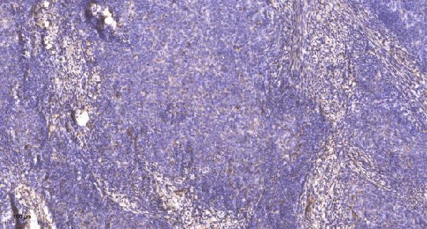
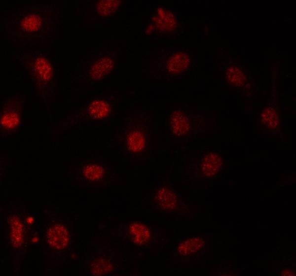
PRPF31 antibodies have IHC data from paraffin-embedded human cervical carcinoma (A30534 image caption) and IF data from A549 cells (A03137-1 image caption); catalog reactivity includes human, mouse, and rat (catalog: A30534 and A03137-1 reactivity).

Real IHC data Immunohistochemical analysis of paraffin-embedded human cervical carcinoma. 1, Antibody was diluted at 1:200(4° overnight). 2, Tris-EDTA,pH9.0 was used for antigen retrieval. 3,Secondary antibody was diluted at 1:200(room temperature, 45min).
Anti-PRP31 Antibody
Cat # A30534
Real IF data IF analysis of PRPF31 using anti-PRPF31 antibody (A03137-1). PRPF31 was detected in an immunocytochemical section of A549 cells. Enzyme antigen retrieval was performed using IHC enzyme antigen retrieval reagent (AR0022) for 15 mins. The cells were blocked with 10% goat serum. And then incubated with 5 μg/mL rabbit anti-PRPF31 Antibody (A03137-1) overnight at 4°C. Cy3 Conjugated Goat Anti-Rabbit IgG (BA1032) was used as secondary antibody at 1:500 dilution and incubated for 30 minutes at 37°C. Visualize using a fluorescence microscope and filter sets appropriate for the label used.
Anti-PRPF31 Antibody ®
Cat # A03137-1

A30534 is listed for IHC with human and mouse reactivity (catalog: A30534 applications/reactivity), and its IHC image shows paraffin-embedded human cervical carcinoma (A30534 image caption). A03137-1 is listed for IF/ICC with human, mouse, and rat reactivity (catalog: A03137-1 applications/reactivity), and its IF image shows A549 cells (A03137-1 image caption).

Which to pick: For paraffin-section IHC, choose A30534, a polyclonal antibody listed for IHC (catalog: A30534 dilution_raw/applications); its human cervical carcinoma image used 1:200 primary antibody and Tris-EDTA retrieval at pH 9.0, while the fixative is unreported (A30534 image caption). For IF/ICC, choose A03137-1, whose A549-cell image used 5 μg/mL primary antibody (A03137-1 image caption). For cross-species planning, A30534 lists human and mouse reactivity, while A03137-1 lists human, mouse, and rat reactivity; their pictured IHC and IF results are human only (catalog: A30534 and A03137-1 reactivity; A30534 and A03137-1 image captions).

Each figure is that product's own IHC / IF validation image from its datasheet.

References

  1. UniProt Consortium. UniProt entry Q8WWY3 (PRP31_HUMAN, U4/U6 small nuclear ribonucleoprotein Prp31).
  2. Human Protein Atlas. PRPF31 tissue IHC expression (reliability: Supported).
  3. Human Protein Atlas. PRPF31 subcellular location (ICC-IF): Localized to the nucleoplasm..
  4. Human Protein Atlas. PRPF31 antibody validation summary (2 antibodies).
  5. Retinal pigment epithelium degeneration caused by aggregation of PRPF31 and the role of HSP70 family of proteins. Molecular medicine (Cambridge, Mass.) 2019 — PMC6938640.
  6. Gene augmentation prevents retinal degeneration in a CRISPR/Cas9-based mouse model of PRPF31 retinitis pigmentosa. Nature communications 2022 — PMC9744804.
  7. Mutant Prpf31 causes pre-mRNA splicing defects and rod photoreceptor cell degeneration in a zebrafish model for Retinitis pigmentosa. Molecular neurodegeneration 2011 — PMC3158551.
  8. Genetic Analysis of Intraductal Carcinoma of the Prostate Detected in High-Grade Prostatic Intraepithelial Neoplasia Cases. Cureus 2024 — PMC11747056.
  9. PubMed PMID:11867543 — UniProt-cited evidence.
  10. PubMed PMID:12747765 — UniProt-cited evidence.
  11. PubMed PMID:11230166 — UniProt-cited evidence.