RHEB / GTP-binding protein Rheb · IHC design guide

Design Immunohistochemistry for RHEB

Plan RHEB chromogenic IHC in paraffin sections around the reported cytoplasmic tissue pattern (HPA tissue IHC) and a starting antibody concentration of 2 μg/mL (datasheet: A00827-1). Use reported stained and unstained cell populations as controls, while accounting for uncertain tissue staining reliability (HPA tissue IHC).

Evidence assembled Oct 2026 · For research use; verify linked source records and product datasheet before use
Immunohistochemistry protocol sheet for RHEB (IHC for RHEB): expected localisation Cytoplasmic staining in several tissues (HPA tissue IHC), antibody A00827-1, validated IHC image, and IHC protocol steps
Printable RHEB IHC protocol sheet — expected localisation Cytoplasmic staining in several tissues (HPA tissue IHC), antibody A00827-1, controls and protocol steps. Open the full RHEB IHC guide →

RHEB Immunohistochemistry Experimental Design Guide

Expected localisation, validated protocols, controls and antibodies — the at-a-glance facts below, then the full design guide.

Must know before staining
Expected localisation Cytoplasmic staining in several tissues (HPA tissue IHC)
Staining pattern Cytoplasmic staining in glandular cells in several tissues (HPA tissue IHC)
Antigen retrieval Citrate pH 6.0 HIER, 95–98 °C, 20 min (rule: cytoplasmic / membrane antigen)
Positive control ⓘ Appendix+4 more · see all
Negative control ⓘ Adipose tissue+4 more · see all
Important caveats
Reasons your staining may differ from the expected pattern.
Fixation Keep fixation consistent across paraffin sections (standard IHC practice; not target-specific)
Caveat Tissue staining reliability is uncertain (HPA tissue IHC)
Regulation No abundance regulator reported (UniProt)
Isoform / epitope No isoforms; residues 182–184 are removed during processing (UniProt)
Section 1

Recommended RHEB IHC & IF Protocols

The catalog antibody’s IHC-P protocol (datasheet) is accompanied by three published RHEB IHC protocols (PMC4960920; PMC11933596; PMC9274486).

Recommended immunohistochemistry (IHC-P) protocol parameters
SampleTissue sections; selected-image fixative not specified (standard IHC workflow)
FixationImage fixative and duration unreported (datasheet A00827-1); verify before use.
Sectioning4–5 µm sections on charged slides (standard)
DeparaffinisationXylene, graded ethanol series to water (standard)
Antigen retrievalHeat-induced epitope retrieval in citrate buffer, pH 6.0, 20 min at 95–98 °C (standard rule: cytoplasmic / membrane antigen)
Peroxidase block3% H2O2, 10 min, room temperature (standard)
Blocking10% normal serum of the secondary host, 30 min, room temperature (standard)
Primary antibodyRabbit anti-RHEB, 2 μg/mL (datasheet A00827-1)
Primary incubationOvernight at 4 °C (standard)
DetectionHRP-polymer secondary, DAB chromogen 5–10 min (standard)
CounterstainHematoxylin, blue, dehydrate and mount (standard)
Expected resultRHEB-positive staining in glandular cells of appendix (HPA tissue IHC: Medium). HPA tissue profile: Cytoplasmic expression in several tissues. No signal in the no-primary control.
💡Decision noteStart with citrate pH 6.0 retrieval at 95–98 °C for 20 min (page antigen-retrieval rule); a published protocol uses microwave retrieval for 16 min (PMC4960920 methods).
Section 2

What Is the Expected RHEB Staining Pattern?

RHEB should appear mainly cytoplasmic in stained cells, consistent with its cytosolic and endomembrane locations and absence of a transmembrane segment (UniProt Q15382 subcellular; UniProt Q15382 topology). HPA reports cytoplasmic staining in several tissues, including glandular and hematopoietic cells, but rates the tissue IHC pattern Uncertain because antibody staining and RNA expression show medium consistency (HPA tissue IHC).

What am I looking at on my slide?
Moderate cytoplasmic staining in appendix, breast, colon, or duodenal glandular cells (HPA tissue IHC).This matches the observed HPA pattern. Score the named cell population and its cytoplasm; HPA's Uncertain reliability limits how strongly one section can confirm specificity (HPA tissue IHC).
Predominantly nuclear staining with little cytoplasmic signal.A nuclear dominant pattern conflicts with the listed cytosolic and endomembrane locations (UniProt Q15382 subcellular). Review controls and staining conditions before calling it RHEB.
Strong staining in adipocytes while expected positive glandular cells remain faint.HPA reports adipocytes as not detected and several glandular populations as medium (HPA tissue IHC). Consider off-target binding or chromogenic detection activity; HPA's uncertain rating makes this a warning, not proof.
Diffuse chromogen across tissue, stroma, and empty areas.A noncellular haze is hard to reconcile with HPA's cell-associated cytoplasmic pattern (HPA tissue IHC). Assess background with a control lacking primary antibody and check blocking and washing (general IHC practice).
No signal in an adequately preserved appendix glandular-cell positive control.HPA reports medium staining there (HPA tissue IHC). First review section and run conditions, including retrieval, antibody dilution, and detection (general IHC practice); absence alone does not establish biological loss.
💡Expected RHEB appearanceA plausible positive is medium cytoplasmic staining in HPA-listed glandular or hematopoietic cells; nuclear dominant or widespread noncellular color calls for investigation (HPA tissue IHC; UniProt Q15382 subcellular; general IHC practice).
How each factor affects the staining
Cell and compartment referenceHPA observes cytoplasmic expression in several tissues, while UniProt lists cytosol and lysosome, Golgi, and ER membranes (HPA tissue IHC; UniProt Q15382 subcellular). A chromogenic section need not resolve each organelle (general IHC practice).
Membrane associationRHEB has no transmembrane segment; farnesylation recruits it to lysosomal membranes (UniProt Q15382 topology; UniProt Q15382 subcellular). Interpret cytoplasmic signal without requiring a continuous cell-surface outline.
Tissue selection and confidenceAppendix glandular cells and bone marrow hematopoietic cells show medium staining; adipocytes are not detected (HPA tissue IHC). The listed antibody, CAB019436, has Uncertain IHC validation (HPA antibodies).
Processing and epitope limitsUniProt lists a 1–181 chain and 182–184 propeptide, but the antibody epitope is unspecified (UniProt Q15382 processing; HPA antibodies). These records cannot predict whether processing changes this stain.
IF/ICC Q&A: what pattern should be expected?A cytoplasmic or endomembrane-associated pattern is biologically plausible (UniProt Q15382 subcellular). HPA supplies no main ICC-IF location or cell-line images, so it does not validate a particular IF appearance (HPA subcellular).
Why is my staining missing, weak or wrong?
SituationLikely causeNext action
Expected glandular cells show no chromogen.The section, retrieval, dilution, or detection run may have failed (general IHC practice); HPA reports medium staining in several glandular populations (HPA tissue IHC).Check tissue integrity and a same-run positive control, then review retrieval and detection settings against the antibody's IHC instructions (general IHC practice).
Nuclei dominate the stain.Nuclear dominance does not fit the documented cytosolic and endomembrane locations (UniProt Q15382 subcellular).Inspect the primary-omission control and compare a positive tissue; reassess antibody concentration and detection background (general IHC practice).
Adipocytes stain more strongly than expected positive cells.HPA lists adipocytes as not detected, while several glandular cell types are medium; its IHC reliability remains uncertain (HPA tissue IHC).Compare the cell types on matched runs and use omission controls before assigning specificity or biological significance (general IHC practice).
Chromogen coats most structures indiscriminately.Broad background may arise from incomplete blocking, residual detection activity, or inadequate washing (general IHC practice).Check a primary-omission section; review blocking, washes, and chromogen development time (general IHC practice).
Signal varies between sections of the same tissue.Target-specific fixation effects are not established by the supplied assay evidence. Verify with a matched IHC source before attributing a result to fixation.Compare section quality and processing records, then repeat with matched positive controls and consistent IHC conditions (general IHC practice).
A fine organelle pattern cannot be resolved in IHC.UniProt assigns RHEB to several endomembranes and cytosol, while HPA reports a broad cytoplasmic tissue pattern (UniProt Q15382 subcellular; HPA tissue IHC).Score the supported cellular and cytoplasmic pattern. Consult an IF/ICC-specific guide for higher-resolution localization; HPA provides no ICC-IF images here (HPA subcellular).

Sample controls for RHEB IHC & IF

🧪Run breast first: its glandular cells should stain (HPA: breast glandular cells, Medium). Use adipose tissue as the comparison negative (HPA: adipocytes, Not detected); if adipocytes are present on the breast slide, they should show background-level staining, providing a potential internal negative (HPA: adipocytes, Not detected).
Positive control tissue: Appendix (Glandular cells, HPA Medium)
Negative control tissue: Adipose tissue (HPA Not detected)
ICC-IF cell lines (HPA subcellular resource): HPA carries no ICC-IF cell line for RHEB; derive a cell-line control from the positive tissue's cell type (Glandular cells) and confirm it by RNA or western blot first.
Technical controls: Include no-primary (secondary-only) and host- and isotype-matched controls, plus RHEB knockout tissue or a peptide-block control where available (standard IHC practice). Check for endogenous peroxidase signal in breast sections before interpreting chromogenic staining (standard IHC practice).
⚠️Feasibility: A target-specific fixation window and retrieval dependency are unreported in the supplied evidence; the selected A00827-1 mouse-brain IHC caption gives 2 µg/mL but does not report a fixative (selected-SKU caption). The evidence does not establish whether frozen sections or IF are easier for RHEB, and the IF/ICC page should guide that application separately. Adipocytes can serve as a tissue-specific low-signal comparison, but their appearance must be assessed against controls because HPA reports them as Not detected without establishing specimen processing (HPA: adipocytes, Not detected).

HPA tissue IHC evidence for RHEB

Comprehensive Human Protein Atlas IHC scoring per tissue (reliability: Uncertain — Medium consistency between antibody staining and RNA expression data.). Rows are taken directly from the HPA tissue chart — click any row's HPA link to view the source.

Positive expression · recommended positive controls

TissueCell typeLevelEvidenceSource
Appendix Glandular cells Medium Protein (IHC) HPA →
Bone marrow Hematopoietic cells Medium Protein (IHC) HPA →
Breast Glandular cells Medium Protein (IHC) HPA →
Cerebral cortex Neuropil Medium Protein (IHC) HPA →
Cervix Squamous epithelial cells Medium Protein (IHC) HPA →

Undetected expression · recommended negative controls

TissueCell typeLevelEvidenceSource
Adipose tissue Adipocytes Not detected Protein (IHC) HPA →
Adrenal gland Glandular cells Not detected Protein (IHC) HPA →
Fallopian tube Glandular cells Not detected Protein (IHC) HPA →
Ovary Follicle cells Not detected Protein (IHC) HPA →
Smooth muscle Smooth muscle cells Not detected Protein (IHC) HPA →
Section 3

Advanced RHEB IHC Tips

Troubleshoot chromogenic RHEB IHC in paraffin sections by checking retrieval, staining pattern, controls and scoring before interpreting biological differences.

What retrieval should I try first when RHEB staining is weak?
Start with citrate buffer at pH 6.0, heated to 95–98 °C for 20 min (page-specified retrieval for a cytoplasmic or membrane antigen). Let sections cool in buffer, then compare them with a reference section processed in the same run so changes in staining are interpretable (standard IHC practice). If staining remains weak, test a different retrieval condition on adjacent sections while holding antibody concentration and detection constant; the payload supplies no product-tested alternative. Excessive retrieval can damage section morphology or increase diffuse staining, so assess tissue preservation alongside signal (standard IHC practice).
Could fixation explain weak or uneven RHEB staining?
Target-specific fixation sensitivity is unknown: the selected mouse brain IHC caption states 2 µg/mL antibody but does not report a fixative (selected-SKU caption: A00827-1). Record the fixative, fixation duration and processing history for each specimen, then compare sections prepared under matched conditions (standard IHC practice). If archival sections differ in staining, evaluate morphology and retrieval performance before attributing the difference to RHEB abundance (standard IHC practice). Use a consistently processed reference tissue in each run, because a staining change alone cannot establish whether fixation, retrieval or detection caused it (standard IHC practice).
Where should convincing RHEB staining appear in paraffin sections?
Assess cytoplasmic staining and possible membrane-associated puncta: RHEB is reported in the cytosol and on lysosomal, Golgi and endoplasmic reticulum membranes (UniProt Q15382 subcellular location). Farnesylation supports recruitment to lysosomal membranes, while the protein has no transmembrane segment (UniProt Q15382 localisation and topology). At light-microscope resolution, avoid assigning individual brown puncta to a particular organelle without an independent compartment marker (standard IHC practice). Predominantly nuclear or extracellular chromogen is inconsistent with the supplied localisation and warrants a review of morphology, blocking and detection controls before biological interpretation (UniProt Q15382 subcellular location; standard IHC practice).
How do I assess epitope effects or apparent RHEB variants?
The supplied record annotates no isoforms and no transmembrane segment for RHEB, so this guide cannot assign differing IHC patterns to a documented splice variant (UniProt Q15382 isoforms and topology). The chain is annotated as residues 1–181, with phosphorylation at 130 and cysteine methylation at 181 (UniProt Q15382 processing and modified residues). Because the antibody epitope is unspecified here, its overlap with those sites and its sensitivity to processing or retrieval are unknown. Compare adjacent sections under controlled retrieval conditions, and seek an epitope map or orthogonal specificity evidence before explaining altered staining through an epitope mechanism (standard IHC practice).
How can I check an IHC pattern with multiplex IF?
Treat IF as a separate assay: the supplied evidence does not establish IF validation for the catalog antibody. For a glandular compartment, pair RHEB with a glandular epithelial marker and verify that the marker identifies the expected cells; glandular cells show medium IHC staining in several listed tissues (HPA tissue IHC). Choose a far-red RHEB fluorophore when shorter wavelengths show tissue autofluorescence, and include single-stain controls to check spectral bleed-through (standard IF practice). If the recognized epitope faces the cytosol, use a titrated mild permeabilisation step to admit antibody while preserving membrane detail; the epitope orientation is not supplied (UniProt Q15382 topology; standard IF practice).
What should I check when brown staining spreads across the section?
First inspect a no-primary control and the section morphology to distinguish detection-system background from cell-associated staining (standard IHC practice). In chromogenic peroxidase IHC, use a peroxidase block before DAB development and assess whether residual enzyme activity contributes to colour; these are general workflow measures, not RHEB-specific evidence (standard IHC practice). If signal is diffuse, titrate the primary antibody and shorten chromogen development while keeping retrieval and counterstain constant (standard IHC practice). Compare the suspicious area with intact internal structures, since the expected RHEB distribution includes cytosol and endomembranes rather than extracellular deposits (UniProt Q15382 subcellular location).
How should I score RHEB IHC across heterogeneous tissue? ⚠ ANSWER MARKED FOR VERIFICATION
Define the cell population and compartment before scoring, then record the percentage of positive cells and staining intensity separately; an H-score can combine those measures (standard IHC practice). For region-based analysis, report positive-cell density per mm² of viable tissue and exclude folds, missing tissue and necrotic areas using a prespecified rule (standard IHC practice). Normalise comparisons to the number of eligible cells or viable area, as appropriate, and apply identical exposure, colour-threshold and counterstain settings across sections (standard IHC practice). Report cytoplasmic and punctate patterns separately when resolvable, because RHEB is associated with both cytosol and endomembranes (UniProt Q15382 subcellular location).
When does a RHEB-positive area look like artefact?
A credible signal follows intact cell boundaries and a cytoplasmic or membrane-associated pattern, consistent with the supplied RHEB localisation (UniProt Q15382 subcellular location). Check the stained cell population against morphology: HPA reports medium staining in colon glandular cells and cerebral-cortex neuropil, but rates its tissue-IHC reliability Uncertain (HPA tissue IHC). Sharp section-edge staining, colour over necrosis or signal in the no-primary control favours a processing or detection artefact (standard IHC practice). Finally, investigate predominantly nuclear staining or widespread extracellular DAB before calling it RHEB, and confirm disputed findings with an independent specificity control (UniProt Q15382 subcellular location; standard IHC practice).
Boster reagents

Best RHEB / GTP-binding protein Rheb IHC Antibodies

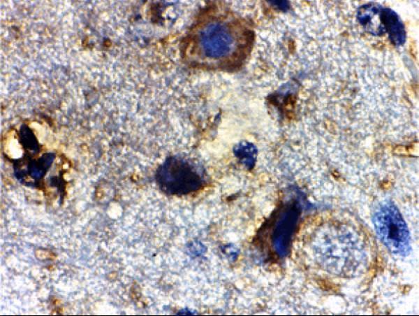
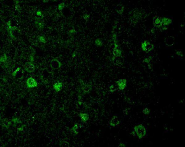
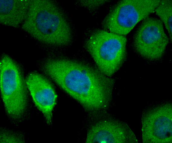
Anti-RHEB antibodies have mouse brain tissue IHC and mouse brain cell IF images (A00827-1 image captions), plus A431 ICC data (A00827-2 image caption); listed reactivity spans human, mouse, and rat (catalog reactivity).

Real IHC data Immunohistochemistry of Rheb in mouse brain tissue with Rheb antibody at 2 μg/mL.
Anti-GTP-binding protein Rheb Rheb Antibody
Cat # A00827-1
Real IF data ICC staining RHEB in A431 cells (green). The nuclear counter stain is DAPI (blue). Cells were fixed in paraformaldehyde, permeabilised with 0.25% Triton X100/PBS.
Anti-GTP-binding protein Rheb RHEB Antibody
Cat # A00827-2

A00827-1 shows mouse brain tissue IHC at 2 μg/mL and mouse brain cell IF at 20 μg/mL (A00827-1 image captions); human, mouse, and rat reactivity is listed (catalog reactivity). A00827-2 shows A431 ICC after paraformaldehyde fixation and Triton permeabilisation (A00827-2 image caption); IHC, IF, and ICC applications and human and mouse reactivity are listed (catalog applications; catalog reactivity).

Which to pick: For paraffin tissue IHC, choose A00827-1: IHC-P is listed and its own IHC image shows mouse brain tissue staining at 2 μg/mL (A00827-1 applications; A00827-1 IHC image caption); the tissue fixative is unreported (A00827-1 IHC image caption). For IF/ICC, A00827-2 offers an A431 ICC example with paraformaldehyde fixation and 0.25% Triton X100/PBS permeabilisation (A00827-2 image caption); its tissue IHC processing is unreported (catalog image alts). For work across the listed species, A00827-1 includes human, mouse, and rat, while A00827-2 includes human and mouse; both are rabbit antibodies with no clone reported (catalog reactivity; catalog host; catalog clone).

Each figure is that product's own IHC / IF validation image from its datasheet.

References

  1. UniProt Consortium. UniProt entry Q15382 (RHEB_HUMAN, GTP-binding protein Rheb).
  2. Human Protein Atlas. RHEB tissue IHC expression (reliability: Uncertain).
  3. Human Protein Atlas. RHEB subcellular location (ICC-IF): Highest expression in GAMG: 398.4 nTPM.
  4. Human Protein Atlas. RHEB antibody validation summary (1 antibodies).
  5. mTORC1 signaling in primary central nervous system lymphoma. Surgical neurology international 2016 — PMC4960920.
  6. miR-200a regulates Rheb-mediated amelioration of insulin resistance after duodenal-jejunal bypass. International journal of obesity (2005) 2016 — PMC4973218.
  7. Identification of autophagy-related immune targets for enhancing immunotherapy in pancreatic cancer aggressiveness. Discover oncology 2025 — PMC11933596.
  8. miR-199a-3p/5p regulate tumorgenesis via targeting Rheb in non-small cell lung cancer. International journal of biological sciences 2022 — PMC9274486.
  9. PubMed PMID:8543055 — UniProt-cited evidence.
  10. PubMed PMID:8661031 — UniProt-cited evidence.
  11. PubMed PMID:14702039 — UniProt-cited evidence.