RNF20 / E3 ubiquitin-protein ligase BRE1A · IHC design guide

Design Immunohistochemistry for RNF20

Plan chromogenic RNF20 IHC in paraffin sections around its widespread nuclear tissue pattern (HPA tissue IHC). High staining in adrenal glandular cells and cortical neurons offers positive tissue references (HPA tissue IHC).

Evidence assembled Oct 2026 · For research use; verify linked source records and product datasheet before use
Immunohistochemistry protocol sheet for RNF20 (IHC for RNF20): expected localisation Nuclear staining in tissue sections (HPA tissue IHC), antibody A03457, validated IHC image, and IHC protocol steps
Printable RNF20 IHC protocol sheet — expected localisation Nuclear staining in tissue sections (HPA tissue IHC), antibody A03457, controls and protocol steps. Open the full RNF20 IHC guide →

RNF20 Immunohistochemistry Experimental Design Guide

Expected localisation, validated protocols, controls and antibodies — the at-a-glance facts below, then the full design guide.

Must know before staining
Expected localisation Nuclear staining in tissue sections (HPA tissue IHC)
Staining pattern Widespread nuclear staining across cell types (HPA tissue IHC)
Antigen retrieval Tris-EDTA pH 9.0 HIER, 95–98 °C, 20 min (rule: nuclear antigen)
Positive control ⓘ Adrenal gland+4 more · see all
Negative control ⓘ None in HPA (detected in all 45 tissues); use no-primary + isotype controls
Important caveats
Reasons your staining may differ from the expected pattern.
Fixation Keep fixation conditions consistent across sections (standard IHC practice; not target-specific)
Caveat Liver cholangiocytes show low staining (HPA tissue IHC)
Regulation Low tissue specificity at the RNA level (HPA tissue RNA)
Isoform / epitope No isoforms annotated; no membrane-sided epitope issue (UniProt)
Section 1

Recommended RNF20 IHC & IF Protocols

The catalog antibody’s IHC-P protocol is accompanied by published RNF20 staining methods for breast tissue microarrays and cervical tumor sections (PMC5007379; PMC12210586).

Recommended immunohistochemistry (IHC-P) protocol parameters
SampleTissue sections; selected-image fixative not specified (standard IHC workflow)
FixationImage fixative and duration unreported (datasheet A03457); verify before use.
Sectioning4–5 µm sections on charged slides (standard)
DeparaffinisationXylene, graded ethanol series to water (standard)
Antigen retrievalHeat-induced epitope retrieval in Tris-EDTA buffer, pH 9.0, 20 min at 95–98 °C (standard rule: nuclear antigen)
Peroxidase block3% H2O2, 10 min, room temperature (standard)
Blocking10% normal serum of the secondary host, 30 min, room temperature (standard)
Primary antibodyRabbit anti-RNF20, 1:100–1:500 starting range (standard)
Primary incubationOvernight at 4 °C (standard)
DetectionHRP-polymer secondary, DAB chromogen 5–10 min (standard)
CounterstainHematoxylin, blue, dehydrate and mount (standard)
Expected resultRNF20-positive staining in glandular cells of adrenal gland (HPA tissue IHC: High). HPA tissue profile: Ubiquitous nuclear expression. No signal in the no-primary control.
💡Decision noteStart with Tris-EDTA pH 9.0 HIER at 95–98 °C for 20 min (page retrieval rule: nuclear RNF20); compare the published pH 6 conditions if needed (PMC5007379; PMC12210586).
Section 2

What Is the Expected RNF20 Staining Pattern?

RNF20 is a nuclear protein with no transmembrane segment (UniProt Q5VTR2: subcellular location and topology). In paraffin-section IHC, expect predominantly nuclear staining across many cell types (HPA tissue IHC: ubiquitous nuclear expression; reliability Supported, with medium consistency between staining and RNA data). High staining is reported in selected neuronal, epithelial and glandular cells (HPA tissue IHC: High in the listed cells); liver cholangiocytes are reported as Low (HPA tissue IHC).

What am I looking at on my slide?
Distinct nuclear stain in cerebellar Purkinje cells or cerebral cortical neurons.This fits the reported compartment and cell pattern (UniProt Q5VTR2: nucleus; HPA tissue IHC: High in Purkinje and cerebral cortical neuronal cells). Judge staining against nuclei on the same section; strong color outside the nucleus should not be counted as a nuclear positive (general IHC scoring practice).
Cytoplasmic color dominates while nuclei remain clear.This conflicts with the expected IHC pattern (HPA tissue IHC: ubiquitous nuclear expression; UniProt Q5VTR2: nucleus). Treat it as suspect until controls and detection background are checked (general IHC practice). The separately reported cytosolic IF location is uncertain and does not establish cytoplasmic IHC positivity (HPA subcellular ICC-IF: cytosol uncertain).
Strong color appears chiefly in cells other than the expected cells of a selected tissue.For example, glandular cells are reported High in colon and duodenum (HPA tissue IHC). A different dominant pattern merits review for cross-reactivity or endogenous detection activity (general IHC practice); it is not proof of either. RNF20 has broad nuclear expression, so staining in additional cell types alone does not make them false positives (HPA tissue IHC: ubiquitous nuclear expression).
Color spreads across nuclei, cytoplasm and tissue spaces with little separation.Diffuse color makes nuclear scoring unreliable (general IHC scoring practice). Examine a no-primary control for detection background and review blocking, washes and antibody concentration (general IHC practice). Do not infer a biological RNF20 distribution from diffuse chromogen (UniProt Q5VTR2: nucleus; HPA tissue IHC: ubiquitous nuclear expression).
Little or no nuclear signal appears in a listed High cell population.Check the staining run before calling RNF20 absent: the selected population is reported High, while tissue IHC reliability is Supported with medium RNA-to-staining consistency (HPA tissue IHC). Verify that the cells are present and review the validated IHC conditions and controls (general IHC practice). A failed run and a true low result cannot be separated by this slide alone.
💡Expected RNF20 appearanceCall a convincing IHC positive when identifiable nuclei stain in the selected High cell population, such as Purkinje cells or colonic glandular cells (HPA tissue IHC: High; ubiquitous nuclear expression); diffuse tissue color or dominant cytoplasmic color is a suspect positive (UniProt Q5VTR2: nucleus; general IHC scoring practice).
How each factor affects the staining
Tissue and cell choiceChoose a documented High population as a positive reference, such as adrenal glandular cells or cervical squamous epithelial cells (HPA tissue IHC: High). Liver cholangiocytes are Low, not a demonstrated negative control (HPA tissue IHC: Low in cholangiocytes; no negative populations listed).
Location and topologyScore RNF20 primarily by nuclear signal (UniProt Q5VTR2: nucleus; HPA tissue IHC: ubiquitous nuclear expression). No transmembrane segment is annotated, so membrane outlining has no support as the expected target pattern (UniProt Q5VTR2: topology).
Evidence strengthThe tissue IHC profile is Supported, with medium consistency between antibody staining and RNA expression (HPA tissue IHC). The listed antibody CAB012478 has Supported IHC validation; the supplied records do not assign IHC status to HPA029442 or HPA051773 (HPA antibodies). Do not describe this profile as Enhanced.
IF/ICC Q: Where should RNF20 appear?A: Mainly in the nucleoplasm (HPA subcellular ICC-IF: nucleoplasm enhanced). Additional cytosolic localization is marked uncertain (HPA subcellular ICC-IF); it should not override the nuclear expectation for paraffin-section IHC (HPA tissue IHC: ubiquitous nuclear expression).
Why is my staining missing, weak or wrong?
SituationLikely causeNext action
A documented High cell population gives no nuclear stain.The run may have insufficient detectable signal (general IHC practice); the population is reported High (HPA tissue IHC).Confirm the expected cells are present, inspect the run controls, then review the IHC-validated antibody's stated retrieval and dilution conditions if available (general IHC practice). No RNF20-specific retrieval requirement is supplied.
Nuclei stain weakly but surrounding color is strong.Background may obscure nuclear signal (general IHC practice); RNF20 is expected in nuclei (UniProt Q5VTR2: nucleus).Compare with a no-primary control and review blocking, washing and antibody concentration (general IHC practice). Score only nuclei that can be distinguished from background.
Cytoplasmic staining is the dominant pattern.The distribution conflicts with nuclear tissue IHC (HPA tissue IHC); cytosolic IF localization is uncertain (HPA subcellular ICC-IF).Check the no-primary control and compare nuclear morphology with the counterstain (general IHC practice). Do not use uncertain IF cytosol as confirmation of cytoplasmic IHC.
Staining centers on unexpected cells rather than the selected High population.Cross-reactivity or endogenous detection activity is possible (general IHC practice); the selected population has a reported High pattern (HPA tissue IHC).Verify cell identity and compare no-primary and positive-reference sections (general IHC practice). Because expression is broadly nuclear, assess compartment and pattern before dismissing additional stained cells (HPA tissue IHC).
Chromogen appears even in the no-primary control.The signal is independent of primary antibody; endogenous detection activity or reagent background is possible (general IHC practice).Review the detection system's endogenous-activity block and reagent controls, then repeat with a clean no-primary control (general IHC practice). Do not score control-associated color as RNF20.
Liver cholangiocytes are faint while other nuclei stain.Cholangiocytes are reported Low; faint staining alone need not indicate a failed run (HPA tissue IHC: Low in liver cholangiocytes).Assess a listed High cell population and the run controls before changing conditions (HPA tissue IHC: listed High populations; general IHC practice). Do not use cholangiocytes as an established negative control.

Sample controls for RNF20 IHC & IF

🧪Run colon first and expect nuclear staining in glandular cells (HPA: High in colon glandular cells; UniProt Q5VTR2: nucleus). HPA detects RNF20 in all 45 scored tissues, so there is no documented negative tissue; use no-primary and isotype controls, and expect any unstained cells on the positive slide to show counterstain alone without treating them as proven RNF20-negative cells (HPA: no negative rows; standard IHC practice).
Positive control tissue: Adrenal gland (Glandular cells, HPA High)
Negative control tissue: None in HPA: RNF20 is detected in all 45 scored tissues. Use a no-primary (secondary-only) and an isotype control instead.
ICC-IF cell lines (HPA subcellular resource): HPA ICC-IF images show RNF20 in A-431, U-251MG, U2OS, SiHa, KOLF2.1J, NIH 3T3, with annotated localisation: Nucleoplasm (enhanced) (HPA subcellular).
Technical controls: Include a no-primary, secondary-only control and an isotype control matched to the primary antibody’s host species and clonality, plus RNF20 knockout material or a validated peptide-block control if available (standard IHC practice). Block endogenous peroxidase and check blood or inflammatory cells in colon for chromogen background (standard chromogenic IHC practice).
⚠️Feasibility: No target-specific fixation window, fixation effect, or retrieval dependency is reported in the supplied evidence; the selected A03457 liver IHC caption does not state a fixative (selected-SKU caption: fixative unreported). The supplied evidence does not establish that frozen sections or IF are easier than paraffin IHC (supplied application evidence: IHC-P protocol; HPA: ICC-IF images). In colon, blood or inflammatory cells can contribute endogenous peroxidase background, so interpret glandular nuclear staining against the controls (standard chromogenic IHC practice; HPA: High in colon glandular cells).

HPA tissue IHC evidence for RNF20

Comprehensive Human Protein Atlas IHC scoring per tissue (reliability: Supported — Medium consistency between antibody staining and RNA expression data.). Rows are taken directly from the HPA tissue chart — click any row's HPA link to view the source.

Positive expression · recommended positive controls

TissueCell typeLevelEvidenceSource
Adrenal gland Glandular cells High Protein (IHC) HPA →
Cerebellum Purkinje cells High Protein (IHC) HPA →
Cerebral cortex Neuronal cells High Protein (IHC) HPA →
Cervix Squamous epithelial cells High Protein (IHC) HPA →
Colon Glandular cells High Protein (IHC) HPA →

Undetected expression · recommended negative controls

TissueCell typeLevelEvidenceSource
None in HPA: RNF20 is detected in all 45 scored tissues. Use a no-primary (secondary-only) and an isotype control instead.
Section 3

Advanced RNF20 IHC Tips

Troubleshoot RNF20 staining in paraffin section IHC by checking retrieval, nuclear localisation, controls and scoring before interpreting differences in signal.

How should I retrieve RNF20 when nuclear staining is weak?
Start with Tris-EDTA at pH 9.0, heated to 95–98 °C for 20 min (page retrieval rule: nuclear antigen). Allow slides to cool consistently, then compare the same tissue and antibody concentration across runs (standard IHC practice). The selected antibody has a human liver IHC image at 2.5 µg/ml, but its caption does not report fixation or retrieval conditions (catalog antibody A03457 caption). If nuclear signal remains weak, trial a milder retrieval condition on matched sections while monitoring tissue damage and background (standard IHC practice). Judge improvement by nuclear signal and preserved morphology, rather than total brown staining (UniProt Q5VTR2 localisation; standard IHC practice).
Could fixation explain variable RNF20 staining between paraffin blocks?
RNF20-specific fixation sensitivity is unknown from the supplied evidence; the selected tissue IHC caption does not state a fixative (catalog antibody A03457 caption). Record fixation and processing history for each block, then compare matched tissue regions processed under the same conditions (standard IHC practice). Variable fixation can alter antigen accessibility and morphology in paraffin IHC, so keep retrieval at Tris-EDTA pH 9.0, 95–98 °C for 20 min during an initial comparison (standard IHC practice; page retrieval rule). Include a reference section in each run and inspect nuclear staining alongside tissue preservation (UniProt Q5VTR2 localisation; standard IHC practice). Do not infer RNF20 fixation sensitivity from its modifications or tissue distribution (UniProt Q5VTR2; HPA tissue IHC).
What staining compartment should count as RNF20 positive?
Score convincing nuclear staining first because RNF20 is annotated as nuclear, and tissue IHC shows ubiquitous nuclear expression (UniProt Q5VTR2 localisation; HPA tissue IHC). HPA places the strongest subcellular signal in the nucleoplasm and labels additional cytosolic localisation uncertain (HPA subcellular). On chromogenic sections, compare nuclear DAB with the counterstain at high magnification and exclude pigment or precipitate that does not follow cell boundaries (standard IHC practice). A predominantly cytoplasmic pattern warrants a review of antibody concentration, retrieval, detection background and tissue preservation before biological interpretation (HPA subcellular; standard IHC practice). Use matched controls when deciding whether faint extranuclear colour is reproducible (standard IHC practice).
Could an isoform or altered epitope explain patchy nuclear staining?
The supplied RNF20 record lists 0 isoforms, so it provides no basis to assign different IHC patterns to specific isoforms (UniProt Q5VTR2 isoforms). Its epitope is not specified here; do not claim that a particular residue is recognised or blocked (supplied catalog antibody evidence). RNF20 has annotated acetylated lysines and phosphorylated serines, but their effect on this antibody's tissue staining is unknown (UniProt Q5VTR2 modified residues). Compare matched sections with consistent retrieval and antibody concentration, then assess whether patchiness follows tissue processing, section folds or viable cell boundaries (standard IHC practice). Treat a reproducible nuclear pattern as evidence of staining, not proof of an epitope mechanism (UniProt Q5VTR2 localisation; standard IHC practice).
How can IF help assess the RNF20 pattern seen by chromogenic IHC?
Use IF as a separate validation experiment and compare its nuclear RNF20 pattern with the chromogenic section, rather than transferring an IF protocol to IHC (UniProt Q5VTR2 localisation; standard IHC practice). Multiplex with a marker for an expected cell population, such as Purkinje cells in cerebellum, and include a nuclear counterstain (HPA tissue IHC; standard IF practice). Choose spectrally separated fluorophores after checking the tissue's autofluorescence and single-label controls (standard IF practice). RNF20 has no transmembrane segment and is mainly nucleoplasmic, so permeabilise fixed cells or sections sufficiently to access the intracellular nuclear epitope (UniProt Q5VTR2 topology; HPA subcellular; standard IF practice). Compare localisation within identified cells, not fluorescence intensity directly with DAB density (standard IF/IHC practice).
How do I reduce diffuse brown staining without losing nuclear RNF20 signal?
First distinguish nuclear signal from diffuse DAB, pigment and edge staining by examining intact cells away from damaged margins (UniProt Q5VTR2 localisation; standard IHC practice). Use a no-primary control to assess detection background and apply a peroxidase block before chromogenic detection (standard IHC practice). If background persists, titrate the primary antibody and optimise blocking and washes on adjacent sections, keeping retrieval and detection time constant (standard IHC practice). The selected liver IHC caption reports 2.5 µg/ml, but does not establish that concentration for every paraffin specimen (catalog antibody A03457 caption). Preserve interpretable nuclear contrast while checking that control sections remain clean (standard IHC practice).
How should I quantify RNF20 IHC across specimens? ⚠ ANSWER MARKED FOR VERIFICATION
Define the scored cell population and region before comparing specimens, because RNF20 tissue IHC is described as ubiquitously nuclear (HPA tissue IHC). Report the percentage of positive nuclei and, when intensity is reliable, an H-score using intensity categories 0–3 across those nuclei (standard IHC scoring practice). Normalise counts to the total evaluable nuclei in the same compartment, or report positive nuclei per mm² with the sampled area stated (standard quantitative IHC practice). Exclude necrotic, folded and edge-damaged regions using the same rules for every section (standard IHC practice). Keep illumination, colour separation and positivity thresholds consistent, and review representative fields manually (standard digital pathology practice).
When is an apparent RNF20 positive result likely to be artefact?
A credible result follows intact nuclei: UniProt places RNF20 in the nucleus, and HPA tissue IHC reports ubiquitous nuclear expression (UniProt Q5VTR2 localisation; HPA tissue IHC). Treat isolated cytoplasmic colour cautiously because HPA calls additional cytosolic localisation uncertain (HPA subcellular). Check whether unexpected cells stain consistently in intact regions; low staining in liver cholangiocytes is reported, but no negative tissue is listed (HPA tissue IHC). Signal confined to section edges, necrosis or folded tissue suggests a processing artefact, while colour in a no-primary control suggests endogenous enzyme or detection background (standard IHC practice). Confirm interpretation on an adjacent section with preserved morphology and appropriate controls (standard IHC practice).
Boster reagents

Best RNF20 / E3 ubiquitin-protein ligase BRE1A IHC Antibodies

RNF20 antibodies have IHC data from human liver tissue and IF data from human liver tissue or cultured cells (catalog image captions); listed reactivity covers human, mouse, and rat (catalog reactivity).

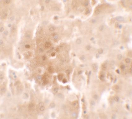
Real IHC data Immunohistochemistry of RNF20 in human liver tissue with RNF20 antibody at 2.5 μg/ml.
Anti-RNF20 Antibody
Cat # A03457
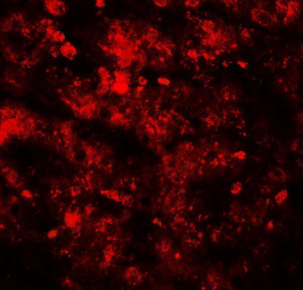
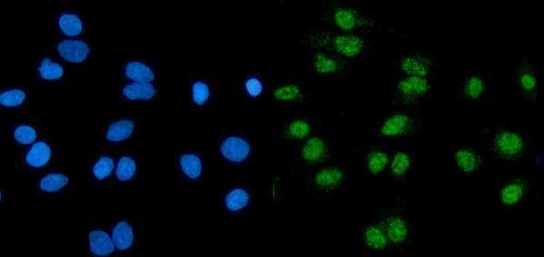
Real IF data IF analysis of RNF20 using anti-RNF20 antibody (A03457-1). RNF20 was detected in an immunocytochemical section of A549 cells. Enzyme antigen retrieval was performed using IHC enzyme antigen retrieval reagent (AR0022) for 15 mins. The cells were blocked with 10% goat serum. And then incubated with 5 μg/mL rabbit anti-RNF20 Antibody (A03457-1) overnight at 4°C. DyLight®488 Conjugated Goat Anti-Rabbit IgG (BA1127) was used as secondary antibody at 1:100 dilution and incubated for 30 minutes at 37°C. The section was counterstained with DAPI. Visualize using a fluorescence microscope and filter sets appropriate for the label used.
Anti-RNF20 Antibody ®
Cat # A03457-1
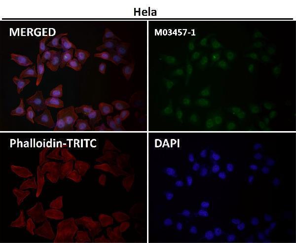
Real IF data Immunofluorescent analysis using the Antibody at 1:50 dilution.
Anti-RNF20 Rabbit Monoclonal Antibody
Cat # M03457-1

A03457 has a human liver IHC image at 2.5 μg/ml and lists IHC-P and IF applications (catalog image caption; catalog applications). A03457-1 has IF data in A549 cells at 5 μg/ml, while M03457-1 has IF data at 1:50 with no sample identified in its caption (catalog image captions).

Which to pick: Choose A03457 for paraffin-section IHC: IHC-P is listed, and its own image shows human liver tissue; the fixative is unreported (catalog applications; A03457 IHC image caption). For IF/ICC, A03457-1 has an A549-cell IF image and lists human and rat reactivity; M03457-1 is a rabbit monoclonal with an IF image and lists human and mouse reactivity (catalog image captions; catalog titles; catalog applications; catalog reactivity). For human and mouse IHC planning, A03457 lists both species and IHC-P, but its IHC image documents human tissue only (catalog reactivity; catalog applications; A03457 IHC image caption).

Each figure is that product's own IHC / IF validation image from its datasheet.

References

  1. UniProt Consortium. UniProt entry Q5VTR2 (BRE1A_HUMAN, E3 ubiquitin-protein ligase BRE1A).
  2. Human Protein Atlas. RNF20 tissue IHC expression (reliability: Supported).
  3. Human Protein Atlas. RNF20 subcellular location (ICC-IF): Mainly localized to the nucleoplasm. In addition localized to the cytosol..
  4. Human Protein Atlas. RNF20 antibody validation summary (3 antibodies).
  5. Ubiquitin ligase RNF20/40 facilitates spindle assembly and promotes breast carcinogenesis through stabilizing motor protein Eg5. Nature communications 2016 — PMC5007379.
  6. RNF20 links the DNA damage response and metabolic rewiring in lung cancer through HIF1α. Nature communications 2025 — PMC12120090.
  7. RNF20/RNF40 supports the aggressive behavior in cervical cancer by regulating a peroxisome-based anti-ferroptotic mechanism. Cell communication and signaling : CCS 2025 — PMC12210586.
  8. H2B ubiquitination regulates meiotic recombination by promoting chromatin relaxation. Nucleic acids research 2016 — PMC5175339.
  9. PubMed PMID:14702039 — UniProt-cited evidence.
  10. PubMed PMID:15164053 — UniProt-cited evidence.
  11. PubMed PMID:15489334 — UniProt-cited evidence.