This website uses cookies to ensure you get the best experience on our website.


- Table of Contents
Plan chromogenic IHC for RPS3 in paraffin sections using the catalog antibody’s documented conditions (datasheet A01542-2). Compare staining with the generally cytoplasmic tissue pattern while accounting for the HPA assessment of uncertain reliability (HPA tissue IHC).

Expected localisation, validated protocols, controls and antibodies — the at-a-glance facts below, then the full design guide.
| Expected localisation | Predominantly cytoplasmic in tissue sections (HPA tissue IHC) | |
| Staining pattern | General cytoplasmic staining across tissues (HPA tissue IHC) | |
| Antigen retrieval | Citrate pH 6 HIER, heat-mediated (datasheet A01542-2) | |
| Positive control | Cerebellum+4 more · see all | |
| Negative control | Adipose tissue+4 more · see all |
| Fixation | Matched tissue-IHC evidence does not establish the fixation claim. Validate the specimen-specific method before use. (selected-SKU IHC image A01542-2) | |
| Caveat | Tissue staining has uncertain RNA concordance (HPA tissue IHC) | |
| Regulation | DNA damage can increase nuclear staining (UniProt) | |
| Isoform / epitope | Two isoforms; check antibody epitope coverage (UniProt) |
Compare the catalog antibody’s citrate pH 6 IHC protocol (datasheet: A01542-2) with published RPS3 IHC protocols (PMC6393244; PMC9280127).
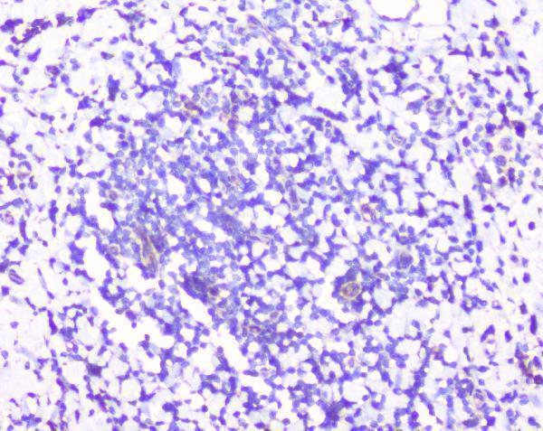
| Sample | Paraffin-embedded human tonsil tissues; fixative not specified (datasheet A01542-2) |
| Fixation | Image fixative and duration unreported (datasheet A01542-2); verify before use. |
| Sectioning | 4–5 µm sections on charged slides (standard) |
| Deparaffinisation | Xylene, graded ethanol series to water (standard) |
| Antigen retrieval | Heat retrieval: Citrate pH 6, 20 min (datasheet A01542-2) |
| Peroxidase block | 3% H2O2, 10 min, room temperature (standard) |
| Blocking | 10% goat serum (datasheet A01542-2) |
| Primary antibody | Rabbit anti-RPS3, 0.5-1μg/ml (datasheet A01542-2) |
| Primary incubation | Overnight at 4 °C (datasheet A01542-2) |
| Detection | Streptavidin-biotin complex (SABC), DAB chromogen (datasheet A01542-2) |
| Counterstain | Hematoxylin, blue, dehydrate and mount (standard) |
| Expected result | RPS3-positive staining in purkinje cells of cerebellum (HPA tissue IHC: High). HPA tissue profile: General cytoplasmic expression. No signal in the no-primary control. |
RPS3 is mainly cytoplasmic in normal cells, with smaller nuclear amounts; it has no transmembrane segment (UniProt P23396). In paraffin sections, expect cytoplasmic staining in several cell types, including cerebellar Purkinje cells, cortical neurons, and esophageal squamous epithelial cells (HPA tissue IHC). Treat tissue comparisons as guides rather than fixed cutoffs: HPA rates the tissue IHC profile Uncertain because antibody staining and RNA expression show medium consistency (HPA tissue IHC).
| Clear cytoplasmic staining in Purkinje cells or cortical neurons, with readable tissue structure. | This fits HPA's High staining in those cells and its general cytoplasmic profile (HPA tissue IHC). Compare the signal with neighboring cells and the negative control on the same staining run (standard IHC practice). |
| Predominantly nuclear staining in otherwise normal tissue, with little cytoplasmic signal. | Review this pattern before calling it RPS3: normal cells contain mainly cytoplasmic RPS3, though some nuclear signal is possible (UniProt P23396). Nuclear accumulation can accompany apoptosis or DNA damage, so the compartment alone cannot establish an artefact (UniProt P23396). |
| Strong staining in adipocytes or skeletal muscle myocytes while expected positive cells are weak. | HPA reports RPS3 as Not detected in those cell types (HPA tissue IHC). Such staining raises concern for cross-reactivity or endogenous detection activity (standard IHC practice); HPA's Uncertain tissue reliability means these observations are checks, not absolute exclusions (HPA tissue IHC). |
| Diffuse color covers cells and extracellular spaces, obscuring cell boundaries. | That distribution is difficult to score as the general cytoplasmic expression reported by HPA (HPA tissue IHC). Review blocking, washing, detection reagents, and the negative control for nonspecific background or endogenous activity (standard IHC practice). |
| No signal in a section containing identifiable Purkinje cells, cortical neurons, or esophageal squamous epithelial cells. | These cells show High staining in HPA tissue IHC, so their absence of signal calls for a run-level check (HPA tissue IHC). Verify tissue identity, reagent performance, and the catalog antibody's IHC-P instructions before interpreting other negative cells (standard IHC practice). |
| Tissue and cell identity (HPA tissue IHC) | HPA reports High staining in Purkinje cells, cortical and hippocampal neurons, cervical and salivary glandular cells, and esophageal and oral squamous epithelial cells; it reports Not detected in adipocytes and skeletal muscle myocytes (HPA tissue IHC). Score the named cells, not an entire tissue as uniformly positive. |
| Strength of the tissue evidence (HPA tissue IHC) | HPA describes general cytoplasmic expression but rates the tissue IHC profile Uncertain, citing medium consistency between antibody staining and RNA expression; its listed antibody, HPA063339, is also Uncertain for IHC (HPA tissue IHC; HPA antibodies). Use the pattern as an expectation requiring controls. |
| Cell state and compartment (UniProt P23396) | RPS3 is mainly cytoplasmic in normal cells, can translocate to nuclei during apoptosis or after DNA damage, and can accumulate in mitochondria with increased ROS (UniProt P23396). These reported changes qualify a compartment call; a nuclear or punctate result needs cellular context before interpretation. |
| Protein features versus assay behavior (UniProt P23396) | RPS3 has 2 isoforms, a KH type-2 domain, reported modified residues, and no transmembrane segment (UniProt P23396). These features do not identify the catalog antibody's epitope or establish isoform selectivity or retrieval requirements; assess those properties from antibody-specific validation. |
| Fixation evidence (HPA tissue IHC; UniProt P23396) | Target-specific fixation effects are not established by the supplied assay evidence. Verify with a matched IHC source before attributing a result to fixation. |
| Situation | Likely cause | Next action |
|---|---|---|
| Expected positive cells are blank. | A run or detection failure is possible when cells reported as High by HPA are unstained (HPA tissue IHC; standard IHC practice). | Confirm cell identity, inspect the run control, and check retrieval, antibody dilution, and detection against the catalog antibody's IHC-P instructions (standard IHC practice). No RPS3-specific retrieval condition is supplied. |
| Signal appears mostly nuclear. | Normal RPS3 is mainly cytoplasmic, but nuclear translocation is reported with apoptosis or DNA damage (UniProt P23396). | Examine cell morphology and cytoplasmic signal, then compare a suitable control section processed in the same run (standard IHC practice). Avoid assigning a cause to nuclear staining from compartment alone. |
| Strong color appears in HPA Not detected cells. | Cross-reactivity or endogenous detection activity may contribute (standard IHC practice); HPA's tissue profile is Uncertain (HPA tissue IHC). | Check a negative reagent control and compare staining in HPA High cells on the run (standard IHC practice; HPA tissue IHC). Treat any disagreement as a validation question rather than automatic proof of absence. |
| Diffuse background makes cytoplasmic scoring difficult. | Incomplete blocking, washing, or control of endogenous detection activity can produce background in chromogenic IHC (standard IHC practice). | Review the negative control, blocking and washing steps, and detection reagents; score only signal resolved within identifiable cells (standard IHC practice). HPA describes a general cytoplasmic pattern (HPA tissue IHC). |
| Staining differs markedly between tissue regions. | Cell composition matters: HPA lists High, Low, and Not detected results for different named cells (HPA tissue IHC). | Identify and score the relevant cell type in each region, then compare sections within the same run (standard IHC practice). Do not apply one intensity threshold to every cell type in the section. |
| Q: Should IF/ICC show the same compartment pattern? | HPA ICC-IF reports supported localization to the cytosol and endoplasmic reticulum, while the tissue IHC profile is Uncertain (HPA ICC-IF; HPA tissue IHC). | A: Use the cytosol and endoplasmic reticulum observation to interpret IF/ICC images (HPA ICC-IF); evaluate chromogenic paraffin sections against their own cell-specific IHC pattern and controls (HPA tissue IHC; standard IHC practice). |


Comprehensive Human Protein Atlas IHC scoring per tissue (reliability: Uncertain — Medium consistency between antibody staining and RNA expression data.). Rows are taken directly from the HPA tissue chart — click any row's HPA link to view the source.
| Tissue | Cell type | Level | Evidence | Source |
|---|---|---|---|---|
| Cerebellum | Purkinje cells | High | Protein (IHC) | HPA → |
| Cerebral cortex | Neuronal cells | High | Protein (IHC) | HPA → |
| Cervix | Glandular cells | High | Protein (IHC) | HPA → |
| Esophagus | Squamous epithelial cells | High | Protein (IHC) | HPA → |
| Hippocampus | Neuronal cells | High | Protein (IHC) | HPA → |
| Tissue | Cell type | Level | Evidence | Source |
|---|---|---|---|---|
| Adipose tissue | Adipocytes | Not detected | Protein (IHC) | HPA → |
| Ovary | Follicle cells | Not detected | Protein (IHC) | HPA → |
| Skeletal muscle | Myocytes | Not detected | Protein (IHC) | HPA → |
| Smooth muscle | Smooth muscle cells | Not detected | Protein (IHC) | HPA → |
| Soft tissue | Chondrocytes | Not detected | Protein (IHC) | HPA → |
Use the matched paraffin-section protocol as the starting point, then interpret RPS3 staining by compartment and cell type (datasheet A01542-2; UniProt P23396; HPA tissue IHC).
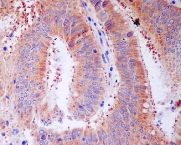
Anti-RPS3 antibodies have paraffin-section IHC images from human tonsil, placenta and colon cancer, and mouse brain (catalog image captions). Both list IF/ICC applications, but neither has an IF image (catalog).
A01542-2 lists IHC and IF/ICC applications and human, mouse and rat reactivity; its IHC captions show paraffin sections of human tonsil and placenta and mouse brain (catalog). M01542 lists the same applications and species reactivity; its IHC caption shows paraffin-embedded human colon cancer (catalog).
Which to pick: For tissue IHC, choose A01542-2 for its documented citrate pH 6 retrieval on paraffin sections; choose M01542 if its rabbit monoclonal format and human colon cancer example suit the study (A01542-2 image captions; M01542 catalog and image caption). Both list IF/ICC, though neither supplies an IF image; the listed IF dilutions are 2 μg/ml for A01542-2 and 1:50 for M01542 (catalog). Both list human, mouse and rat reactivity, but A01542-2 has the mouse tissue IHC example; neither product’s IHC caption reports the fixative (catalog; image captions).